Luís Pedro Gafeira Gonçalves
Dissertation presented to obtain the Ph.D degree in Biochemistry
Instituto de Tecnologia Química e Biológica | Universidade Nova de Lisboa
Oeiras,
Osmo- and thermo-adaptation
in hyperthermophilic Archaea:
Subtitle
Subtitle
O OH O O OH OH P O O--O 3P O OH O O OH OH O OH OH P O O--O 3Pc c c
OH OH OH
CDP c c c
OH OH OH
CDP
C C C
OH OH OH
O
O
P O
O
-C C C
OH OH OH
O O P O O -CTP PPi CTP PPi CMP CMP O OH OH O OH OH P
O O
-O OH OH O OH OH O OH OH O OH OH P
O O
-O OH O O OH OH P
O O
--O 3P O OH O O OH OH O OH OH P
O O
--O 3P
c
c
c
OH OH OH
CDP
c
c
c
OH OH OH
CDP
Instituto de Tecnologia Química e Biológica
Osmo- and thermo-adaptation in
hyperthermophilic
Archaea
:
identification of compatible solutes, accumulation
profiles, and biosynthetic routes in Archaeoglobus
spp.
Luís Pedro Gafeira Gonçalves
C C C
OH OH OH
O
O
P O
O
-C C C
OH OH OH
O O P O O -CTP PPi CTP PPi CMP CMP Pi Pi O OH OH O OH OH P
O O
-O OH OH O OH OH O OH OH O OH OH P
O O
-O OH O O OH OH P
O O
--O 3P O OH O O OH OH O OH OH P
O O
--O 3P
c
c
c
OH OH OH
CDP
c
c
c
OH OH OH
CDP
Instituto de Tecnologia Química e Biológica
Osmo- and thermo-adaptation in
hyperthermophilic
Archaea
:
identification of compatible solutes, accumulation
profiles, and biosynthetic routes in Archaeoglobus
spp.
Luís Pedro Gafeira Gonçalves
C C C
OH OH OH
O
O
P O
O
-C C C
OH OH OH
I nst it ut o de Tecnologia Quím ica e Biológica
Osm o- and t herm o- adapt at ion in hypert herm ophilic
Archaea
: ident ificat ion of com pat ible solut es,
accumulation profiles, and biosynthetic routes in
Archaeoglobus
spp.
This dissertation was presented to obtain a Ph. D. degree in Biochemistry
at the Instituto de Tecnologia Química e Biológica, Universidade Nova de Lisboa.
By
Luís Pedro Gaf eira Gonçalves
Supervised by
Prof. Dr. Helena Sant os
Ap o io fina nc e iro d a Fund a ç ã o p a ra a C iê nc ia e Te c no lo g ia (PO C I 2010 – Fo rma ç ã o
Ava nç a d a p a ra a C iê nc ia – Me d id a IV.3) e FSE no â mb ito d o Q ua d ro C o munitá rio d e
The work presented in this thesis, would not have been possible without the help, in terms of time and knowledge, of many people, to whom I am extremely grateful.
Firstly and mostly, I need to thank my supervisor, Prof. Helena Santos, for her way of thinking science, her knowledge, her rigorous criticism, and her commitment to science. Her attitude as a scientist taught me what a scientist should be. Her investment, with time and knowledge, together with her ability of providing financial support, allowed the fulfilment of this work.
To Dr. Paula Fareleira, who was always available with an enthusiastic word to say, for her deep knowledge about the tricky Archaea and her priceless metabolic maps, and above all for her friendship, which was absolutely essential for the completion of this thesis.
To Prof. R. Huber of Regensburg University, who provided us with some of the organisms studied in the present work, I thank his scientific interest in my work.
To Prof. Milton da Costa, I thank his interest in my work and our stimulating and useful scientific discussions.
To Dr. Nuno Borges, for his availability to help, his concern with the evolution of the work and his critic spirit; his support was essential for this thesis.
To Dr. Pedro Lamosa, for his NMR knowledge and his availability to teach and help; his fluent way of thinking was extremely important for this work.
To Isabel Pacheco e Ana Mingote, I thank their technical support in purification procedures and for teaching me some of their expertise.
To Marta Rodrigues, for her youth, working capability and her continuous search for new challenges, that was of great help in this work.
logistic and informatics support, and their friendship and joy.
To Fundação para Ciência e Tecnologia for the financial support provided by the PhD grant, and to the Instituto de Tecnologia Química e Biólogica, for providing conditions to pursue scientific excellence. I wish it can continue as dreamt by his relentless creator, Prof. António Xavier.
Ao Nuno, Pedro, Tiago e Paula, pois algumas das minhas mais gratas recordações neste longo caminho foram no Beer Hunter ou num jantar de quarta-feira.
Aos meus amigos pois sem eles a vida não tinha o mesmo significado, temos de nos encontrar nem que seja no Biotretas. Em especial ao Vítor, pelos quase vinte anos de amizade; ao João, que me acompanhou nos primórdios desta jornada; e as meninas do IGC, Sofia e Patrícia, por tornarem os dias mais alegres.
À minha família, obrigado por todo o amor. Joana, apesar da distância e ausência de telefonemas estás sempre no meu coração. Mãe e Pai, obrigado.
À minha família em construção. O amor incondicional do Freud. Inês, o teu sorriso vale uma vida inteira. Jacinta, pelo teu amor e pela nossa vida junta, que me faz uma melhor pessoa.
Hyperthermophilic organisms have optimum growth temperatures above 80°C and belong to genera that are placed near the root of the Tree of Life, in short phylogenetic branches within the domains Bacteria or Archaea. Although hyperthermophiles have been isolated from a variety of hot environments, most species originate from marine geothermal areas, hence they are slightly halophilic. The accumulation of low-molecular mass organic solutes, i. e., compatible solutes, is one of the most common strategies developed by cells to cope with fluctuations of the salinity of the medium. Interestingly, in marine hyperthermophiles, compatible solute accumulation occurs not only in response to an increase in the external salt concentration, but also in response to supraoptimal growth temperatures. Moreover, microorganisms adapted to grow optimally at elevated temperatures tend to use negatively charged solutes that are not present or rarely encountered in mesophilic organisms.
In this work, the organic solute pool of several members of the genus Archaeoglobus as well as of the extreme hyperthermophile Pyrolobus fumarii was investigated by Nuclear Magnetic Resonance (NMR). The profiles of solute accumulation as a function of the growth temperature and the NaCl concentration were determined; moreover, the pathways for the synthesis of two major compatible solutes, diglycerol phosphate and mannosylglycerate, were elucidated.
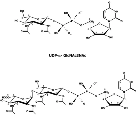
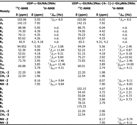
Pyrolobus fumarii belongs to the crenarchaeote branch of the domain Archaea. It is one of the most hyperthermophilic organisms known, being able to grow at temperatures up to 113°C. P. fumarii accumulates di-myo-inositol phosphate as the major solute (0.21 μmol/mg protein), a finding that reinforces the correlation between di-myo-inositol phosphate accumulation and hyperthermophily. The ethanol extracts of P. fumarii cells also contain several UDP-sugars (total concentration 0.11 μmol/mg protein). The structures of the two major UDP-sugars were identified as UDP-α-GlcNAc3NAc and UDP-α-GlcNAc3NAc-(4←1)-β -GlcpNAc3NAc. Interestingly, the latter compound appears to be derived from the first one by addition of a 2,3-N-diacetylglucoronic acid unit, suggesting that these UDP-sugars are intermediates of an N-linked glycosylation pathway.
and shifting the higher limit for growth from 4.5% to 6.3% NaCl. The optimal NaCl concentration was, however, the same for both strains, around 1.8% NaCl. Surprisingly, the total pool of compatible solutes was lower than in the parental strain, but increased notably at salinities above 4.5% NaCl, reaching a value of 3.34 μmol/mg protein at 6.0% NaCl. In response to salt stress, A. fulgidus VC-16S accumulated mainly diglycerol phosphate, an osmolyte thus far confined to the Archaeoglobales. Furthermore, the level of diglycerol phosphate increased approximately 28-fold in cells grown in medium containing 6.0% NaCl, when compared with cells grown at low salinity (0.9% NaCl). At supra-optimal growth temperatures, di-myo-inositol phosphate was the predominant solute, increasing 11-fold from the optimal to the maximal growth temperature (83°C to 89°C). At the highest temperature examined, di-myo-inositol phosphate and diglycerol phosphate represented 75% (molar percentage) of the total solute pool.
An unknown solute, initially designated as “unknown di-myo-inositol phosphate isomer” was present in low amounts in extracts of strains A. fulgidus VC-16 and VC-16S. The structure of this compound was firmly identified as glycero-phospho-myo-inositol by using a combination of NMR and Mass Spectrometry. Thus far, this newly discovered solute has been found only in A. fulgidus (strains VC-16, VC-16S and 7324) and in two hyperthermophilic bacteria of the genus Aquifex (Aquifex aeolicus and Aquifex pyrophilus). The level of glycerol-phospho-myo-inositol responded primarily to the combination of supra-optimal temperature and supra-optimal NaCl concentration. The remarkable structural relationship between diglycerol phosphate, di-myo-inositol phosphate, and the newly identified solute is disclosed in this work. The unit glycero-phospho-myo-inositol is part of the polar head in phosphatidylinositol, an important constituent of lipid membranes, but this is the first report on the occurrence of glycero-phospho-myo-inositol as a cell metabolite.
phosphate, mannosylglycerate and α-glutamate, and a similar set of solutes was observed in extracts of A. profundus, except that diglycerol phosphate was absent.
In summary, the major solutes found in members of the genus Archaeoglobus are di-myo-inositol phosphate, diglycerol phosphate and mannosylglycerate. Di-myo-inositol phosphate was found in all the strains examined; diglycerol phosphate was also a common solute being present in all strains except for A. profundus; mannosylglycerate accumulated in A. profundus, A. veneficus and A. fulgidus 7324, but was not detected in A. fulgidus VC-16 and VC-16S. The newly identified compound, glycero-phospho-myo-inositol, has only been found in the strains of the species A.fulgidus.
The pathways for the synthesis of mannosylglycerate and diglycerol phosphate were established based on the detection of relevant enzymatic activities in cell extracts. The synthesis of mannosylglycerate involved the condensation of GDP-mannose with D-3-phosphoglycerate by the action of mannosyl-3-phosphoglycerate synthase (MPGS) to form a phosphorylated intermediate, mannosyl-3-phosphoglycerate (MPG), which was subsequently dephosphorylated by a specific phosphatase, mannosyl-3-phosphoglycerate phosphatase (MPGP). Using degenerated PCR and inverse PCR, it was possible to identify two contiguous open reading frames homologous to mpgS (encoding MPGS) and mpgP (encoding MPGP) in A. profundus and A. veneficus. Downstream of these genes, an open reading frame coding for a putative mannosidase was found. This organization of the mannosylglycerate gene cluster, comprising a putative mannosidase, has not been observed elsewhere. The mpgS and mpgP genes of A. fulgidus 7324 have been partially sequenced.
The synthesis of diglycerol phosphate in A. fulgidus strains VC-16 and 7324was also elucidated: the enzymatic activities were detected in cell extracts and the intermediate metabolite was characterized by NMR. Addition of glycerol 3-phosphate and CTP to cell-free extracts led to the formation of diglycerol phosphate; a transient accumulation of CDP-glycerol was also observed. Maximum diglycerol phosphate production was obtained when glycerol 3-phosphate and CDP-glycerol were used as substrates. It was verified that glycerol and other nucleotides (ATP, UTP and GTP) were not precursors for the synthesis of CDP-glycerol and diglycerol phosphate.
and the intensity of the diglycerol phosphate resonance increased by the corresponding amount. After purification using column chromatography this new metabolite was identified as diglycerol phosphate phosphate by 2D-NMR techniques.
Os organismos hipertermófilos apresentam temperaturas óptimas de crescimento acima dos 80°C e pertencem a géneros dos domínios Bacteria e Archaea, que estão dispostos em ramos curtos, próximo da origem da Árvore da Vida. Apesar destes organismos terem sido isolados a partir de uma grande variedade de biótopos quentes, a maioria das espécies hipertermofílicas conhecidas foi isolada de áreas geotermais marinhas, apresentando portanto, halotolerância ou halofilia moderada. A acumulação de solutos orgânicos de baixa massa molecular, i. e., solutos compatíveis, é uma das estratégias celulares mais comuns para fazer face a variações da salinidade do meio. Curiosamente, nos hipertermófilos marinhos a acumulação de solutos compatíveis ocorre não só em resposta a um aumento da salinidade, mas também em resposta a condições de agressão térmica. Além disso, estes microrganismos que proliferam optimamente a temperaturas elevadas, tendem a acumular solutos carregados negativamente, nunca, ou raramente, encontrados em organismos mesofílicos.
Neste trabalho, investigou-se por Ressonância Magnética Nuclear (NMR) a acumulação de solutos orgânicos em vários membros do género Archaeoglobus e no hipertermófilo extremo Pyrolobus fumarii. Foram determinados os perfis de acumulação de solutos em função da temperatura de crescimento e da concentração de NaCl presente no meio de cultura; para além disso, elucidou-se a biossíntese de dois solutos compatíveis em Archaeoglobus: o fosfato de diglicerol e o manosilglicerato.
O arqueão Pyrolobus fumarii pertence ao reino Crenarchaeota e é um dos organismos conhecidos que apresenta maior grau de termofilia, sendo capaz de crescer até 113°C. P. fumarii acumula como soluto maioritário o fosfato de di-myo-inositol (0,21 μmol/mg proteína), um facto que reforça a correlação entre hipertermofilia e acumulação deste composto. Os extractos etanólicos realizados a partir de massa celular de P. fumarii também revelaram a presença de vários derivados glicosilados de UDP (concentração total de 0,11
μmol/mg proteína). Utilizando técnicas de NMR bidimensional, foi possível identificar a estrutura de dois destes compostos como sendo: UDP-α-GlcNAc3NAc e UDP-α-GlcNAc3NAc-(4←1)-β -GlcpNAc3NAc. O segundo destes compostos aparenta ser derivado do primeiro através da adição de uma unidade de ácido 2,3-N-diacetilglucorónico, sugerindo a possibilidade destes compostos fazerem parte de uma via de N-glicosilação.
para 6,3% (p/v) de NaCl. Quando comparada com a estirpe parental, a nova estirpe demonstrou possuir uma maior tolerância salina, apresentando taxas de crescimento mais elevadas em toda a gama de salinidades testada e desviando o limite superior de salinidade que ainda permite o crescimento de 4,5% para 6,3% de NaCl. No entanto, a concentração de NaCl a que corresponde crescimento óptimo não se alterou, sendo cerca de 1,8% para ambas as estirpes. Surpreendentemente, a quantidade total de solutos compatíveis acumulada pela estirpe variante era inferior à da estirpe parental, aumentando notavelmente apenas a concentrações de NaCl superiores a 4,5%, e atingindo um valor de 3,34 μmol/mg proteína a 6,0% de NaCl. Como resposta a agressão salina, A. fulgidus VC-16S acumulou principalmente fosfato de diglicerol, um osmólito cuja ocorrência parece confinada à ordem Archaeoglobales. Comparando com células cultivadas em meio de baixa salinidade (0,9% NaCl), a presença de 6,0% NaCl no meio causou um aumento de cerca de 28 vezes na concentração intracelular de fosfato de diglicerol. A valores de temperatura acima da óptima, o soluto predominantemente acumulado foi o fosfato de di-myo-inositol, cujos níveis aumentaram 11 vezes em resposta a um aumento de 6oC (da temperatura óptima de crescimento de 83°C para 89°C). À mais elevada temperatura estudada, os fosfatos de di-myo-inositol e de diglycerol constituíram 75% (em percentagem molar) da quantidade total de solutos presente.
Em extractos das estirpes de A. fulgidus VC-16 e VC-16S encontraram-se quantidades relativamente baixas de um soluto desconhecido, designado previamente como “um isómero desconhecido de fosfato de di-myo-inositol”. Usando uma combinação de técnicas de NMR bidimensional e Espectrometria de Massa foi possível elucidar a estrutura deste composto, tratando-se de gliceril-fosfo-myo-inositol. Este composto apenas foi observado em A. fulgidus (estirpes VC-16, VC-16S e 7324) e em duas bactérias hipertermofílicas classificadas no género Aquifex, nomeadamente, Aquifex aeolicus e Aquifex pyrophilus. Os níveis de gliceril-fosfo-myo -inositol responderam principalmente a uma combinação de agressões térmica e salina. Este é um facto curioso tendo em conta as relações estruturais do composto agora identificado com os fosfatos de diglicerol e de di-myo-inositol, que respondem primariamente à salinidade e à temperatura, respectivamente. A unidade gliceril-fosfo-myo-inositol está presente na constituição da cabeça polar do fosfatidilinositol, um constituinte importante das membranas lipídicas; no entanto, esta foi a primeira vez que a ocorrência do gliceril-fosfo-myo-inositol como metabolito celular foi relatada.
radicalmente quando o ácido láctico é substitudo por amido, sendo manosilglicerato o soluto maioritariamente acumulado nestas condições de crescimento. Na presença de amido, o crescimento desta estirpe em meio contendo 4,5% de NaCl, uma concentração bem acima do valor óptimo para o crescimento (1,8% NaCl), conduziu a um aumento dos níveis intracelulares de manosilglicerato em cerca de 5 vezes, tornando-se este soluto o único detectável nestas condições. O manosilglicerato também foi detectado nas outras duas espécies estudadas de Archaeoglobus, nomeadamente, A. veneficus e A. profundus, que utilizam acetato como fonte de carbono preferencial. O conjunto dos solutos acumulados por A. veneficus compreende fosfato de diglycerol, fosfato de di-myo-inositol, manosilglicerato e α-glutamato. Um conjunto semelhante de solutos foi também observado em extractos de A. profundus, com a única excepção da ausência de fosfato de diglycerol.
Em resumo, os solutos maioritários encontrados em membros do género Archaeoglobus são fosfato de di-myo-inositol, fosfato de diglicerol e manosilglicerato. Encontrou-se fosfato de di-myo-inositol em todas as estirpes examinadas; o fosfato de diglicerol era também um soluto comum, estando presente em todas as estirpes à excepção de A. profundus; observou-se acumulação de manosilglicerato em A. profundus, A. veneficus e A. fulgidus 7324, mas não em A. fulgidus VC-16 e VC-16S. O composto identificado neste trabalho, gliceril-fosfo-myo-inositol, apenas foi detectado em estirpes da espécie de A.fulgidus.
As vias de síntese de manosilglicerato e de fosfato de diglicerol foram determinadas com base na detecção das actividades enzimáticas relevantes em extractos celulares. A síntese de manosilglicerato envolveu a condensação de GDP-manose com D-3-fosfoglicerato através da actividade da sintase de manosil-3-fosfoglicerato (MPGS), produzindo um intermediário fosforilado, manosil-3-fosfoglicerato (MPG), que foi subsequentemente desfosforilado por uma fosfatase específica, a fosfatase de manosil-3-fosfoglicerato (MPGP). Usando técnicas de PCR degenerado e PCR inverso, foi possível identificar dois genes contíguos com homologia a mpgS (que codifica para MPGS) e a mpgP (que codifica para MPGP) em A. profundus e A. veneficus. A jusante destes genes, encontrou-se um gene putativamente atribuído a uma manosidase. Esta organização do conjunto de genes que codifica para a síntese do manosilglicerato, incluindo o gene codificante da manosidase, nunca tinha sido observada. Os genes mpgS e mpgP de A. fulgidus 7324 foram sequenciados parcialmente neste trabalho de tese.
Obteve-se uma produção maximizada de fosfato de diglicerol quando se utilizaram 3-fosfoglicerol e CDP-glicerol como substratos. Verificou-se que a síntese de CDP-glicerol e fosfato de diglicerol não ocorria quando glicerol ou outros nucleótidos (ATP, UTP e GTP) foram usados como substratos.
Ao incubar um extracto celular com 3-fosfoglicerol e CDP-glicerol na presença de fluoreto de sódio, um conhecido inibidor de fosfatases, obteve-se evidência sólida para a existência de um intermediário fosforilado na síntese de fosfato de diglicerol. Nestas condições, foi detectada uma nova ressonância na região dos fosfodiésteres no espectro de 31P-NMR da
mistura reaccional. Ao tratar esta preparação com fosfatase alcalina, a nova ressonância desapareceu, com aumento concomitante de intensidade do sinal correspondente ao fosfato de diglicerol. Após a purificação por cromatografia em coluna, este novo metabolito foi identificado como fosfato de diglicerol fosfato por técnicas de NMR bidimensional.
C
xiv,
Thesis Outlinexvi
,
Abbreviations
0 0 1 ,
Cha pt er 1 | General Introduction0 3 1 ,
Cha pt er 2 | Di-myo-inositol phosphate and novel UDP-sugars accumulatein the extreme hyperthermophile Pyrolobus fumarii
0 4 7 ,
Cha pt er 3 | Compatible Solutes of Archaeoglobus spp.0 7 1 ,
Cha pt er 4 | Mannosylglycerate biosynthesis in Archaeoglobus spp.8 9 ,
Cha pt er 5 | Diglycerol phosphate biosynthesis in Archaeoglobus fulgidus1 1 3 ,
Cha pt er 6 | General Discussion
T
HESI S
O
UTLI NE
(Hyper)thermophilic organisms living in aqueous environments are periodically confronted with fluctuations in osmolarity and/or temperature, to which they must adapt to survive and proliferate. The knowledge of the mechanisms developed by these organisms to cope with osmotic and/or heat stress is an important scientific issue with high biotechnological potential.
The work presented in this thesis was planned to determine the organic solute pool of several members of the genus Archaeoglobus as well as of the extreme hyperthermophile Pyrolobus fumarii. The three species of the genus Archaeoglobus were chosen as targets for the determination of the profile of solute accumulation as a function of the growth temperature and NaCl concentration in the growth medium. In addition, the pathways for the synthesis of two major compatible solutes, diglycerol phosphate and mannosylglycerate, were elucidated.
Chapter 1, starts with an overview of the domain Archaea and the extremophilic organisms, highlighting the adaptive mechanisms developed by these organisms to cope with high temperature and salinity. This is followed by a general overview on distribution, accumulation and roles of compatible solutes in osmoadaptation and thermoadaptation, specially in (hyper)thermophiles. The pathway for the synthesis of mannosylglycerate, one of the most common compatible solutes in (hyper)thermophiles, is explained as model case. Finally, the genus Archaeoglobus is introduced with special attention given to the metabolic features of Archaeoglobus fulgidus.
The organic solute pool of the extreme hyperthermophile Pyrolobus fumarii, that is able to grow up to 113°C, is depicted in Chapter 2. P. fumarii accumulates di-myo-inositol phosphate as the major solute along with several UDP-sugars that probably do not serve as compatible solutes. The structures of the two major UDP-sugars were identified as UDP-α -GlcNAc3NAc and UDP-α-GlcNAc3NAc-(4←1)-β-GlcpNAc3NAc. Interestingly, the latter compound appears to derive from the first one by addition of a 2,3-N-diacetylglucoronic acid unit, suggesting that these UDP-sugars are intermediates of an N-linked glycosylation pathway.
fulgidus VC-16S, that was isolated from cultures adapted to high salinity was also studied. Moreover, an unknown compatible solute accumulating to minor amounts in A. fulgidus strains was firmly identified as glycero-phospho-myo-inositol. The occurrence of this compound as a cell metabolite is described here for the first time. The major solutes found in members of the genus Archaeoglobus are di-myo-inositol phosphate, mainly accumulating at supra-optimal temperatures, and diglycerol phosphate and mannosylglycerate, two solutes preferentially accumulating in response to high salinity. Di-myo-inositol phosphate was found in all the strains examined; diglycerol phosphate was also a common solute being present in all strains except for A. profundus; mannosylglycerate accumulated in A. profundus, A. veneficus and A. fulgidus 7324, but was not detected in A. fulgidus VC-16 or VC-16S. The novel compound, glycero-phospho-myo-inositol, has only been found in the strains of the species A.fulgidus. The effect of the carbon source used for growth in the pattern of solute accumulation was studied in A. fulgidus 7324; when lactate was utilised, mannosylglycerate was absent; growth on starch, however, led to the accumulation of mannosylglycerate, which became the major solute in the response to salt stress.
In Chapter 4, the activity of mannosyl-3-phosphoglycerate synthase (MPGS), the synthase of the two-step pathway for mannosylglycerate synthesis, was detected in cell extracts of A. profundus and A. fulgidus 7324. Based on degenerated and inverse PCR, two contiguous genes, mpgS (encoding MPGS) and mpgP (encoding mannosyl-3-phosphoglycerate phosphatase), were identified in A. profundus and A. veneficus. Downstream of mpgP, a gene coding for a putative mannosidase was found. This organization of the mannosylglycerate gene cluster, including a putative mannosidase, was observed here for the first time.
The biosynthetic pathway of diglycerol phosphate (DGP) in A. fulgidus strains VC-16 and 7324is proposed in Chapter 5. The synthesis of DGP proceeds from glycerol 3-phosphate via three steps: (1) glycerol 3-phosphate activation to CDP-glycerol at the expense of CTP; (2) CDP-glycerol condensation with glycerol 3-phosphate yielding the phosphorylated intermediate, diglycerol phosphate phosphate; (3) finally, dephosphorylation of the intermediate into diglycerol phosphate by the action of a yet unknown phosphatase.
cBPG Cyclic-2,3-bisphosphoglycerate COSY Homonuclear shift COrrelation SpectroscopY
DGP Diglycerol phosphate
DGPP Diglycerol phosphate phosphate
DGPPS Diglycerol phosphate phosphate synthase DIP Di-myo-inositol phosphate
DIPP Di-myo-inositol phosphate phosphate
DIPPS Di-myo-inositol phosphate phosphate synthase FPLC Fast protein liquid chromatography
GCT CTP:L-glycerol-3-phosphate cytidylyltransferase
GDH Glutamate dehydrogenase
GPI Glycero-phospho-myo-inositol
GPIP Glycero-phospho-myo-inositol phosphate
GPIPS Glycero-phospho-myo-inositol phosphate synthase Glu Glutamate
Gly 3-P Glycerol 3-phosphate
HMBC Heteronuclear Multiple Bond Connectivity by 2D multiple quantum NMR HMQC 1H detected Heteronuclear Multiple Quantum Coherence via direct coupling
Ino 1-P myo-inositol 1-phosphate
IPTG Isopropyl-1-thio-β-D-galactopyranoside
MG Mannosylglycerate
MGS Mannosylglycerate synthase
M1P-GT Mannose 1-phosphate guanylyl transferase
MPG Mannosyl-3-phosphoglycerate
MPGP Mannosyl-3-phosphoglycerate phosphatase MPGS Mannosyl-3-phosphoglycerate synthase NAD+ Nicotinamide adenine dinucleotide, oxidised form
NADH Nicotinamide adenine dinucleotide, reduced form
NADPH Nicotinamide adenine dinucleotide phosphate, reduced form NMR Nuclear magnetic resonance
NOESY Nuclear Overhauser Effect SpectroscopY PAGE Polyacrylamide gel electrophoresis
3PGA 3-phosphoglycerate
ORF Open reading frame
Pi Inorganic phosphate
PMI Phosphomannose isomerase
PMM Phosphomannose mutase
PPi Pyrophosphate
UGNN-(4←1)β-GlcpNAc3NAc UDP-N-2,3-diacetamino-2,3-dideoxy-D-glucopyranose-(4← 1)-β-2,3-diacetamido-2,3-dideoxy-β-glucoronic acid TLC Thin layer chromatography
C
HAPTER
1
C ha pte r
|
1
|
C o nte nts
3
, Archaea6
, Extremophiles7
, Hyperthermophilic and thermophilic organisms8
, Adaptation to high temperature12
, Osmoadaptation14
, Compatible solutes of (hyper)thermophiles18
, Uptake of compatible solutes in (hyper)thermophiles19
, Mannosylglycerate synthesis and regulation23
, Overview on the physiology of Archaeoglobus spp.23
, The genus Archaeoglobus24
, Archaeoglobus fulgidus24
, Genetics25
, MetabolismArchaea
considers the division of the natural world into three domains, Bacteria, Archaea and Eukarya, based on the comparison of 16S rRNA sequences (Woese et al. 1990; Kates 1993). The differences between these three domains run very deep at different levels of cell organisation. The most representative, distinctive, characteristics are summarised in Table I.1.
Table I.1 Some representative characteristics of the three natural domains (adapted from Woese et al.
1990).
Bacteria Archaea Eukarya
Nucleus - - +
Membrane lipids Ester Ether Ester
Cell wall with muranic acid + - -
Ribosome 70S 70S 80S
tRNA Formylmethionine Methionine Methionine
Operons + + -
(+) presence and (-) absence
Life is no longer near the root of the tree, but is now located as a phylogenetic arm at the same level of Bacteria and Eukarya (Figure I.1).
Figure I.1 Universal phylogenetic tree based on 16S rRNA. Red branches indicate hyperthermophilic
lineages (Reproduced from Stetter (2006), History of discovery of the first hyperthermophiles,
Extremophiles vol. 10, with permission of the publisher. License number: 1863710404742).
the method and the protein chosen as model, the resulting phylogenetic tree is very similar (Gribaldo and Brochier-Armanet 2006). Crenarchaeota was formerly a more homogeneous group, constituted entirely by thermoacidophilic organisms belonging to the class Thermoprotei, which comprises five orders (Caldisphaerales, Thermoproteales, Cenarchaeales, Sulfolobales and Desulfurococcales). However, DNA sequences obtained recently from marine plankton, freshwater samples, deep sub-surfaces, and soil environments revealed the existence of Crenarchaeota in these habitats (Schleper et al. 2005), hence extending the representatives of this kingdom beyond hyperthermophiles.
Nanoarchaeum equitans, an obligate hyperthermophilic symbiont of the crenarchaeon Ignicoccus hospitalis, is the smallest organism known to date and has the shortest genome sequenced thus far (490 Mb) (Huber et al. 2002; Huber et al. 2003; Waters et al. 2003). This remarkable discovery led to the proposal of a third phylum in Archaea, the Nanoarchaeota (Huber et al. 2002). However, this view is not consensual, with some authors considering that Nanoarchaeum represents an euryarchaeal fast-evolving lineage, distantly related to the Thermococcales (Brochier et al. 2005). Genomic sequences from two other groups of archaeons, Korarchaeota (Barns et al. 1996) and the Ancient Archaeal Group (Takai and Horikoshi 1999), were found in environmental hyperthermophilic samples (uncultivated organisms).
Although members of the domain Archaea are found in almost all biotopes, their presence is more frequent in environments with conditions that are inhospitable for animals and many other organisms.
Extremophiles
By definition, extremophiles are organisms requiring, or able to endure, extreme environmental conditions. Obviously, the term “extreme” has different meanings for different subjects. A pool of boiling water is an extreme environment for humans, but quite comfortable for Pyrolobus fumarii, an organism unable to grow at 30oC, which is a moderate temperature
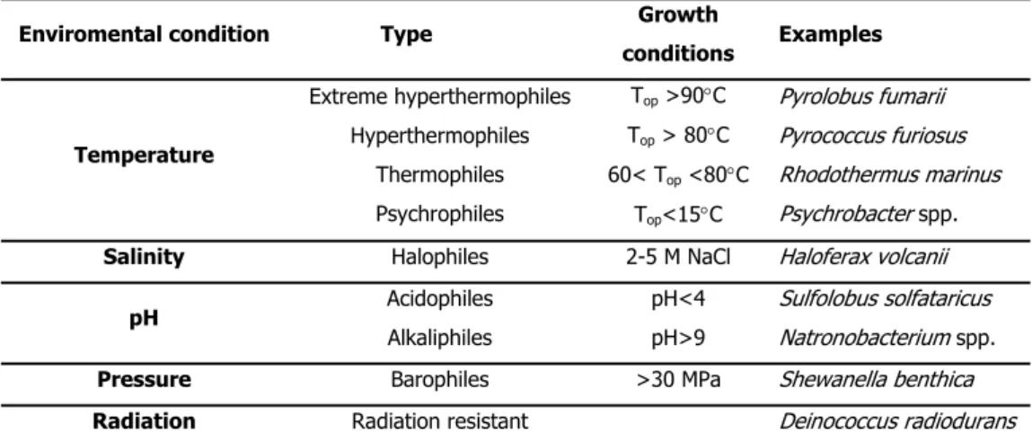
below 2 or above 10 are approaching the limits for life, as far as this parameter is concerned. Naturally, this concept can be extended to other parameters to include high or low temperature, high salinity, elevated levels of gamma radiation or high hydrostatic pressure, among others (da Costa et al., 1998). Table I.2 presents some environmental conditions that can be considered extreme and in which life can be found.
Table I.2 Extremophiles and growth conditions (adapted from Rothschild and Mancinelli 2001).
Enviromental condition Type Growth
conditions Examples
Temperature
Extreme hyperthermophiles
Hyperthermophiles
Thermophiles
Psychrophiles
Top >90°C
Top > 80°C
60< Top <80°C
Top<15°C
Pyrolobus fumarii
Pyrococcus furiosus
Rhodothermus marinus
Psychrobacter spp.
Salinity Halophiles 2-5 M NaCl Haloferax volcanii
pH Acidophiles
Alkaliphiles
pH<4
pH>9
Sulfolobus solfataricus Natronobacterium spp.
Pressure Barophiles >30 MPa Shewanella benthica
Radiation Radiation resistant Deinococcus radiodurans
It is not unusual to find organisms living in environments characterised by more than one extreme condition. This is the case for many hyperthermophiles isolated from hydrothermal submarine areas with high salinity or sulfurous hot springs, with pH in the range of 1 to 5 (Stetter et al. 1990).
Hyperthermophilic and thermophilic organisms
The placement of hyperthermophiles near the root of the Tree of Life in short phylogenetics branches within the Bacteria and Archaea (Fig. I.1) indicates an early separation and a slow rate of evolution of these organisms. This has been interpreted as evidence for a hyperthermophilic origin of life (Stetter 2006). However, this is a very controversial topic and this view has been often challenged (Lazcano and Miller 1996).
Hyperthermophiles have been isolated from different biotopes: terrestrial (hot springs, solfataric fields, and geothermally heated oil stratifications) and submarine (hydrothermal systems situated at shallow to abyssal depths). With this variety of biotopes, hyperthermophiles are adapted to distinct environmental factors, like different pH, redox potential, salinity and temperature. For example, the salt tolerance of hyperthermophiles isolated from terrestrial hot springs is quite different from that of isolates from marine hydrothermal vents. Within the genus Thermococcus, species thriving in freshwater hot springs, such as Thermococcus zilligii and Thermococcus waiotapuensis, grow optimally in medium containing less than 0.6% NaCl and tolerate a maximum of 1.4% NaCl; in contrast, species isolated from marine environments, like Thermococcus marinus, Thermococcus profundus and Thermococcus hydrothermalis, grow optimally in salt concentrations between 2 and 4% NaCl, with a maximum of 8% for some species (Gonzalez et al. 1999; Kobayashi 2001; Jolivet et al. 2004).
The physiology of hyperthermophiles is diverse. Most species are chemolithoautotrophic, using inorganic redox reactions for energy production (chemolithotrophy) and CO2 as the only carbon source for growth (autotrophy). The most
common electron donor is molecular hydrogen, but some species can use sulfide, sulfur and ferrous iron. Diverse types of respiration can be found in hyperthermophiles, both anaerobic (e.g., nitrate, sulfate, and sulfur respiration) and aerobic; in the latter case, hyperthermophiles are generally microaerophilic, requiring low concentrations of oxygen for optimal growth. Several chemolithoautotrophic hyperthermophiles also show heterotrophic growth and some are obligate heterotrophs, requiring organic substrates as carbon and energy sources (Stetter 2006).
Adaptation to high temperature
aggression can not be transferred to the extracellular space. This is certainly the case of temperature, since it is impossible for any microorganism to have an internal temperature different from that of the environment. Therefore, (hyper)thermophiles had to develop mechanisms to protect all their cellular components against the damaging effects of high temperature. These mechanisms are not fully understood yet, but several features related to the stabilisation of cellular structures in these organisms have been identified (Ladenstein and Antranikian 1998; Daniel and Cowan 2000).
Another difficulty that hyperthermophiles have to overcome is the low thermal stability of some commonly used metabolites, such as ATP and NAD+. To circumvent this problem,
hyperthermophiles developed different strategies, which include microenvironmental protection, metabolite channelling, high catalytic efficiency, and substitution by other metabolites that are more stable at high temperature (Daniel and Cowan 2000). An example of metabolite substitution is the utilisation of pyrophosphate or ADP instead of ATP, a more thermolabile phosphate donor, in the catabolic reactions catalysed by hexokinase and phosphofructokinase (Selig et al. 1997; Siebers et al. 1998; Tuininga et al. 1999; Koga et al. 2000; Xavier et al. 2000; Labes and Schönheit 2001; Sakuraba et al. 2002; Dorr et al. 2003; Hansen and Schönheit 2004; Sakuraba et al. 2004). Additionally, some redox reactions are catalysed by ferredoxin-linked oxidoreductases instead of NAD(P) dependent enzymes (Mescher et al. 1976).
The tRNAs of (hyper)thermophiles have a higher GC content and an increased number of post-transcriptional modifications that seem to confer extra thermostability (Sundaran 1986). However, the same trend is not observed at the DNA level. Some hyperthermophiles, such as Methanococcus igneus and Pyrococcus furiosus, have a GC content between 30 and 40 %, which is near the lower limit allowing for preservation of information (Stetter 1998). A unique reverse gyrase, present in all hyperthermophiles studied so far and absent in mesophiles, has been associated to DNA stabilisation (Kikuchi and Asai 1984; Bouthier de la Tour et al. 1990). The enzyme introduces positive supercoils in DNA, which becomes more compact, and therefore more resistant to thermal denaturation. Reverse gyrase has been considered a marker of hyperthermophilia (Bouthier de la Tour et al. 1990). However, this view was contested by a study involving the genetic manipulation of the hyperthermophile Thermococcus kodakaraensis, showing that mutants with disruption of the gene coding for reverse gyrase were able to grow up to 90°C (Atomi et al. 2004).
mechanism likely to be involved in thermoadaptation of hyperthermophiles is the preferential usage of some codons in detriment of others that are more frequent in mesophiles; this approach would lead to error minimisation at the translation level by preventing mutations that could harm protein thermostability (De Farias and Bonato 2002; van der Linden and Farias 2006).
(Hyper)thermophilic Archaea have also developed strategies to increase the thermal resistance of the cytoplasmic membranes. Some members of the thermoacidophiles have bipolar phytanyl chain lipids fused in C40-44 tetraether core, forming a monolayer, instead of the
bilayer commonly found in mesophilic Bacteria (Kates 1993; Daniel and Cowan 2000). Also, the ether bond of the lipidic chain to the glycerol unit contributes to a higher membrane stability, since it is more resistant to hydrolysis than the ester bond (Daniel and Cowan 2000). In some cases, cyclopentane rings are present in the biphytanyl chains, a feature that enhances membrane packing and reduces membrane fluidity (Daniel and Cowan 2000). Among (hyper)thermophilic bacteria, diether lipids are present only in Thermotoga maritima, as components of the cytoplasmic membrane (Huber et al. 1986). In the others cases, the thermostability of the membrane is achieved by an increase in the length and degree of saturation of the acyl chain and the ratio iso/anteiso branching (Tolner et al. 1998).
Surprisingly, there are no striking differences between proteins from (hyper)thermophilic organisms and those from mesophiles. The amino acid sequences of homologous (hyper)thermophilic and mesophilic proteins present an identity of 40 to 80%; they have overlapping three-dimensional structures and share the same catalytic mechanisms (Vieille et al. 1996; Vieille and Zeikus 2001). No new amino acids, covalent modifications or structural motifs were found in proteins from (hyper)thermophiles that could explain their ability to function at higher temperatures (Li et al. 2005). Nevertheless, it was observed that enzymes from (hyper)thermophilic organisms are usually more thermostable (exhibit a higher intrinsic stability) and thermophilic (with higher optimal temperature) than their mesophilic counterparts (Li et al. 2005). This increased stability may be the result of the combination of small structural modifications that are achieved with exchange of amino acids and modulation of the canonical forces (e.g. hydrogen bonds, ion-pair interactions, hydrophobic interactions) (Li et al. 2005).
al. 1980). The ionic pair networks between several amino acid residues are uncommon in typical proteins of mesophiles, though they are frequently found in hyperthermophilic proteins. The increased packing density in the hydrophobic cores of thermophilic proteins has also been related to their higher thermostability. An optimised packing reduces the protein/water contacts, increasing the intermolecular van der Waals forces, and consequently increasing the compactness of the protein (Jaenicke 1991; 1996). Other features are a decrease in the degree of cavity formation (Yamagata et al. 2001), an increase in the number of disulfide bonds (Littlechild et al. 2004), of hydrophobic interactions at subunit interfaces (Roitel et al. 2002), and a higher number of subunits when compared with mesophilic homologues (Thoma et al. 2000). In respect to amino acid composition, hyperthermophilic proteins have a lower number of residues that are susceptible to undergo covalent modifications induced by heat, namely cysteine, asparagine and glutamine (Hensel 1993), and show a preference for charged over neutral amino acids, specially glutamate and lysine over glutamine and histidine (De Farias and Bonato 2002; Farias and Bonato 2003). However, a universal strategy for protein stabilisation has not been identified, because individual proteins, with particular sequence and structural characteristics, utilise differently the palette of mechanisms available.
Surprisingly, some proteins from hyperthermophilic organisms exhibit a thermostability that is lower than expected from its optimal growth temperature (Hensel and König 1988), indicating that protein stability is not exclusively derived from factors intrinsic to the protein structure. A higher protein turnover is one of the mechanisms suggested to be used by hyperthermophiles to overcome the problem of protein denaturation at high temperatures. Another possible mechanism involves the presence of extrinsic stabilising factors. In fact, several hyperthermophiles possess high levels of chaperones, whose concentration increases in response to heat shock. For example, 80% of the soluble protein content in cells of Pyrodictium occultum, grown at the supra-optimal temperature of 108°C, is composed of thermosome, a heat inducible molecular chaperone (Phipps et al. 1991). In vitro assays demonstrated the protein thermostabilising effect of a recombinant chaperonin derived from Pyrococcus sp. strain KOD1 (later designated Thermococcus kodakaraensis) (Yan et al. 1997). Natural substrates or co-factors are also described as enzyme stabilisers, specifically by stabilising their active site structure (Takai et al. 1997). Additionally, the presence of compatible solutes, also designated chemical chaperones, could play an important role in protein stability.
(Hensel and König 1988; Martins and Santos 1995). In vitro studies designed to evaluate the ability of compatible solutes to protect proteins against several stresses confirmed a positive effect, not only on proteins isolated from thermophiles and hyperthermophiles, but also from mesophiles (Ramos et al. 1997; Lamosa et al. 2000; Borges et al. 2002; Lamosa et al. 2003; Pais et al. 2005)
Osmoadaptation
All microorganisms must adapt to fluctuations in the water activity of the environment to maintain cell turgor. When the osmotic pressure of the external medium is lower than that of the intracellular space (<0.15 M), water enters the cell, leading to turgidity. If the net internal osmotic pressure exceeds the limits of the cell wall resistance, the cell will lyse. In contrast, when the osmotic pressure is higher in the external medium, water leaves the cell, leading to plasmolysis (da Costa et al. 1998). For these reasons, organisms must develop mechanisms of adaptation that enable them to avoid cell damage induced by fluctuations in water activity of the external medium. The strategies to maintain the osmotic equilibrium through the cell membrane can be categorised in two groups and involve: i) influx of high amounts of inorganic ions (K+, Na+, Cl-) to the cytoplasm; or ii) intracellular accumulation of organic solutes (Brown
1976; da Costa et al. 1998) (Fig I.2).
water
Accumulation of organic solutes
Influx of ions
Figure I.2 Schematic representation of the strategies used by cells to cope with an osmotic
up-shock: influx of inorganic ions from the medium to the cytoplasm (•); and the accumulation of small organic
The osmoadaptation strategy using solely the influx of inorganic ions is confined to halophiles, such as those belonging to the archaeal extreme halophilic family Halobacteriaceae, the moderate halophilic anaerobic bacteria of the order Haloanaerobiales, and the extreme halophilic bacterium Salinibacter ruber (Rainey et al. 1995; Kamekura 1998; Anton et al. 2002). The influx of ions leads to increased ionic strength in the cytoplasm, implying that the whole cellular machinery has to be adapted to high salinity. In fact, these organisms exhibit a predominance of proteins with a negative, global charge, thought to support the protein hydratation layer by attracting water to the protein surface in the form of hydrated potassium and sodium cations. Also, the stability and activation of most enzymes from these organisms are dependent on salt (da Costa et al. 1998). This strategy allows the colonisation of highly saline environments (near NaCl saturation) where biological diversity is relatively low and competition for available resources less demanding; however, it also restricts these organisms to such type of environments.
The accumulation of low molecular mass organic compounds in the cytoplasm is the most common strategy of osmoadaptation. The organic compounds that serve as osmolytes can accumulate to high levels without interfering with cell metabolism, hence the term “compatible solutes” coined by Brown in the 70’s (Brown 1976). When the water activity in the environment decreases the microorganism responds by accumulating compatibles solutes in the cytoplasm, thus preventing water efflux and maintaining cell turgor. This strategy allows the ionic strength of the cytoplasm to be kept approximately constant, while adapting to changes in the environmental salinity. Also, no drastic alterations in the cellular machinery are required, since the intracellular conditions are roughly the same throughout the colonisable salinity range (Galinski 1995; da Costa et al. 1998). Nevertheless, the flexibility allowed by this strategy comes with an energetic cost. Accumulation of compatible solutes in the intracellular space is accomplished either by de novo synthesis, or uptake from the medium. When suitable solutes are available in the medium, the second way is preferred, since the energetic cost of uptake is significantly lower than de novo synthesis (Pfluger and Muller 2004). Therefore, mesophilic, halophilic Bacteria and Archaea generally have active transport systems for compatible solutes (Lai et al. 1991; Horlacher et al. 1998; Lai et al. 2000; Robert et al. 2000; Grammann et al. 2002; Tetsch and Kunte 2002; Pfluger and Muller 2004; Vermeulen and Kunte 2004; Silva et al. 2005). These uptake systems must be efficient scavengers because the concentration of compatible solutes in the external medium is usually low. Therefore, they have high affinity for the substrates (KM values in the μM range) and are osmotically regulated (Lai et al. 2000;
that de novo synthesis is favoured since uptake systems for solutes typically associated with thermophily, such as mannosylglycerate and di-myo-inositol-phosphate, have not been found (unpublished results obtained in our team).
The chemical diversity of compatible solutes is not high and these compounds generally belong to the following categories: amino acids and derivatives, polyols and derivatives, sugars, betaines and ectoines (da Costa et al. 1998). Classically, the concept of compatible solute was restricted to molecules with low molecular mass, with polar functional groups (therefore, highly soluble in water), without global charge at physiological pH, and that accumulate in response to salt stress. More recently, with the isolation of new Extremophiles, the concept of compatible solute was extended to contemplate compounds that accumulate in response to stress types other than osmotic, namely high temperature and desiccation (da Costa et al. 1998). Here, the term “compatible solute” will be used to designate any small organic molecule that accumulates in response to heat or salt stress.
Compatible solutes of (hyper)thermophiles
In many halophilic or halotolerant (hyper)thermophiles, accumulation of compatible solutes occurs not only in response to an increased salinity, but also in response to supraoptimal growth temperatures. Interestingly, besides solutes that are frequently found in non-thermophilic organisms, (hyper)thermophiles accumulate solutes that have not been or are rarely encountered in mesophilic organisms (Table I.4). Trehalose, α-glutamate, aspartate, proline, and glycine betaine are some of the compatible solutes that can be found both in (hyper)thermophiles and mesophiles. On the other hand, di-myo-inositol phosphate and diglycerol phosphate have never been found outside the (hyper)thermophilic world; mannosylglycerate is widely distributed among (hyper)thermophiles and rarely occurs in mesophiles. Solutes whose accumulation is restricted to (hyper)thermophiles or rarely encountered in mesophiles have been denominated “thermolytes” (Santos et al.). Here, this term will be used for the convenience of a short designation. One of the characteristics common to most thermolytes studied thus far is the negative charge at physiological pH, mannosylglyceramide being the only exception (Fig. I.3).
of the most widespread thermolyte. Among (hyper)thermophiles, mannosylglycerate was firstly reported in the thermophilic bacterium Rhodothermus marinus (Nunes et al. 1995). Since then it has been found in diverse organisms: the thermophilic bacteria Thermus thermophilus and Rubrobacter xylanophilus, the crenarchaeota Aeropyrum pernix and Stetteria hydrogenophila, the euryarchaeota Methanothermus fervidus, and in the three genera of the order Thermococcales: Thermococcus, Pyrococcus, and Palaeococcus (Table I.3) (Martins and Santos 1995; Martins et al. 1996; Martins et al. 1997; Lamosa et al. 1998; Silva et al. 1999; Neves et al. 2005). In these organisms, mannosylglycerate accumulation occurs primarily in response to osmotic stress; an exception to this behaviour was found in Rhodothermus marinus, an organism that accumulates mannosylglycerate also at supraoptimal growth temperatures (Silva et al. 1999).
Diglycerol phosphate OH OH O OH HO O P O -O O O O O O O O P P OOC Cyclic-2,3-bisphosphoglycerate Mannosyl-glucosylglycerate HO OH
CH2OH O
O CH2OH
COO
-O HO
HO
CH2OH
HO
O
Di-myo-inositol phosphate
O -O O O P HO OH OH OH HO HO OH OH OH OH Mannosylglycerate O HO OH
CH2OH
HO
O
CH2OH
COO
-O HO
OH
CH2OH
HO
O
CH2OH
C
O
NH2
Mannosylglyceramide
CH2OH
O HO
CH2 O
COO
-HO HO
CH2OH
HO O O OH OH Glucosyl-glucosylglycerate O P O -O HO OH OH OH HO OH OH O
Glycero-phospho-myo-inositol mannosyl-di-myo-inositol phosphate
OH
O
-OH
CH2OH
OH O O O P HO OH OH OH HO HO OH OH O OH O Diglycerol phosphate OH OH O OH HO O P O -O Diglycerol phosphate OH OH O OH HO O P O -O O O O O O O O P P OOC O O O O O O O P P OOC Cyclic-2,3-bisphosphoglycerate Mannosyl-glucosylglycerate HO OH
CH2OH O
O CH2OH
COO
-O HO
HO
CH2OH
HO
O HO
OH
CH2OH O
O CH2OH
COO
-O HO
HO
CH2OH
HO
O
Di-myo-inositol phosphate
O -O O O P HO OH OH OH HO HO OH OH OH OH O O P HO OH OH OH HO HO OH OH OH OH Mannosylglycerate O HO OH
CH2OH
HO
O
CH2OH
COO
-Mannosylglycerate O
HO
OH
CH2OH
HO
O
CH2OH
COO
-O HO
OH
CH2OH
HO
O
CH2OH
C O NH2 MannosylglyceramideO HO OH
CH2OH
HO
O
CH2OH
C
O
NH2
Mannosylglyceramide
CH2OH
O HO
CH2 O
COO
-HO HO
CH2OH
HO O O OH OH Glucosyl-glucosylglycerate
CH2OH
O HO
CH2 O
COO
-HO HO
CH2OH
HO O O OH OH Glucosyl-glucosylglycerate O HO
CH2 O
COO
-HO HO
CH2OH
HO O O OH OH Glucosyl-glucosylglycerate O P O -O HO OH OH OH HO OH OH O
Glycero-phospho-myo-inositol
O P O -O HO OH OH OH HO OH OH O O P O -O HO OH OH OH HO OH OH O
Glycero-phospho-myo-inositol mannosyl-di-myo-inositol phosphate
OH
O
-OH
CH2OH
OH O O O P HO OH OH OH HO HO OH OH O OH O
mannosyl-di-myo-inositol phosphate
OH
O
-OH
CH2OH
OH O O O P HO OH OH OH HO HO OH OH O OH O O P HO OH OH OH HO HO OH OH O OH O
Tabela I.3 Distribution of thermolytes among (hyper)thermophiles (adapted from Santos et al. 2007).
Solutes
Organisms TOpt.
(ºC) cBPG Tre MG DIP α-Glu β-Glu Asp Other Ref
Archaea
Pyrodictium occultum 105 + + 1
Pyrobaculum aerophilum 100 + 1
Pyrobaculum islandicum 100 1
Pyrococcus furiosus 100 ↑(S) ↑(T) + 2
Pyrococcus horikoshii 98 + ↑(S) + + 3
Methanopyrus kandleri 98 + + 1
Stetteria hydrogenophila 95 + + + 4
Aeropyrum pernix 90 + + 5
Methanotorris igneus 88 ↑(T) ↑(S) ↑(S) 6, 7
Thermoproteus tenax 88 + 1
Thermococcus stetteri 87 ↑(S) ↑↑(T) (S) + ↑(S) 8
Thermococcus celer 87 ↑(S) ↑(T) + ↑(T) 8
Thermococcus litoralis 85 ↑(S) ↑(S) ↑(T) + ↑(S) GalHl 8
Thermococcus kodakaraensis 85 + + + 9
Methanocaldococcus
jannaschii 85 ↑(S) 7
Palaeococcus ferrophilus 83 ↑↑(T);
(S) ↑(T) + 4
Methanothermus fervidus 83 + + 1
Archaeoglobus fulgidus VC-16 83 ↑(T) + GPI DGP ↑↑(T, S) (S), 1
Acidianus ambivalens 80 + 2
Thermococcus zilligii 75 8
Sulfolobus sulfataricus 75 + 2
Metallosphaera sedula 75 + 2
Methanothermobacter
Thermoautotrophicus 70 + + TCH 10
Methanothermococcus
okinawensis 70 + + 9
Methanococcus
thermolithotrophicus 65 + + + NAL 7, 11 Bacteria
Aquifex pyrophilus 80 ↑(T) GPI mDIP ↑(T, S) ↑(S) 17 Thermotoga maritima 80 ↑(S) mDIP ↑ (T) 12 Thermotoga neapolitana 80 ↑(S) + + mDIP ↑(T) 12
Thermosipho africanus 75 + Pro 12
Thermotoga thermarum 70 12
Marinitoga piezophila 70 + Pro 9
Fervidobacterium islandicum 70 12
Persephonella marina 70 + + GG,GGG 9
Thermus thermophilus 70 ↑(S) ↑(S) + GB 13 Rhodothermus marinus 65 + ↑↑(S) (T) + MGA ↑(S) 14, 15
Petrotoga miotherma 55 + MGG, Pro 16
Abbreviations: cBPG, cyclic 2,3-bisphosphoglycerate; Tre, trehalose; MG, α-mannosylglycerate; MGA,
α-mannosylglyceramide; DIP, di-myo-inositol-1,3’-phosphate; mDIP, mannosyl-di-myo-inositol-phosphate; DGP, di-glycerol phosphate; GPI, glycerol-1,3-inositol phosphate TCH, 1,3,4,6-tetracarboxyhexane; α-Glu, α-glutamate; β-Glu, β -glutamate; Asp, aspartate; GalHl, β-galactopyranosyl-5-hydroxylysine; GB, glycine betaine; Pro, proline; NAL, Nε -acetyl-β-lysine; GG, glucosylglycerate; GGG, glucosylglucosylglycerate; MGG, mannosylglucosylglycerate.
Symbols: The plus sign indicates the presence of the solute in cases for which the response to environmental conditions
References: 1- Martins et al. 1997; 2- Martins and Santos 1995; 3- Empadinhas et al. 2001 4- Neves et al 2005; 5- Santos and da Costa 2001; 6- Ciulla et al. 1994; 7- Robertson et al. 1990 8- Lamosa et al. 1998; 9-our unpublished results; 10- Gorkovenko et al. 1993 1- Martins et al. 1999; 13- Martins et al. 1996; 14- Nunes et al. 1995; 15- Silva et al. 1999; 16- Jorge et al. 2007; 17- Lamosa et al. 2006.
In contrast to the frequent occurrence of mannosylglycerate, the other hexose derivatives are less common. Mannosylglyceramide, which resembles mannosylglycerate but has the carboxylic group of glycerate replaced by an amide group, was found only in a few strains of R. marinus, where it accumulates in response to salt stress and is absent at supra-optimal growth temperatures (Silva et al. 1999). The patterns of mannosylglycerate and mannosylglyceramide accumulation during R. marinus osmo-adaptation are different, the first solute being accumulated immediately after salt up-shock, whereas mannosylglyceramide accumulation presents a lag time (Borges 2004). Other compounds chemically related to mannosylglycerate were found in (hyper)thermophiles: Persephonella marina accumulates glucosylglucosylglycerate and glucosylglycerate (Santos et al. 2007). While the first was never found in any other organism, the latter is relatively common in halotolerant mesophilic bacteria (Robertson et al. 1992; Goude et al. 2004). In the thermophilic bacterium Petrotoga miotherma, mannosylglucosylglycerate was found to play a role during low-level osmotic adaptation (Jorge et al. 2007).
members of the genus Archaeoglobus, and glycerophospho-inositol, a structural chimera of di-myo-inositol phosphate and di-glycerol phosphate that was found only in Archaeoglobus fulgidus and Aquifex spp. (Martins et al. 1997; Lamosa et al. 2006).
Other organic solutes have been found in hyperthermophiles and do not fall into either of these two categories. These include cyclic-2,3-bisphosphoglycerate, which is restricted to methanogens; β-galactosylhydroxylysine, found in Thermococcus litoralis (Lamosa et al. 1998); 1,3,4,6-tetracarboxyhexane, found in Methanothermobacter thermoautotrophicus (Ciulla et al. 1994), and Nε-acetyl-β-lysine, present in Methanococcus thermolithotrophicus (Ciulla and Roberts 1999).
An interesting feature of (hyper)thermophiles is the differential pattern of solute accumulation in response to different stress factors. Mannosylglycerate, di-glycerol phosphate and amino acids accumulate preferentially in response to salt stress, while the accumulation of di-myo-inositol phosphate and derivatives occurs mainly in response to heat stress. This behaviour is particularly evident in Pyrococcus furiosus, in which the pool of mannosylglycerate increases 10-fold when the salinity of the growth medium is increased from 2.8 to 5% NaCl, while di-myo-inositol phosphate levels increase 20-fold for a temperature up-shift of 6°C, being the only solute present at supra-optimal growth temperatures (Martins and Santos 1995).
This tendency of solute specialisation, however, is not always observed. In fact, organisms that do not accumulate di-myo-inositol phosphate tend to use mannosylglycerate during both thermo and osmo adaptation, as is the cases of Rhodothermus marinus and Palaeococcus ferrophiluse (Silva et al. 1999; Neves et al. 2005). Conversely, Thermococcus kodakaraensis and Thermotoga spp., that do not synthesise mannosylglycerate, accumulate di-myo-inositol phosphate in response to both types of stress (Martins et al. 1996; Santos et al. 2007). So, it seems that mannosylglycerate and di-myo-inositol phosphate can replace each other’s typical function in osmo- and thermo-adaptation.
Uptake of compatible solutes in (hyper)thermophiles
1998). A binding protein dependent ABC transporter with a high affinity for trehalose (Km 17
nM) was found in Thermococcus litoralis. This transport system is induced by trehalose and probably is the responsible system for trehalose uptake and accumulation in Thermococcus litoralis (Xavier et al. 1996).
The low availability of suitable compatible solutes in (hyper)thermophilic biotopes and/or the unique nature of some of them, can imply the need for de novo synthesis instead of uptake. In the last years, the knowledge of the biosynthetic pathways of compatible solutes in (hyper)thermophiles increased significantly. Besides the compatible solutes commonly found in mesophiles, such as trehalose and glucosylglycerate, the biosynthetic pathways of some typical thermolytes, like mannosylglycerate and di-myo-inositol phosphate, have been elucidated.
Mannosylglycerate synthesis and regulation
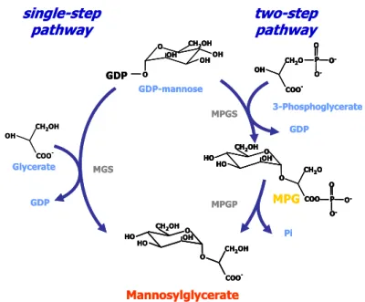
The biosynthesis of mannosylglycerate has been characterised in depth in a variety of (hyper)thermophiles. Two distinct pathways were identified: a single-step and a two-step pathway. In the single-step pathway the direct condensation of GDP-mannose with D-glycerate occurs to yield mannosylglycerate through the action of the mannosylglycerate synthase (MGS). This pathway was found only in the thermophilic bacterium Rhodothermus marinus (Martins et al. 1999) and in the mesophilic red alga Caloglossa leprieurii (Santos et al. 2007) (Fig I.4). In the second pathway, GDP-mannose is condensed with D-3-phosphoglycerate to form a phosphorylated intermediate, mannosyl-3-phosphoglycerate (MPG), in a reaction catalysed by mannosyl-3-phosphoglycerate synthase (MPGS); this intermediate is subsequently dephosphorylated by a specific phosphatase, mannosyl-3-phosphoglycerate phosphatase (MPGP). This pathway is present in all (hyper)thermophiles known to accumulate mannosylglycerate.
et al. 2005). GDP-glucose, UDP-mannose, and UDP-glucose are used as substrates at 37°C but not at 90°C; at this temperature, GDP-mannose and D-glycerate are specific substrates for the reaction (Flint et al. 2005).
Mannosylglycerate
O
HO OH
HO O
COO
-CH2OH
CH2OH
COO
-CH2OH
OH
Glycerate
3-Phosphoglycerate
COO
-CH2O
OH O -P O O -Pi OH OH OH
O CH2OH
O GDP GDP-mannose MGS MPGP GDP
single-step
pathway
MPGtwo-step
pathway
MPGS GDP O HO OH HO O COOCH2OH
CH2O
O -P O O -Mannosylglycerate O HO OH HO O COO
-CH2OH
CH2OH
O
HO OH
HO O
COO
-CH2OH
CH2OH
COO
-CH2OH
OH
Glycerate
3-Phosphoglycerate
COO
-CH2O
OH O -P O O -COO
-CH2O
OH
COO
-CH2O
OH O -P O O -O -P O O -Pi OH OH OH
O CH2OH
O
GDP
OH OH
OH
O CH2OH
O GDP GDP-mannose MGS MPGP GDP
single-step
pathway
MPGtwo-step
pathway
MPGS GDP O HO OH HO O COOCH2OH
CH2O
O -P O O -O HO OH HO O COO
CH2OH
CH2O
O
HO OH
HO O
COO
CH2OH
CH2O
O -P O O -O -P O O
-Figure I.4. The two pathways for the synthesis of mannosylglycerate. The single-step pathway is catalysed
by mannosylglycerate synthase (MGS), while the two-step pathway involves the actions of
mannosyl-3-phosphoglycerate synthase (MPGS) and mannosyl-3-mannosyl-3-phosphoglycerate phosphatase (MPGP).
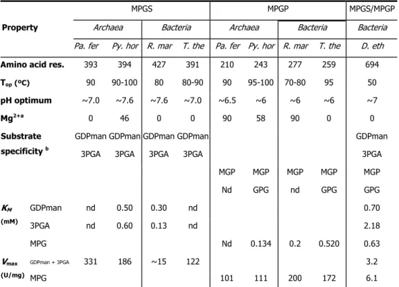
Unlike the single-step pathway, the two-step pathway is widespread among (hyper)thermophiles. It was firstly identified in Rhodothermus marinus (Martins et al. 1999) and subsequently detected in Pyrococcus horikoshii (Empadinhas et al. 2001), Thermus thermophilus (Empadinhas et al. 2003), Palaeococcus ferrophilus and Thermococcus litoralis (Neves et al. 2005). All MPGSs present similar biochemical properties and kinetic parameters (Table I.4): high specificity for GDP-mannose and 3-phosphoglycerate at the temperature examined (83oC), an elevated optimal temperature (80-100°C) and an optimum pH around 7.3.
The KM values for both substrates are similar in all MPGSs, but the Vmax of the MPGS from
Rhodothermus marinus is 10 times lower than that observed for the other MPGSs. Interestingly, the R. marinus MPGS has 30 additional amino-acids in the C-terminus region; when this extension was deleted, the Vmax of the enzyme became similar to that of other MPGSs.