Khadija Shabbiri1*; Waqar Ahmad1; Quratulain Syed 2; Ahmad Adnan1
1
GC University, Lahore, Pakistan; 2Pakistan Council of Scientific and Industrial Research Labs Complex, Lahore, Pakistan.
Submitted: May 21, 2009; Approved: March 16, 2010.
ABSTRACT
A respiratory complex was isolated from plasma membrane of pathogenic Proteus mirabilis strain ATCC
29245. It was identified as complex II consisting of succinate:quinone oxidoreductase (EC 1.3.5.1)
containing single heme b. The complex II was purified by ion-exchange chromatography and gel filtration.
The molecular weight of purified complex was 116.5 kDa and it was composed of three subunits with
molecular weights of 19 kDa, 29 kDa and 68.5 kDa. The complex II contained 9.5 nmoles of cytochrome b
per mg protein. Heme staining indicated that the 19 kDa subunit was cytochrome b. Its reduced form
showed absorptions peaks at 557.0, 524.8 and 424.4 nm. The -band was shifted from 557.0 nm to 556.8
nm in pyridine ferrohemochrome spectrum. The succinate: quinone oxidoreductase activity was found to
be high in this microorganism.
Key words: Proteus mirabilis, complex II, cytochrome b
INTRODUCTION
Proteus mirabilis belongs to family Enterobacteriaceae
and is facultative anaerobic, rod shaped, and gram negative
bacterium (14). It is mainly found in GI tract, soil, infections of
bladder, lung, urinary tract and wounds. It infects and persists
for a long period of time (26). It also causes pneumonia, chills,
fever, cough and chest pain (4, 12, 27).
In bacterial cells, the generation of energy in the form of
adenosine triphosphate (ATP) is mainly motivated by the
activity of respiratory chain enzymes of inner plasma
membrane (5). The respiratory chain of bacteria usually
composed of enzyme complexes I to IV, ubiquinone,
cytochromes, and ATP synthase (complex V), transfers
electrons from NADH and succinate at one end to molecular
oxygen at the other (2). Van Wielink et al. (22) observed the
presence of cytochrome b, a1 and d in spectral investigation of
the plasmic membrane of Proteus mirabilis. Van Wielink et al.
also (20) characterized the presence of cytochrome b and c in
plasmic membranes of Proteus mirabilis by means of 77K
spectra in both aerobic and anaerobic conditions. The function
of cytochrome b was investigated in Proteus mirabilis (21)
revealed the presence of cytochrome b in Q or b-cycle between
the two HQNO inhibition sites. Thus a few types of respiratory
components of Proteus mirabilis have been studied with their
enzymatic and structural features. However the presence of the
succinate: quinine oxidoreductase (complex II) of Proteus
mirabilis has not so far been completely studied.
Complex II of the bacterial electron transport chain is of
special interest. It functions as a dehydrogenase in the
respiratory system and plays an important role in the
tricarboxylic acid cycle (TAC) (17). This membrane associated
complex catalyses the oxidation of succinate to fumarate and
reduce ubiquinone to ubiquinol, and has been characterized in
bacteria and heterophilic eukaryotes (19). Complex II
(succinate:quinone oxidoreductase) of aerobic respiratory chain
oxidizes succinate to fumarate and passes electrons directly
into the quinone pool. It serves as the only direct link between
activity in the citric acid cycle and electron transport in the
membrane (1). Membrane-bound succinate dehydrogenases
(succinate:quinone reductases, SQR) couples the oxidation of
succinate to fumarate to the reduction of quinone to quinol and
also catalyzes the reverse reaction. SQR (respiratory complex
II) is involved in aerobic metabolism as part of the citric acid
cycle and of the aerobic respiratory chain (11). Iron sulfur
clusters and flavin adenine dinucleotide (FAD) are located in
the water soluble moiety, while cytochrome b is present in
membrane-bound moiety. The purified succinate quinine
oxidoreductase from Bacillus subtilis contains two hemes b and
the other gram negative bacteria such as E. coli (6, 9) and P.
denitrificans (15) contains one molecule of heme b per FAD
which showed that cytochrome b was generally present with
the association of succinate: quinine oxidoreductase.
In present study we report the isolation and purification of
complex II found in the plasmic membrane of Proteus
mirabilis strain ATCC 29245 and its activity in vitro.
MATERIALS AND METHODS
Materials
DEAE-Bio-Gel and Bio-Gel-P100 were purchased from
Bio-Rad Pakistan. DEAE-Sephadex and Sodium succinate
from Sigma, Triton-X100 from Acros (USA), Commassie
Brilliant Blue dye from Fischer, DCPIP and Tris salt from
Fluka and Pyridine and Sodium dithionite from BDH (U.K)
and Nutrient Broth, Nutrient agar, Potassium dichromate, SDS
and EDTA were purchased from Merck. All other chemicals
used in this study were of the extra pure grade.
Organism and culture conditions
The gram –ve facultative bacterium Proteus mirabilis
strain (ATCC 29245) was used in the present study. The strain
was provided by PCSIR Laboratories Lahore, Pakistan. The
bacterium was aerobically grown on nutrient medium (Merck)
in conical flasks at 37oC on shaker (C24 KC Refrigerated
Incubator shaker USA) at 250 rpm. Growth of Proteus was
studied after the regular interval of 2 hours by taking its optical
density by spectrophotometer (CICIL/UV-Visible
Spectrophotometer HitachiU-2001) at 595 nm. The cultivation
of Proteus mirabilis was performed in 200 liters of the above
nutrient medium with a stainless steel fermenter of 500 L
volume. The culture was harvested at the early exponential
phase by centrifugation at 4000 rpm for 25 min and suspended
in 50mM Tris HCl buffer (pH: 8.4) containing 50mM EDTA.
Purification of membrane bound complex- II
Frozen cells (about 25gm in a centrifugally packed state)
were suspended in 100ml of 50mM Tris HCl buffer (pH: 8.4)
and 20ml of 50mM EDTA (buffer A). The suspension was
sonicated with a sonic oscillator (Soniprep 150 SANYO UK) at
12-14 KHz for total period of 15 min with intervals of 1 min at
4oC. After sonication the suspension was subjected to
centrifuge by using centrifuge machine (HITACHI-CP 80 MX)
at 15000 rpm at 4oC for 15 min. Supernatant contained
membrane fraction and cytoplasmic fraction where as pellets
were of unbroken cells . The supernatant then ultra centrifuged
at 35000 rpm and 4oC for 60 min. The reddish pellet obtained
was of cell membrane whereas the supernatant was of
cytoplasm. Then cell membrane pellet was suspended in 25ml
of 50mM Tris-HCl buffer (pH 8.4) with 2.5ml of 50mM EDTA
and 4.2 ml of 20% (wt/vol) Triton-X 100 (with final
concentration of detergent is 3%). The resulting mixture was
stirred using a magnetic stirrer for 1 hour to solubilize the total
cell membrane proteins. This suspension was then ultra-
centrifuged at 45000 rpm and 4oC for 45 min. Reddish
supernatant thus obtained was total membrane proteins and
subjected to a first ion-exchange chromatography (Biologic LP
System, BIO-RAD) on a DEAE-Sephadex column( 4.0 by 16.0
cm) equilibrated with buffer A. The column was washed with
400ml of buffer A containing 1% of Triton X-100, then
enzyme was eluted by using linear gradient solution of 600ml
each of buffer A containing 1% of Triton X-100 and buffer B
containing 1% Triton X-100 and 1.0 M NaCl. The eluates
2110 Fraction collector) and dialyzed against 1 liter of buffer A
and then subjected to a second ion exchange chromatography
on DEAE-Bio-Gel (1.5 by 8.0 cm) equilibrated with buffer A.
The adsorbed enzyme was eluted with a linear gradient of NaCl
(0-1.0 M NaCl) produced in 600ml of buffer A containing 1%
Triton X-100. Thus the fractions 30-34 (5ml each) with the
enzymatic activity as shown in (Figure 2) were collected and
concentrated by lyophilization to an appropriate size and
subjected to gel filtration with a Bio-Gel P-100 column
equilibrated with 50mM Tris-HCl buffer (pH:8.4) containing
1% Triton X-100 and 0.5 M NaCl. The yellowish-red complex
II fraction was collected and used as purified preparation.
Spectroscopy
Absorption spectra of respiratory proteins were studied by
using a quartz cuvette with the CICIL/UV-Visible
Spectrophotometer Hitachi U-2001 at room temperature in
visible range. Cytochrome b from Proteus mirabilis strain
ATCC 29245 was suspended in 50mM Tris-HCl (pH: 8.4)
containing 1% Triton X-100. Cytochrome b was oxidized with
1M Potassium dichromate solution and reduced by adding few
pellets of sodium dithionite. For obtaining ferrohemochrome
spectra, the 0.7mL enzyme was suspended in 0.5mL of 0.2 N
NaOH, 1.8mL of distilled water and 0.5ml of pyridine and then
reduced with a small amount (500mg)of sodium dithionite.
Polyacrylamide Gel Electrophoresis
Native polyacrylamide gel electrophoresis was performed
in the presence of 0.4% Triton X-100 and Sodium dodecyl
sulfate polyacrylamide gel electrophoresis (SDS-PAGE) was
performed under denaturing conditions at room temperature by
the method of Schagger and von Jagow (18). In the estimation
of the apparent molecular weight by SDS-polyacrylamide gel
electrophoresis, a set of protein markers (PAGE ruler
pre-stained protein ladder, SM0671 Fermentas) with known
molecular weights were used. The presence of cytochrome b
in the native gel was detected by heme staining reagent (3).
Assay of Redox Activity
Cytochrome b oxidoreductase activity was measured at
room temperature spectrophotometrically (CICIL/UV-Visible
Spectrophotometer. Hitachi U-2001) in time scanned mode.
The oxidation of succinate to fumarate with concomitant
transport of electron to DCPIP dye (which acts as electron
accepter) was determined by monitoring the decrease in
absorbance at 600nm for 5 min. The reaction mixture contained
9µL of 0.1M EDTA, 3mL of 50mM Tris-HCl buffer (pH 8.4),
159µL of 1mM (dichlorophenolindophenol) DCPIP dye, and
60µL of 0.1M sodium succinate. Addition of the enzyme
(40µL of Complex II) initiated the reaction.
RESULTS
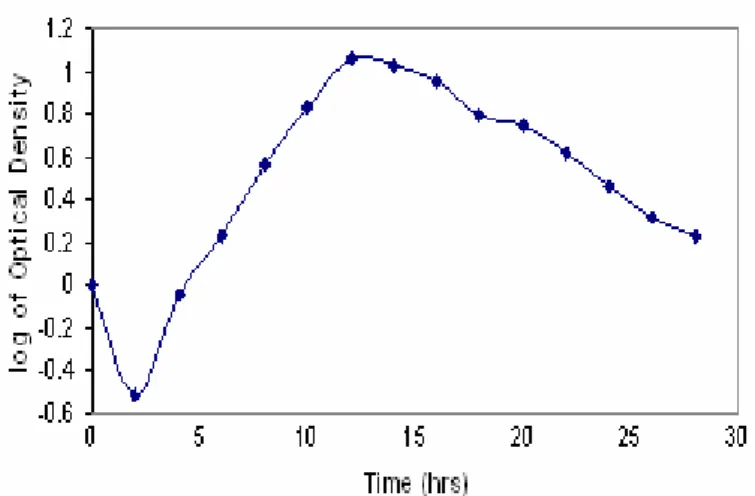
Growth profile of Proteus mirabilis strain ATCC 29245 Growth profile of Proteus mirabilis showed that maximum
growth could be achieved after 12-14 hours of incubation at
37oC (Fig.1). Biomass was collected after 12 hours of
incubation to ensure maximum activity of complex II.
Figure 1. Growth profile of Proteus mirabilis strain ATCC 29245. It shows that maximum active biomass can be achieved
in 12-14 hrs after incubation.
Purification of Complex II from Proteus mirabilis strain ATCC 29245
The membrane bound respiratory complex II was
solublized with Triton X-100 and purified by ion-exchange
chromatography and gel filtration. Purification factors and
solublized membranes containing a total amount of 235mg of
protein and 310 nmol of total heme b. The final preparation
appeared to be homogenous as revealed by nondenaturing
polyacrylamide gel electrophoresis (Native PAGE) since a
single band was observed after heme staining.
Figure 2. Anion exchange chromatography of complex II. The enzyme was eluted by using linear gradient solution of 600ml each of buffer A containing 1% of Triton X-100 and buffer B containing 1% Triton X-100 and 1.0 M NaCl.
Table 1. Purification of complex II from Proteus mirabilis strain ATCC 29245
Step Total Vol.
(ml)
Total Protein (mg)
Total heme b (nmol)
Heme b protein
nmol/mg Yield %
Solublized membranes 80.0 235 310 1.3 100
DEAE- Sephadex 25.0 39 119 3.05 38.34
DEAE- Bio Gel 10.0 8.7 41.3 4.08 13.3
Gel Filtration 8.0 4.2 35.6 8.5 11.48
Purified Enzyme 1.0 2.9 27.4 9.5 8.7
Spectral properties of Complex II from Proteus mirabilis ATCC 29245
Absorption spectrum of complex II is shown in the Figure
3. Figure 3A showed the oxidized spectrum of the membrane
proteins and showed a single absorption peak at 411.0 nm.
When the enzyme was reduced with the addition of sodium
dithionite it showed peaks at 557, 524.8 & 424 nm as shown in
figure 3B. The characteristic -peak at 557 nm showed the
presence of heme b. The figure 3C showed peaks at 556.8,
524.8 & 420 nm of pyridine ferrohemochrome spectrum, which
further confirmed the presence of cytochrome b in complex II.
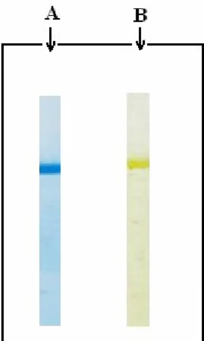
Polyacrylamide Gel Electrophoresis
The purified enzyme was subjected to Native PAGE (7)
followed by Coomassie brilliant blue and the heme staining. A
single band showed that the enzyme was purified to
homogeneity (Fig. 4).
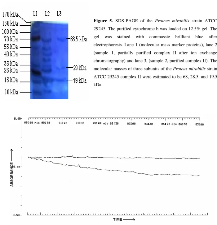
Apparent molecular weights of proteins were estimated by
polyacrylamide gel electrophoresis in the presence of SDS by
the method of Laemmli (10). Purified sample of solubilized
membrane proteins was loaded upon 12.5% SDS-PAGE along
with a standard marker proteins ladder (Fig. 5). The sample
19 kDa.
Assay of Redox activity
The enzymatic properties of the Proteus mirabilis
succinate: quinone oxidoreductase was analyzed by using the
artificial electron acceptor DCPIP. When succinate was added
to the oxidized enzyme under aerobic conditions, the heme b
moiety of enzyme was fully reduced in 5 min (Fig.6) at 660nm.
Figure 3. Spectral properties of the Proteus mirabilis strain ATCC 29245.
Absorption spectra of cytochrome b from
Proteus mirabilis strain ATCC 29245 at
room temperature. The cytochrome b was
suspended in 50 mM tris-HCl (pH 8.4)
containing 1% Triton X-100. A: Oxidized
absorption spectrum. The cytochrome b
was oxidized by adding 1M potassium
dichromate. B: Reduced spectrum of
membrane-bound cytochrome b. The
cytochrome b was reduced with sodium
dithionite. C: Pyridine ferrohemochrome
spectrum. The cytochrome b was
suspended in 0.2 N NaOH and pyridine
and reduced with a small amount of
sodium dithionite.
Figure 4. NATIVE PAGE of the Proteus mirabilis strain ATCC 29245. The purified cytochrome b was
run on NATIVE PAGE and stained. Column A:
Commassie brilliant blue staining of heme b,
Figure 6. Enzyme assay of complex II from Proteus mirabilis strain ATCC 29245. The enzymatic reaction was time-scanned for 5 min at 660nm to observe the decrease in absorption of DCPIP dye which acts as artificial electron acceptor in vitro.
. DISCUSSION
Proteus mirabilis is a pathogenic microorganism so the
study of its respiratory proteins is important. Growth profile of
Proteus mirabilis was studied in shake flasks, incubated at
37oC at 250rpm (diameter 2.5cm). The culture was found to
grow exponentially between 8-14 hours. Maximum biomass
was accumulated after 12 hours as shown in Figure 1. Decline
phase started after 14 hours was due to the exhaustion of
nutrients and accumulation of toxic by-products particularly
release of H2S gas (23).
Cytochrome b is a component of Complex II and is
required for electron transfer from succinate to ubiquinone (8).
The b-type cytochromes in bacteria function as essential
Figure 5. SDS-PAGE of the Proteus mirabilis strain ATCC 29245. The purified cytochrome b was loaded on 12.5% gel. The
gel was stained with commassie brilliant blue after
electrophoresis. Lane 1 (molecular mass marker proteins), lane 2
(sample 1, partially purified complex II after ion exchange
chromatography) and lane 3, (sample 2, purified complex II). The
molecular masses of three subunits of the Proteus mirabilis strain
ATCC 29245 complex II were estimated to be 68, 28.5, and 19.5
components of the respiratory complexes. The cytochrome b
functions between two HQNO sites or more probably in a Q-
or b-cycle (22). Presence of succinate: quinone oxidoreductase
and many types of cytochrome b of complex II present in
bacterial electron transport chain have been reported and
confirmed in Proteus mirabilis but its isolation from plasma
membrane could not be achieved. In the present study, we
have reported for the first time the isolation of cytochrome b
and succinate: quinone oxidoreductase in complex II from
plasma membrane of Proteus mirabilis ATCC 29245. The
nonionic detergent Triton X-100 was found to be the best
suitable detergent for solubilization and stabilization of
cytochrome b and succinate: quinone oxidoreductase of this
bacterium. The enzyme is stable in the presence of Triton
X-100 and has showed high succinate: quinone oxidoreductase
activity. When the enzyme was oxidized with K2Cr2O7, the
spectra showed that the Soret absorption peak at 411nm; upon
reduction with sodium dithionite, the Soret absorption maxima
shifted at 424nm and the -absorption peak appeared at 557nm
while -absorption peak appeared at 525nm as shown in figure
3. These absorption peaks were attributed to the reduced form
of cytochrome b and are very close to the values observed in
isolated cytochrome b from other bacteria as given in Table 2.
The presence of single form of cytochrome b is indicated by
the symmetrical -absorption peak at 557nm in reduced form.
This result suggests that in the isolated complex II from
Proteus mirabilis, only one type of cytochrome b is involved in
electron transfer.
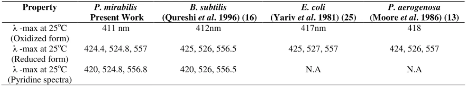
Table 2. Comparison of soluble cytochrome b-557 from different bacteria Property P. mirabilis
Present Work
B. subtilis (Qureshi et al. 1996) (16)
E. coli
(Yariv et al. 1981) (25)
P. aerogenosa (Moore et al. 1986) (13) -max at 25oC
(Oxidized form)
411 nm 412nm 417nm 418
-max at 25oC (Reduced form)
424.4, 524.8, 557 425, 526, 556.5 425, 527, 557 424, 526, 557
-max at 25oC (Pyridine spectra)
420, 524.8, 556.8 420, 526, 556.5 N.A N.A
A prominent protein stained bands in the region of about
68.5, 29, and 19 kDa of covalently bound succinate: quinone
oxidoreductase in the membrane fraction of Proteus mirabilis
appeared when the membrane proteins from Proteus mirabilis
run on 12.5% SDS-PAGE along with standard marker
proteins (Fig.5). The 19 kDa subunit of complex II from the
Proteus mirabilis strain ATCC 29245 stained with
heme-staining reagent, considering that the succinate: quinone
oxidoreductase seems to combine heme b component in
molecule which strongly suggests that the enzyme isolated
with Triton X-100 maintains its intact structure.
Bacterial succinate: quinone oxidoreductase may have one
or two hemes b in the molecule. Spectral properties of the
succinate: quinone oxidoreductase enzyme from Proteus
mirabilis strain ATCC 29245 also suggests the presence of
only one type of heme b in the molecule. On the basis of total
heme b content, the recovery in the purification was calculated
to be about 9.5 nmol /mg of heme b, which yields 8.7% of the
total solublized proteins. For further investigation and
conformation of the presence of cytochrome b in Proteus
mirabilis, energy-linked reduction of cytochrome b has been
studied when complex II is fully reduced with a substrate such
as succinate. It is generally accepted that succinate: quinone
oxidoreductase (Complex II) is a membrane bound respiratory
complex and is associated with cytochrome b (24). The
enzymatic properties of the Proteus mirablis succinate:
quinone oxidoreductase was analyzed by using the artificial
electron acceptor DCPIP. When sodium succinate (an electron
donor) was added to the oxidized enzyme, the heme b moiety
decrease in absorbance of DCPIP dye .The overall activity was
quite prominent and notable. It looks like; the succinate:
quinone oxidoreductase enzyme in this bacterium is highly
active.
In the present study we have found that the complex II of
Proteus mirabilis strain ATCC 29245 has a single heme b in
the molecule which was confirmed by spectroscopic studies.
REFERENCES
1. Ackrell, B.A. (2000). Progress in understanding structure-function relationships in respiratory chain complex II. FEBS. Lett., 466, 1-5. 2. Anraku, Y. (1988). Bacterial electron transport chains. Annu. Rev.
Biochem., 57,101-132.
3. Connelly, J.L.; Morrison, M.; Stotz, E. (1958). Hemins of beef heart muscle. J. Biol. Chem., 233, 743-747.
4. Decaro, N.; Campolo, M.; Desario, C.; Cirone, F.; D'Abramo, M.; Lorusso, E.; Greco, G.; Mari, V.; Colaianni, M.L.; Elia, G.; Martella, V.; Buonavoglia, C. (2008). Respiratory disease associated with bovine coronavirus infection in cattle herds in Southern Italy. J. Vet. Diagn. Invest., 20, 28-32.
5. Goldberg, A.L.; Menon, A.S.; Goff, S.; Chin, D.T. (1987). The mechanism and regulation of the ATP-dependent protease La from Escherichia coli. Biochem. Soc. Trans., 15, 809-811.
6. Hagerhall, C.; Aasa, R.; Wachenfeldt, C.V.; Hederstedt, L. (1992). Two hemes in Bacillus subtilis succinate:menaquinone oxidoreductase (complex II). Biochemistry., 31, 7411–7421.
7. Hames, B.D.; Rickwood, R. (1989). Gel Electrophoresis of Proteins: A Laboratory Manual. IRL Press, NY.
8. Hirawake, H.; Taniwaki, M.; Tamura, A.; Kojima, S.; Kita, K. (1997). Cytochrome b in human complex II (succinate-ubiquinone oxidoreductase): cDNA cloning of the components in liver mitochondria and chromosome assignment of the genes for the large (SDHC) and small (SDHD) subunits to 1q21 and 11q23. Cytogenet. Cell Genet., 79, 132-138.
9. Kita, K.; Vibet, C.R.T.; Meinhardt, S.; Guest, J.R.; Gennis, R.B. (1989). One step purification from Escherichia coli of complex II (succinate:ubiquinone oxidoreductase) associated with succinate reducible cytochrome b556. J. Biol. Chem., 264, 2672–2677.
10. Laemmli, U.K. (1970). Cleavage of Structural Proteins during the Assembly of the Head of Bacteriophage T4. Nature., 227, 680-685. 11. Lancaster, C.R.; Kröger, A. (2000). Succinate: quinone oxidoreductases:
new insights from X-ray crystal structures. Biochim. Biophys. Acta., 1459, 422-431.
12. Lima, A.; Zunino, P.; D'Alessandro, B.; Piccini, C. (2007). An iron-regulated outer-membrane protein of Proteus mirabilis is a haem receptor
that plays an important role in urinary tract infection and in in vivo growth. J. Med. Microbiol., 56, 1600-1607.
13. Moore, G.R.; Mann, S.; Bannister, J.V. (1986). J. Inorg. Biochem., 28, 329-336.
14. O’Hara, C.M.; Brenner, F.W.; Miller, J.M. (2000). Classification, identification and clinical significance of Proteus, Providencia and Morganella. Clin. Microbiol. Rev., 13, 534-546.
15. Pennoyer, J.D.; Ohnishi, T.; Trumpower, L.B. (1988). Purification and properties of succinate-ubiquinone oxidoreductase complex from Paracoccus denitrificans. Biochim. Biophys. Acta., 935,195–205.
16. Qureshi, M.H.; Fujiwara, T.; Fukumori, Y. (1996). Succinate:Quinone Oxidoreductase (Complex II) Containing a Single Heme b in Facultative Alkaliphilic Bacillus sp. Strain YN-2000. J. Bacteriol., 178, 3031-3033. 17. Sambongi, Y.; Iko, Y.; Tanabe, M.; Omote, H.; Iwamoto-Kihara, A.;
Ueda, I.; Yanagida, T.; Wada, Y.; Futai, M. (1999). Mechanical rotation of the c subunit oligomer in ATP synthase (F0F1): direct observation. Science., 286, 1722-1734.
18. Schagger, H.; von Jagow, G. (1987). Tricine-sodium dodecyl sulfatepolyacrylamide gel electrophoresis for the separation of proteins in the range from 1 to 100 kDa. Anal. Biochem., 166, 368–379.
19. Simon, C.B.; Stuart, J.F.; Bernd, L.M.; Dudley, P.; Oliver-Matthias, H.R.; Rob, J.M.; van Spanning. (1988). Molecular Genetics of the Genus Paracoccus: Metabolically Versatile Bacteria with Bioenergetic Flexibility. Microbiol. Mol. Biol. Rev., 62, 1046–1078.
20. Van Wielink, J.E.; Reijnders, W.N.; Oltmann, L.F.; Leeuwerik, F.J.; Stouthamer, A.H. (1983). The membrane-bound b and c-type cytochromes of Proteus mirabilis grown under different conditions. Characterization by means of coupled spectrum deconvolution and potentiometric analysis. Arch. Microbiol., 134, 118-122.
21. Van Wielink, J.E.; Reijnders, W.N.; Oltmann, L.F.; Stouthamer, A.H. (1983). Electron transport and cytochromes in aerobically grown Proteus mirabilis. Arch. Microbiol., 136, 152-157.
22. Van Wielink, J.E.; Reijnders, W.N.; Van Spanning, R.J.; Oltmann, L.F.; Stouthamer, A.H. (1986). The functional localization of cytochromes b in the respiratory chain of anaerobically grown Proteus mirabilis. Antonie. Van. Leeuwenhoek., 52,105-16.
23. Van Woert, M.H. (1967). Proteus mirabilis enterocolitis following abdominal irradiation. Report of a case. Am. J. Dig. Dis., 12, 737-741. 24. Waldeck, A.R.; Stowell, M.H.; Lee, H.K.; Hung, S.C.; Matsson, M.;
Hederstedt, L.; Ackrell, B.A.; Chan, S.I. (1997). Electron paramagnetic resonance studies of succinate:ubiquinone oxidoreductase from Paracoccus denitrificans. Evidence for a magnetic interaction between the 3Fe-4S cluster and cytochrome b. J. Biol. Chem., 272, 19373- 19382. 25. Yariv, J.; Kalb, A.J.; Sperling, R.; Bauminger, E.R.; Cohen, S.G.; Ofer,
S. (1981). J. Biol. Chem., 259, 112-123.
resistance phenotypes for bacteria isolated from outpatients in urine cultures. Roum. Arch. Microbiol. Immunol., 65, 93-99.
27. Zunino, P.; Sosa, V.; Schlapp, G.; Allen, A.G.; Preston, A.; Maskell, D.J.