REVIEW
PEDIATRIC CARDIAC POSTOPERATIVE CARE
F r o m t h e A n e s t h e s i a a n d S u r g i c a l Intensive Care Unit, Heart Institute (INCOR), Hospital das Clínicas, Faculty of Medicine, University of São Paulo.
RHCFAP/3081
José Otávio Costa Auler Jr., Alessandra Costa Barreto, Solange Coppola Gimenez and Deipara Monteiro Abellan
AULER Jr. JOC et al. - Pediatric cardiac postoperative care. Rev. Hosp. Clín. Fac. Med. S. Paulo 57(3): 2002.
The Heart Institute of the University of São Paulo, Medical School is a referral center for the treatment of congenital heart diseases of neonates and infants. In the recent years, the excellent surgical results obtained in our institution may be in part due to modern anesthetic care and to postoperative care based on well-structured protocols.
The purpose of this article is to review unique aspects of neonate cardiovascular physiology, the impact of extracorporeal circulation on postoperative evolution, and the prescription for pharmacological support of acute cardiac dysfunction based on our cardiac unit protocols. The main causes of low cardiac output after surgical correction of heart congenital disease are reviewed, and methods of treatment and support are proposed as derived from the relevant literature and our protocols.
DESCRIPTORS: Pediatrics patients. Heart congenital disease. Cardiac surgery. Postoperative care.
The Heart Institute of University of São Paulo Medical School is a refer-ral center for the treatment of neonate and pediatric congenital cardiopathies for the state of São Paulo, as well as for other regions of Brazil that do not have access to specialized centers in this type of care. Over the past 5 years, 2298 children have undergone cardiac surgery here, including 1592 who un-derwent surgeries with cardiopulmo-nary bypass (CPB) and 706 without CPB. In the last 12 months, for example, the following surgeries were per-formed: correction of an atrial septal defect (ASD) (52), correction of a ven-tricular septal defect (VSD) (59), cor-rection of tetralogy of Fallot (94), trans-position of the great arteries (TGA) (23), and correction of atrioventricular canal defects (AV canal ) (38).
Over the years, several protocols especially concerning human re-sources and intensive care have been
adopted in our ICU that have improved postoperative results. Progress de-pends on harmony among several fac-tors such as accurate diagnosis, effi-cient planning and scheduling of sur-gery, and postoperative support. The challenge for the multi-professional team is to achieve the child’s full re-integration into the family with pre-served neurological, affective, psy-chological, and social capacity. The purpose of this study was to review some unique aspects of neonatal car-diovascular physiology, since some of the children with heart disease are neonates, as well as to review spe-cific aspects of postoperative cardiac care of neonates.
Neonatal heart
The neonatal heart presents physi-ological characteristics (Table 1) that differ significantly from children of other ages or from adults, and these should be considered in the peri-operative ap-proach1.
storage of catecholamines. Moreover, myocardial contractility during the first week of life occurs because of circulat-ing catecholamines, especially epineph-rine, and adequate cardiac output is maintained primarily by an elevated heart rate. Newborns are more depen-dent on the action of circulating cat-echolamines for determining heart rate than on preload for maintenance of ad-equate cardiac output. In contrast, para-sympathetic enervation is similar to that of the adult, making the neonate more susceptible to parasympathetic stimuli. Second, the myocardial fibers have greater length at baseline conditions, resulting in less diastolic reserve for volume overload. Therefore, contractil-ity and ventricular compliance are ef-fectively reduced, so the neonate myo-cardium is near its functional limit at baseline conditions.
Third, neonatal and fetal myocardial function is characterized by ventricu-lar interdependency2. An overload in volume or pressure imposed on one of the ventricles influences filling charac-teristics of the other ventricle. In this way, the dilated right ventricle increases the filling pressure of the left one, and this increased pressure then generates elevated pressure on the right side in order to maintain transatrial flow.
Fourth, immature fetal and neonatal myocardium uses the metabolites of carbohydrates and amino acids (glyco-gen, glutamate, pyruvate, and lactate) for contraction. Elevated stores of gly-cogen and reduced numbers of mito-chondria reflect adaptation to
anaero-bic conditions, with greater recovery capacity and tolerance for hypoxic and ischemic insults. Therefore, the neonate is more vulnerable to hypoglycemia and reacts to stress situations with rapid alterations in pH, lactic acid, glycemia, and temperature. These characteristics are perpetuated beyond the neonatal period. Gradual transformations occur during all the first year of life, and com-plete maturity of the myocardium oc-curs only after 2 years of age3,4.
Cardiopulmonary Bypass
The development of cardiopulmonary bypass (CPB) has been essential for the treatment of the great majority of con-genital heart diseases. Management of CPB for congenital heart disease dif-fers from that for adults because of the problems of aortopulmonary shunts, the immature cardiovascular system, and the eventual use of deep hypoth-ermic circulatory arrest. During deep hypothermic circulatory arrest, the blood available in the systemic veins (generally the cavas vein) is drained to the oxygenator, which provides oxygen, removes carbon dioxide, and reduces the blood temperature. In the process, the oxygenated blood returns to the aorta by a system of pumps that gener-ates continuous flow from the CPB to the patient.
The plastic polyvinyl circuits of the CPB system are full of a balanced solu-tion made of colloids and crystalloids, the composition of which varies accord-ing to the age and weight of the
pa-tient, maintaining a pH and electrolyte composition similar to that of the plasma.
To reduce the metabolic rate and for protection of tissues during CPB, mod-erate (25oC to 32oC) or profound (15oC to 20oC) hypothermia is used. Profound hypothermia permits reduction of flow through the CPB, reduces trauma to blood elements, and allows total circula-tory arrest, usually during aortic arch repair or in very small children. Hypoth-ermia also preserves the highly energetic phosphate stores and reduces liberation of toxic cerebral neurotransmitters.
Different methods (eg, pH-stat, alpha-stat) have been evaluated in the past several decades for correction of acid-base equilibrium during hypothermia, primarily by comparing the neurologi-cal evolution of patients. According to du Plessis et al5, children undergoing surgery using the alpha-stat method have poorer cognitive development in the late postoperative follow-up period compared to those undergoing the pH-stat method, probably because of the acidification that the pH-stat method provides, favoring vasodilatation and homogeneous hypothermic cerebral protection5.
In general, CPB alters all physiologi-cal processes of the organism and may lead to organic dysfunction of differ-ent magnitudes. The morbidity associ-ated with CPB is largely associassoci-ated with damage to blood elements and proteins caused by blood gas alterations and the prosthetic surface interface.
The exposure of blood to various abnormal conditions is implicated in a systemic inflammatory response. This response includes interstitial edema, coagulation cascade activation that generates kallikrein and bradykinin, and platelet consumption. Neutrophils and monocytes are also activated to express reactive cellular receptors and release vasoactive and cytotoxic substances. Activated complement proteins and products from neutrophils and mono-cytes mediate much of the inflamma-tory response to CPB. Hemodilution used during CPB, on the other hand, reduces microcirculation resistance Table 1 - Neonatal heart physiological considerations.
• Decreased myocardial contractile force.
• 50% reduction in myofibers and a greater quantity of nonconnective tissue. • Myofibers present in a chaotic or nonlinear arrangement.
• Small number of sarcomeres, and mitochondria being immature for calcium storage. • Immature sympathetic nervous system with reduced catecholamine storage. • Cardiac output dependent on elevated heart rate and catecholamines rather than
preload.
• Parasympathetic enervation developed.
• Reduced diastolic reserve due to low ventricle compliance.
• Straight ventricular interdependency: overload in volume or pressure imposed on one of the ventricles influences filling characteristics of the other ventricle.
• Myocardium utilizes carbohydrates and amino acids for contraction.
• Elevated stores of glycogen and reduced number of mitochondria suggestive of adaptation to anaerobic conditions.
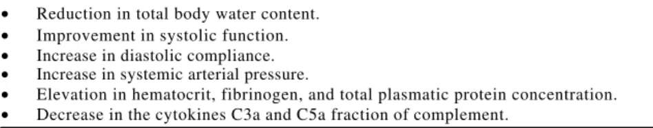
while exacerbating interstitial edema. The formation of edema can be ex-plained by the hemodilution (Table 2) and by the inflammatory process un-leashed by the passage of blood and elements through the nonendothelial surfaces of the CPB circuits. Permeabil-ity of the capillary vessels is greater in immature individuals than in adults. Potent vasoactive substances are acti-vated, including the anaphylatoxins C3a and C5a, tumor necrosis factor, and free radicals derived from oxygen, and neutrophils are activated (Table 3). Con-ventional ultrafiltration has been used in an attempt to prevent this edema; however, its effectiveness seems lim-ited. In 1993, Naik and Elliot6,7 modified the ultrafiltration system, placing the hemoconcentrator between the aorta and the right atrium immediately follow-ing CPB (Table 4). With this modifica-tion, the following have been noted: a reduction in total body water content, improved systolic function, increased diastolic compliance, increased sys-temic arterial pressure, hematocrit, fi-brinogen, and total plasmatic protein, and decreased levels of endothelial se-rum cytokines and the C3a and C5a frac-tions of complement6,7.
The more prolonged the CPB pro-cedure is, the greater are the deleteri-ous effects on these factors, since CPB imposes an overload on the systemic circulation that is often already com-promised by the previous heart failure.
Immediate postoperative care and monitoring
Adequate monitoring during the postoperative period involves a
combination of clinical or auxiliary meth-ods for evaluating the surgical correc-tion, myocardial funccorrec-tion, and the rela-tionship between systemic and pulmo-nary blood flow8. Standard monitoring in the postoperative period is similar to that during anesthesia and surgery. Sometimes, depending on the clinical evolution, more sophisticated monitor-ing may be added to facilitate clinical diagnosis and treatment. Standard monitoring consists of ECG, direct ar-terial pressure, temperature probe, and central venous pressure. Pulse oximeters and sometimes capnography are useful for accompanying mechani-cal ventilation. To obtain direct arterial pressure, radial catheters are generally preferable. Peripheral arterial catheters may not precisely reflect aortic pres-sure, particularly when placed in in-fants’ feet and are even less accurate in the presence of hypothermia. Tem-perature monitoring is very important for assessing the metabolism. Probes can be placed in the rectum or near the esophagus to provide adequate as-sessment of body and core tempera-ture. A nasopharyngeal probe gives an estimate of brain temperature. The cen-tral venous line is generally established by insertion of a catheter utilizing the Seldinger technique into the internal jugular vein of choice. Depending on the degree of severity and type of car-diac disease, transthoracic or transvenous measurement of pulmo-nary and left atrial pressures may give
useful physiologic information. Cardiac output can be also be determined by thermodilution. In small children, the traditional Swan-Ganz 7 or 5F catheter cannot be used, but cardiac output can be measured by means of a thermodilu-tion 2F probe inserted by the surgeon into the pulmonary artery. Newer tech-niques including echocardiography and on-line monitoring of arterial and mixed venous saturation obtained by fiberoptic catheters are increasingly used in postoperative care.
In postoperative care of the pediat-ric patient, clinical evaluation must be complete and systematic. Conse-quently, complications can be foreseen, and catastrophic situations can be avoided. Care should be initiated while the child is still in the operating room, with special attention to rewarming to 36.5ºC, control of bleeding, ventilation, and acid-base and electrolyte balance. It is very important during this phase to stabilize cardiac function through maintaining correct intravascular vol-umes, adequate heart rate, and ad-equacy of myocardial contractility.
The information concerning the an-esthetic and surgical procedure that should be transmitted in detail to the multi-professional ICU team includes surgical technique, type of anesthesia, perfusion and aortic clamping time, wa-ter and colloid balance, diuresis, venous and arterial catheter placement, pace-maker wire and mediastinal or thoracic drainage tube positioning, ventilatory Table 2 – Excessive hemodilution
during and after cardiopulmonary bypass may cause:
_____________________________________ • More requirement for blood
transfusion.
• Greater use of inotropic agents. • Undesirable hemodynamic
performance.
• Depressed myocardial contractility. ___________________________________________
Table 3 - Causes of excessive edema in neonates and small children under cardiopulmonary bypass:
• Permeability of the capillary vessels is greater in immature individuals than for adults. • Potent vasoactive substances are activated during CPB including anaphylatoxins, C3a
and C5a, TNF, and other cytokines that contribute to capillary leaking.
Table 4 - Modified ultrafiltration in pediatric open heart operation results in:
• Reduction in total body water content. • Improvement in systolic function. • Increase in diastolic compliance. • Increase in systemic arterial pressure.
conditions, acid-base balance, heart rate and arrhythmias, and coagulation and vasoactive medication in use9.
Supplementary monitoring, includ-ing left atrial and pulmonary arterial pressure and serial echocardiography, is indicated in all children undergoing more serious or complex cardiopathy correction that present: a) significant myocardial dysfunction (left coronary anomaly, transposition of the great ar-teries, hypoplastic left heart); b) pul-monary arterial hypertension during or prior to the operative period (total anomalous drainage of the pulmonary veins, aortic arch interruption, truncus arteriosus communis); c) valvoplasty
or correction of an atrioventricular ca-nal defect with a residual defect of mi-tral insufficiency.
Cardiovascular function
The cardiovascular function of a child can be considered adequate in the postoperative period when cardiac out-put is sufficient to supply the demands for cellular oxygen.
The main determining factors of car-diac output are preload, afterload, con-tractility, heart rate, and diastolic func-tion1. Other indirect factors, such as anxiety, pain, temperature, hemoglobin level, endogenous, and exogenous cat-echolamines, and blood biochemical composition, can alter the myocardial oxygen supply-demand relationship in addition to affecting the cardiac out-put10.
During the postoperative period, cardiovascular dysfunction repre-sented by low cardiac output can de-velop for a series of reasons. First are all of the pre-operative conditions, in-cluding the structural defect itself and the functional cardiac state, the gen-eral birthing conditions, transfer from hospital to specialized care centers, the detail and efficiency of planning for the surgery, and associated complications such as respiratory insufficiency, infec-tion, and metabolic and electrolyte dis-turbances. Secondly, the dysfunction is dependent on the surgery performed, degree of ventricle muscle incision,
placement of corrective patches, myo-cardial protection, CPB time and aortic clamping, level of hypothermia during CPB, use of total circulatory arrest, type of anesthesia, and intra-operative com-plications.
Cardiovascular Evaluation
Postoperative cardiovascular func-tion can be evaluated by clinical ex-amination, related tissue oxygen in-dexes, echocardiography and hemody-namic and/or radioisotope evalua-tion10.
Important clinical signals for the evaluation of cardiac output are per-spiration, adequate level of conscious-ness, coloring and temperature of ex-tremities, thermal gradient between knees and feet, central and peripheral thermal gradient, amplitude of periph-eral pulse, capillary filling, arterial pres-sure, and urinary output. Accordingly, cardiac output is considered adequate when there is no cold perspiration or psychomotor agitation, the members of extremities are warm and colored, the feet are hotter than the knees, the central-peripheral thermal gradient is less than 4oC, the peripheral pulse is easily palpable, capillary filling is sat-isfactory, arterial pressure is within the normal limits for the age group, and urinary output is greater than l mL/kg/ hour. It is important to remember that adequate peripheral vasodilatation only occurs after the fourth postop-erative hour, with normal re-establish-ment of tissue perfusion around the sixth postoperative hour10.
Among the related tissue oxygen indexes, lactate is a marker of anaero-bic metabolism and tissue energy defi-cit. It reflects hypoxia, ischemia, or aggressive action of a toxic tissue agent. Values for serum lactate above 2.0 – 3.0 mmol/L reflect significant evi-dence of tissue hypoxia. Following total circulatory arrest, profound hy-pothermia (< 20oC), or low cardiac out-put, serum lactate levels commonly exceed 6-10 mmol/L. Also regarding tissue oxygenation, the calculation of the arterial-venous oxygen differential,
as well as the consumption, transport, and peripheral extraction of oxygen can be used in evaluation of low car-diac output in small children. Clinical outcome can be related to the relation-ship of these indexes to normal levels. Measurement of troponin could be a specific marker for the evaluation of myocardial injury, according Taggart et al11. They found that infants under-going cardiac surgery with CPB had elevated levels of troponin during the postoperative period that remained el-evated for 72 hours. Infants undergo-ing cardiac surgery without CPB, did not have elevated levels of troponin. On the other hand, infants from both groups had elevated creatine kinase MB isoenzyme levels that progres-sively decreased over 72 hours.
Two-dimensional and Doppler echocardiography are valuable tools for postoperative cardiac functional and structural evaluation. These meth-ods permit analysis of cardiac cham-bers and operative results, detection of residual defects, evaluation of po-sition and function of valvar prosthe-sis, segmental and global myocardial analysis, calculation of shortening and the ventricular ejection fraction, and estimation of pressures inside cardiac chambers.
using other methods of diagnosis. Direct measurement of cardiac out-put can be obtained by invasive and noninvasive methods. The noninvasive method uses a relationship between expected oxygen consumption and the arterial-venous oxygen differential. However, the method may be incorrect when used to determine the expected consumption in the postoperative pe-riod. Noninvasive methods for deter-mining oxygen consumption calculated by nomograms are dependent on heart rate, age, sex, and body temperature; some of these factors are quite variable in the first hours following CPB12.
As previously mentioned, the ther-modilution method for determining car-diac output is most frequently used in postoperative care; in pediatrics, the data are indexed to body surface area. Accordingly, the cardiac index is con-sidered normal in children when it is above 3 L/min/m2, moderately reduced between 2.0 to 3.0 L/min/m2, and se-verely reduced when lower than 2.0 L/ min/m2. In general, the cardiac index tends to be lower in the fourth postop-erative hour in relation to the immedi-ate postoperative period and increases after the 9th or 12th hours. Kirklin et al12 correlated a greater number of hos-pital deaths with a cardiac index below 2.0 L/min/m2 in a postoperative study of 174 children under the age of 3 months.
Low cardiac output
Low postoperative cardiac output is primarily caused by reduction in myo-cardial contractility caused by one of the mechanisms or factors above men-tioned. Severe myocardial dysfunction can be observed, for example, in more complex congenital heart disease that demands a lengthy CPB procedure and aortic clamping. Left anomalous coro-nary artery in the pulmocoro-nary trunk, hy-poplastic left heart syndrome, trans-position of the great arteries, severe tetralogy of Fallot, and severe pulmo-nary hypertension are all associated with significant risk of poor cardiac func-tion after surgery.
Secondarily, hemodynamic instabil-ity is caused by inadequate intravas-cular volume due to a series of factors including an endothelial inflammatory process resulting from the CPB proce-dure, which transfers fluids to the in-terstitial area (primarily in the first 24 hours); vasodilatation during the re-warming period after CPB (4 to 6 hours postoperatively); urinary output; and active blood loss.
Control of intravascular volume and indirectly of preload should promote more adequate systolic volume, accord-ing to the Frank-Starlaccord-ing law. In the ab-sence of more severe valvar injury, the final diastolic pressure of the ventricles corresponds to the mean pressure of the atrium; therefore, volume can be controlled via right and left atrial pres-sures. During the postoperative period, the atrial pressure should remain around 15 mmHg, although it can reach 18 mmHg in the right atrium and 20 mmHg in the left when there is hyper-trophy or hypocontractility, partial ob-struction in the ventricular outflow, or pulmonary artery hypertension.
Afterload is a third factor influenc-ing cardiac output. It can be elevated by vasoconstriction secondary to CPB, hypothermia, excessive endogenous catecholamines, or administration of vasoactive amines. The heart with any degree of myocardial dysfunction can experience significant reduction of myo-cardial fiber length and consequently its systolic volume when subjected to any increase in afterload.
Heart rate is dependent on factors such as use of digital or beta-blocking agents in the preoperative period, type of surgery, perioperative rhythm distur-bances, volume, temperature, pain, anxi-ety, anemia, metabolic disturbances, and use of vasoactive agents with chro-notropic action.
Alternatively, postoperative myo-cardial edema could be responsible for ventricular diastolic restriction.
Treatment
Therapeutic measures for low post-operative cardiac output include 3
con-comitant and related procedures: 1) di-agnosis, 2) reduction in metabolic de-mand, 3) adequate tissue perfusion and oxygen transport1.
Suspected cardiac dysfunction should be promptly investigated for etiological diagnosis by clinical or supplemental methods so specific and effective conduct can be adopted.
Reduction in metabolic demand re-quires the use of measures that favor adequate temperature and reduction in respiratory workload. Initial measures, even during diagnostic investigation can be adopted to maintain body tem-perature around 36.5oC 10.
Ventilation and respiratory mechan-ics play a primary role in the improving the hemodynamic state of the postop-erative patient through use of positive end expiratory pressure and adjustment of PaCO2 and pH. Factors that influence the hemodynamic state include general anesthesia, pulmonary retraction, edema due to residual injury, reduced functional residual capacity leading to alterations in gas exchange, pulmonary compliance, abnormalities in the venti-lation-perfusion ratio, and intrapulmo-nary shunts10.
Mechanical ventilation is main-tained until bleeding is controlled, he-modynamics are stabilized, adequate body temperature attained, metabolic disturbances are corrected, and acid-base balance is achieved.
in-creasing liberation of norepinephrine in the myocardial presynaptic sympa-thetic storage sites and decreasing en-zymatic degradation and reabsorption of norepinephrine. Dopamine also acts as a pulmonary and systemic vasocon-strictor through action in alpha 1- and alpha 2-postsynaptic receptors. Be-cause of its action on dopamine adren-ergic receptors, dopamine acts as a re-nal, spleen, coronary, and cerebral va-sodilator. In animal research, dopamine has been shown to be less efficient in neonates than in adult animals, prob-ably because of the greater clearance, reduced myocardial adrenergic enerva-tion, lower density of myocardial beta 1-adrenergic receptors, and differing maturation among peripheral alpha re-ceptors and myocardial beta 1 and dopaminergic receptors. In neonates, dopamine increases cardiac output, heart rate, and systemic arterial pres-sure10. Dopamine is indicated in moder-ately low cardiac output, especially when there is water retention from the cardiac insufficiency itself or because of the CPB procedure. In doses of 1 to 2 µg/kg/min, dopamine acts as vasodi-lator on the mesenteric and renal circu-lation. In doses between 2 and 10 µg/ kg/min, it acts on myocardial beta 1 re-ceptors producing increased contrac-tility and coronary flow with conse-quent improvement in mean arterial pres-sure and cardiac index. However, Driscoll et al.14, observed that the
neo-natal myocardium seems less sensitive to the action of dopamine than does the myocardium of older children, and more elevated doses are necessary to obtain the same effects. In another study, Lang et al.15 observed that dopamine increases heart rate, cardiac index, and systemic arterial pressure after pediatric cardiac surgery when given in doses greater than 15 µg/kg/ min but does not have significant ac-tion on systemic vascular resistance. In higher doses, dopamine raises pul-monary resistance, which is why it is not used in cases of previous pulmo-nary artery hypertension.
Dobutamine is a synthetic sym-pathomimetic amine, beta 1-adrenergic agonist. It differs from dopamine by not liberating endogenous norepinephrine and has little peripheral action. Dobutamine increases cardiac output and reduces systemic vascular resis-tance and ventricular filling pressure. Since it exerts a positive chronotropic effect, elevating heart rate, its use could be limited in the postoperative cardiac surgery period in children and neo-nates12. Dobutamine is indicated for low cardiac output when not accompanied by severe hypotension. In sepsis with-out cardiac failure, dobutamine is the therapy of choice, and when cardiac compromise already exists, it can be in-dicated when associated with a vaso-constrictor like norepinephrine. In-crease in cardiac output and reduction
in systemic vascular resistance can be obtained by starting dobutamine at low doses such as 2.5 µg/kg/min. In gen-eral, doses of 2 to 20 µg/kg/min are ef-fective.
Isoproterenol is a synthetic ana-logue of norepinephrine. It is an ago-nist that stimulates myocardial beta 1-adrenergic receptors, with direct inotro-pic and chronotroinotro-pic action, and pro-motes peripheral vasodilatation medi-ated by beta 2-adrenergic receptors. It increases cardiac output and systolic arterial pressure, and it vasodilates re-nal, spleen, and skeletal muscle tissue with a reduction in diastolic arterial pressure and systemic vascular resis-tance13. Isoproterenol is indicated mainly in the presence of sinusal brady-cardia or transitory atrioventricular block. In the neonatal population, it can be indicated for controlling low cardiac output secondary to persistent arterial hypertension in the newborn. The ini-tial dose of isoproterenol is 0.01 to 0.05 µg/kg/minfor symptomatic bradycardia and 0.05 to 0.1 µg/kg/min as a positive inotropic agent. With the increase in heart rate, an accentuated increase in myocardial oxygen consumption and transitory myocardial ischemia may oc-cur. Sinus tachycardia ventricular arrhythmias due to compromised coro-nary flow can appear with more elevated doses.
Epinephrine is an endogenous cat-echolamine that is liberated from the adrenal medulla and derived from nore-pinephrine. It acts on alpha-, beta 1- and beta 2-adrenergic receptors. Depending on the dose, it increases heart rate and systolic arterial pressure, decreases di-astolic arterial pressure, and relaxes the peripheral vascular bed. At high doses the alpha-adrenergic effect predomi-nates, producing skin, gastrointestinal, and renal perfusion10. Epinephrine is indicated for low cardiac output accom-panied by severe systemic arterial hy-potension that compromises coronary perfusion, especially in postoperative cardiac surgery with cardiogenic or sep-tic shock that is not responsive to dopamine and dobutamine. It is also effective in cardiac arrest. At doses of Table 5 - Agents utilized in pharmacological manipulations for increasing right
ventricular inotropism, and/or controlling pulmonary vascular resistance: • Sodium nitroprusside.
• Nitroglycerin.
• Phosphodiesterase inhibitors. • Prostaglandin E1 (PGE1). • Prostacyclin (PGI2).
• Ultra-short-acting intravenous vasodilators: adenosine and adenosine triphosphate. • Inhaled nitric oxide.
Table 6 - Phosphodiesterase inhibitors—cardiovascular effects: • Increase myocardial contractility.
• Increased lusitropic activity (energy-dependent diastolic relaxation). • Vasodilating properties.
• Reduce ventricular wall stress. • Reduce filling pressures.
0.03 to 0.1 µg/kg/min, beta 1 and beta 2 effects are predominant. At doses of 0.1 to 0.2 µg/kg/min, there is a mixed alpha and beta effect. At doses above 0.2 up to 1 µg/kg/min, the alpha re-sponse is predominant. Ventricular ar-rhythmia possibly caused by subendocardic ischemia and peripheral vasoconstriction may be observed with this agent.
Norepinephrine it is a local adrener-gic neurotransmitter. It has beta 1 and alpha action, which increases the sys-tolic arterial pressure as much as the diastolic pressure. Cardiac output can increase or decrease depending on the myocardial reserve. A starting dose of 0.05 to 0.1 µg/kg/min has been recom-mended.
Another possibility for therapy in the control of low cardiac output is the use of phosphodiesterase inhibitors, introduced into clinical practice in 1984. Phosphodiesterase agents, such as amrinone and milrinone inhibitors, are non-glycoside non-sympathomimetic agents that selectively inhibit the cy-clic phosphodiesterase nucleotide, in-creasing myocardial and vascular cAMP, independent of the beta recep-tors. Elevation of cAMP increases con-traction through calcium regulation by 2 mechanisms: first, by activation of the kinase protein that facilitates the rapid entry of calcium through calcium chan-nels, and second, by activation of the calcium stores of the sarcoplasmic reticulum. Accordingly, the phosphodi-esterase inhibitors have 3 actions: in-creasing inotropism and contractility, increasing arteriolar and venous vasodi-latation, and increasing ventricular re-laxation during diastole (Table 6). Amrinone increases the cardiac index and decreases left ventricular end dias-tolic pressure, pulmonary capillary pres-sure, and right atrial pressure in con-gestive heart failure and in postopera-tive low cardiac output. Additionally, when associated with dobutamine, it has greater action on the elevation of the cardiac index and on the reduction in systemic vascular resistance, and it does not significantly increase heart rate16.
Milrinone is considered to be 10 to 30 times more potent than amrinone. It does not elevate myocardial consump-tion and is a coronary vasodilator. Milrinone is metabolized primarily in the kidneys, necessitating dosage correc-tion in the presence of renal failure. Amrinone is metabolized by the liver and excreted primarily by the kidneys.
Phosphodiesterase inhibitors can be indicated for low cardiac output with myocardial dysfunction and elevated systemic vascular resistance, but with-out severe arterial hypotension. Pedi-atric dosages of amrinone are variable in the literature but have been cited as: 0.75 µg/kg bolus dose (over time for 2-3 doses) followed by infusion of 5-10 µg/kg/min for maintenance. In neo-nates, bolus doses of 3 to 4.5 µg/kg are used, followed by 5 to 15 µg/kg/min of continuous infusion. Since the half-life of this agent is relatively long (3 to 15 hours), special attention must be given to those children that already have sys-temic arterial hypotension. In adults with normal renal function, the bolus of milrinone is 50 µg/kg over 10 minu-tes, followed by 0.375 to 0.75 µg/kg/min infusion. Side effects can include hy-potension caused by vasodilatation, necessitating administration of fluids; thrombocytopenia, usually 7 to 10 days after its administration; and elevation of hepatic enzymes, as described for amrinone infusion (Table 7).
Vasodilators for low cardiac out-put are adjuvants to inotropic therapy, reducing systemic arterial resistance and enhancing ventricular ejection. Sodium nitroprusside in doses of 0.5 to 8 µg/kg/min acts to increase vascu-lar relaxation through cGMP, causing arteriolar and venous vasodilatation.
The start of action after endovenous administration occurs in a few minutes. The primary metabolites of nitroprus-side are thiocyanate and cyanide, which have been detected during prolonged infusion13.
Sodium nitroprusside is the vasodi-lator most used, not only for control-ling systemic and pulmonary hyperten-sive states, but also for reducing afterload in low cardiac output, prima-rily after cardiac surgery. Nowadays for pediatric postoperative cardiac pa-tients, sodium nitroprusside has pro-gressively been replaced by phos-phodiesterase inhibitors, which are not only inotropic but also are arteriolar and venous vasodilators.
Nitroglycerine in doses of 0.5 to 20 µg/kg/min is a vasodilator that acts by releasing nitric oxide. The main he-modynamic effect is vasodilation with reduction in ventricular filling pres-sures. Nitroglycerine is indicated for situations of increased preload and signs of pulmonary and systemic venous congestion.
The utilization of inhaled nitric oxide in postoperative period
Nitric oxide (NO) produced by the endothelial cells exerts important func-tions on cardiovascular system. In lung tissue, the maintenance of blood ves-sels in a relaxed status is fundamentally important, especially in the presence of previous pulmonary hypertension and/ or right ventricle dysfunction. Right ven-tricular (RV) dysfunction is frequently observed in infants and children in the postoperative period. Pharmacological and ventilatory manipulations are di-rected at increasing RV inotropism,
uti-Table 7 - Pharmacology of amrinone and milrinone.
Variable measured Amrinone (IV) Milrinone(IV) Loading dose, mg/kg (per approved product labeling) 1.5 (0.72x2) 0.050
Loading dose (maximally effective) 3.0 0.075
Infusion rate, mg/kg/min (labeling) 5-10 0.375-0.75
Incompatible with dextrose solutions Yes No
Average half-life in CHF patients (h) 5-8 2-3
Incidence of thrombocytopenia 2.6% 0.4%
lizing inotropic drugs as outlined above, by optimizing RV preload, and by con-trolling pulmonary vascular resistance (PVR). In the presence of RV dysfunc-tion, control of PVR is extremely impor-tant, since RV output is very sensitive to the variations of afterload. As previ-ously discussed, the most commonly used agents for controlling PVR are so-dium nitroprusside, nitroglycerin, and phosphodiesterase inhibitors17. Prostag-landin E1 (PGE1) and prostacyclin (PGI2) both have a pulmonary vasodilating
ef-fect. The problem is that the effects of these drugs are not limited to the pulmo-nary circulation. Because of the lack of specificity of vasodilator drugs on the pulmonary bed, newer pharmacological methods of controlling elevated PVR are being recommended. Inhaled NO and ultra-short-acting intravenous vasodila-tors, such as adenosine and adenosine triphosphate, represent new modalities for treating acute pulmonary hyperten-sion in postoperative pediatric cardiac patients (Table 8). Although nonselec-Table 8 - Inhaled nitric oxide in post-op after cardiac surgery: Main indication: Pulmonary hypertension accompanied of right ventricular dysfunction
• Correct metabolic disturbances.
• Provide sedation and adequate ventilation. • Consider using phosphodiesterase inhibitors first. • Start inhaled NO with low doses.
• Closely monitor with echocardiography. • Remove NO carefully.
tive, NO is rapidly inactivated by hemo-globin; therefore, when NO is inhaled, the systemic circulation is protected from its vasodilating properties. When we administered inhaled NO to a group of patients after cardiac surgery, we ob-served a significant pulmonary vasodi-latation and no clinically significant ef-fects on systemic pressure18. These same effects have also been observed in chil-dren after cardiac surgery19. In more se-vere situations of RV dysfunction ac-companied by pulmonary hypertension, we have used small doses of inhaled NO (3 to 5 parts per million) in our postop-erative children. Although several stud-ies have demonstrated the vasodilating properties of NO on pulmonary circula-tion, recommendation for its use as a routine agent during cardiac surgery in pediatric patients suffering pulmonary hypertension is still under investiga-tion20.
RESUMO RHCFAP/3081
AULER Jr. JOC e col. - Cuidados pediátricos pós-operatórios de cirur-gia cardíaca. Rev. Hosp. Clín. Fac. Med. S. Paulo 57(3): 2002.
O Instituto do Coração da Faculda-de Faculda-de Medicina da UniversidaFaculda-de Faculda-de São Paulo é centro de excelência para trata-mento das cardiopatias congênitas que abranjam o período neonatal e pediátrico. Nos últimos anos, parte dos
excelentes resultados cirúrgicos da Ins-tituição devem-se ao progresso da anestesia e a estruturação dos cuida-dos pós-operatórios por meio de pro-tocolos bem definidos.
Este artigo se propõe a revisar aspec-tos de interesse da fisiologia cardiovascular do neonato, as repercus-sões orgânicas da circulação extra-corpórea, o pós-operatório imediato, in-cluindo avaliação cardiovascular e a
te-rapia farmacológica desta Unidade. Será apresentada uma revisão das causas mais comuns de baixo débito; cirurgias para a correção das cardiopatias congênitas e propostas de métodos diagnósticos e terapêuticos, baseados na literatura es-pecializada e protocolos da Unidade.
1. FELTES T – Postoperative recovery from congenital heart disease. In: GARSON Jr A. - The science and practice of pediatric cardiology. Baltimore, Williams & Wilkins, 1998. p. 2387-413. 2. KLEINMAN CS – Abnormal fetal cardiovascular physiology. In: POLIN RA, FOX WW - Fetal and neonatal physiology. Philadelphia, Saunders, 1992. p. 666-70.
3. FISHER DJ, HEYMAN MA & RUDOLPH AM – Myocardial oxygen and carbohydrate consumption in fetal lambs in adult sheep. Am J Physiol 1980; 238:H399.
4. FISHER DJ, HEYMAN MA & RUDOLPH AM – Myocardial consumption of oxygen and carbohydrates in newborn sheep. Pediatr Res 1981; 15:843-6.
5. DU PLESSIS AJ, JONAS RA, WYPIJ D et al. – Perioperative effects of alpha-stat versus pH-stat strategies for deep hypo-thermic cardiopulmonary bypass in infants. J T h o r a c Cardiovasc Surg 1997; 114:990-1001.
6. NAIK SK & ELLIOTT MJ – Ultrafiltration and pediatric car-diopulmonary bypass. Perfusion 1993; 8:101-12.
7. ELLIOTT MJ – Ultrafiltration and modified ultrafiltration dur-ing pediatric open-heart operations. Ann Thorac Surg 1993; 56:1518-22.
8. STANLEY TE & NEWMAN MF – Monitoring of the cardiac surgery patient. In: BARASH PG, REVES JG - Cardiac anes-thesia: principles and clinical practice. Philadelphia, Fawzy G.Estafanous, 1994. p.185-220.
9. REICH DL & KAPLAN JA – Hemodynamic monitoring. In: KAPLAN JA - Cardiac Anesthesia. Philadelphia, Saunders, 1993. p.261-98. 10. WERNOVSKY G, CHANG AC & WESSEL DL – Intensive care. In: EMMANOUILIDES GC, ALLEN HD, REIMEN-SCHNEIDER TA et al. - Heart disease in infants, children and adolescents. 5th ed. Baltimore, Willians & Wilkins, 1995.
p.398-439.
11. TAGGART DP, HADJINIKOLAS L, HOOPER J et al. – Effects of age and ischemic time on biochemical evidence of myocar-dial injury after pediatric cardiac operation. J Thorac Cardiovasc Surg 1997; 113:728-35.
12. KIRKLIN JW & BARRATT-BOYES BG – Postoperative care. In: KIRKLIN JW & BARRATT-BOYES BG. - Cardiac sur-gery. New York, Churchill Livingstone, 1993. p.195-248. 13. TALNER NS – Heart failure. In: EMMANOUILIDES GC, ALLEN
HD, REIMENSCHNEIDER TA et al. - Heart disease in in-fants, children and adolescents. 5th ed. Baltimore,
Will-iams & Wilkins, 1995. p.1746-73.
14.DRISCOLL DJ, GILETTE PC & MCNAMARA DG – The use of dopamine in children. J Pediatr 1978;92:309-14. 15. LANG P, WILLIAMS RG, NORWOOD WI et al. – The hemody-namic effects of dopamine in infants after corrective cardiac surgery. J Pediatr 1980; 96:630-34.
16. WESSEL DL, TRIEDMAN JK, WERNOVSKY G et al. – Pulmo-nary and systemic hemodynamics of amrinone in neonates following cardiopulmonary bypass. C i r c u l a t i o n 1989; 80(suppl II):488.
17.PRIELIPP RC, BUTTERWORTH JF, ZALOGA GP et al. – Ef-fects of amrinone on cardiac index, venous oxygen saturation, and venous admixture in patients recovering from cardiac sur-gery. C h e s t 1991; 99:820-5.
18.CARMONA MJC & AULER JR JOC – Effects of inhaled nitric oxide on respiratory system mechanics, hemodynamics, and gas exchange after cardiac surgery. J Cardiothorac Vasc Anesth 1998; 12:157-61.
19.JOURNOIS D, PUARD P, MAURIAT P et al. – Inhaled nitric oxide as a therapy for pulmonary hypertension after opera-tions for congenital heart defects. J Thorac Cardiovasc Surg 1994; 107:1129-35.
20. AZEKA E, AULER JR JOC, KAJITA L et al. Effects of low doses of inhaled nitric oxide combined with oxygen. Pediatric Cardiol 2002; 23(1)20-6.