Analysis of the use of fasciocutaneous flaps for immediate vulvar
Analysis of the use of fasciocutaneous flaps for immediate vulvar
Analysis of the use of fasciocutaneous flaps for immediate vulvar
Analysis of the use of fasciocutaneous flaps for immediate vulvar
Analysis of the use of fasciocutaneous flaps for immediate vulvar
reconstruction
reconstruction
reconstruction
reconstruction
reconstruction
Análise do emprego de retalhos fasciocutâneos para reconstrução vulvar
Análise do emprego de retalhos fasciocutâneos para reconstrução vulvar
Análise do emprego de retalhos fasciocutâneos para reconstrução vulvar
Análise do emprego de retalhos fasciocutâneos para reconstrução vulvar
Análise do emprego de retalhos fasciocutâneos para reconstrução vulvar
imediata
imediata
imediata
imediata
imediata
DIOGO FRANCO, TCBC-RJ1; GUTEMBERG ALMEIDA2; MARCIO ARNAUT JR, ACBC-RJ3; GUILHERME ARBEX3;YARA FURTADO4;
TALITA FRANCO, ECBC-RJ5
A B S T R A C T A B S T R A C T A B S T R A C T A B S T R A C T A B S T R A C T
Objective Objective Objective Objective
Objective: To analyze the use of immediate reconstruction techniques of the vulva after surgical resection, with fasciocutaneous flaps of the medial and/or posterior thigh. MethodsMethodsMethodsMethodsMethods: We conducted a transversal, retrospective study to analyse the outcome of immediate surgical reconstruction with fasciocutaneous flaps in nine patients who underwent vulvectomy from May 2009 to August 2010. ResultsResultsResultsResultsResults: Mean age was 61 years (range 36-82). In 56% of cases, diagnosis was vulvar intraepithelial neoplasia (VIN), usual type. Radical vulvectomy was performed in 45% of patients, simple vulvectomy in 33% and wide resections in 22%. Eleven fasciocutaneous flaps were made, of which 36.3% were flap transpositions from the posterior thigh, 18.2% from the medial thigh, 18.2% were in advancement flaps, 18.2% simple advancement flaps and 9.1% flap rotation from the posterior thigh. There were no major losses of the flaps made. ConclusionConclusionConclusionConclusionConclusion: Thigh fasciocutaneous flaps are currently the best options for immediate reconstruction after resection of vulvar cancer due to the preservation of sensibility and tissue availability in the donor areas. The association of the Plastic Surgeon with the Gynecologist offers tranquility for patients and provides good postoperative results.
Key words Key words Key words Key words
Key words: Vulva. Vulva/surgery. Vulvar diseases. Vulvar diseases/rehabilitation. Vulvar neoplasms.
Work performed at the Department of Plastic Surgery, Clementino Fraga Filho University Hospital (UFRJ-HUCFF) and Vulvar Pathology Service of the Institute of Gynecology (IG-UFRJ) - Federal University of Rio de Janeiro (UFRJ), Rio de Janeiro – RJ, Brazil.
1. Associate Professor, Plastic Surgery, Faculty of Medicine, Federal University of Rio de Janeiro (UFRJ-HUCFF)-RJ-BR; 2. Associate Professor, Gynecology, Faculty of Medicine, Federal University of Rio de Janeiro (UFRJ-IG), RJ-BR; 3. Resident, Plastic Surgery, Clementino Fraga Filho University Hospital(UFRJ-HUCFF)-RJ-BR; 4. Assistant Professor, Gynecology, Federal University of Rio de Janeiro State (UNIRIO)-RJ-BR; 5. Professor, Plastic Surgery, Faculty of Medicine, Federal University of Rio de Janeiro (UFRJ-HUCFF)-RJ-BR.
INTRODUCTION
INTRODUCTION
INTRODUCTION
INTRODUCTION
INTRODUCTION
C
ancer of the vulva accounts for approximately 2% to 4% of cases of malignant tumors in the lower genital tract, affecting two of every 100,000 women in developing countries. The most frequent histological type is the squamous cell carcinoma (SCC)1.The vulvar intraepithelial neoplasia (VIN) has been considered the main cause of pre-neoplastic vulvar lesion for which surgical procedures are indicated. This lesion has been observed in seven out of every 100,000 women per year, with a mean age at diagnosis around 46 years, though it most often occurs in the seventh decade of life 2.
Although the natural history of VIN regarding the regression, persistence or progression to invasive lesion is indefinite, the appropriate surgical resection is necessary. Eight to 19% hidden invasion has been found in extensive resection of samples for the treatment of VIN and a nine percent incidence of invasive carcinoma has been identified
in non-resected VIN 3. Furthermore, the incidence of VIN and invasive vulvar cancer has increased over the past three decades in developing countries 2.
The vulvar reconstruction after surgical treatment of vulvar lesions started at the beginning of the twentieth century, when women with malignant tumors of the vulva were submitted to perineal resection of soft tissue and the reconstruction occurred by second intention healing of the wound.
More defined oncological principles, with en bloc resection of the inguinal region with the injury, developed in the decades of 30 and 40. However, this procedure, which aimed at the synthesis of the primary lesion caused by the resection, was rarely effective due to excessive tension and contamination.
reconstruction gained real progress with skin flaps based on vascular territories, particularly the flaps, pioneered by McCraw et al. 5.
After this time, fasciocutaneous flaps based on the internal pudendal vessels proved to be advantageous for partial or total reconstruction of the vulva. Therefore, there were variable donor sites, usually available, and lesser excess of tissue in the reconstructed area.
Based on the foregoing, the goal of this paper is to analyze the use of fasciocutaneous flaps for immediate reconstruction after vulvar cancer resection.
METHODS
METHODS
METHODS
METHODS
METHODS
We conducted a transversal, retrospective study at the Institute of Gynecology, Federal University of Rio de Janeiro (UFRJ) and at the Clementino Fraga Filho University Hospital (UFRJ-HUCFF). We selected 16 patients who underwent surgical procedures for treatment of vulvar lesions from May 2009 to August 2010. The data necessary for making the study were extracted from archived medical records. Seven patients were excluded because they were subject to immediate vulvar reconstruction by primary synthesis, without the use of flaps, performed by the Gynecology team.
Patients were previously submitted to biopsy and, in cases of confirmation of an invasive lesion, staging was defined according to criteria of the International Federation of Gynecology and Obstetrics (FIGO - 2009)6. Where the histopathologic diagnosis was vulvar intraepithelial neoplasia (VIN), the classification followed the criteria of the International Society for the Study of Diseases of Vagina and Vulva (ISSVD - 2004)7. The histopathological study was performed by the Pathology Institute of Gynecology of UFRJ. Surgical planning was based on the previous biopsy, favoring the possibility of performing a surgical program by the medical staff, allowing immediate reconstruction by the Plastic Surgery team.
The following types of procedures were performed by the gynecology team: wide resection of the lesion with free surgical margins of at least 5mm; simple vulvectomy (resection of large and minora labia, vestibule, including the clitoral region, and the fat pad to the depth of the aponeurosis of the superficial muscles of the perineum); and radical vulvectomy (resection of the pubic region, genito-femoral sulci and the entire perineal region until the anal margin, associated with bilateral inguinal-femoral lymphadenectomy).
All reconstructions were performed with the use of fasciocutaneous flaps.
We used flaps with two well-established vascular pedicles to enable the production of long flaps of the medial and / or posterior thigh. The first vascular pedicle arises from the descending branch of the inferior gluteal artery, after crossing the lower border of the gluteus maximus
muscle, between the greater trochanter and the ischial spine and takes the subfascial plane toward the popliteal region, following the posterior cutaneous nerve of the thigh. It enables the fabrication of a large innervated fasciocutaneous flap from posterior thigh. The second comes from branches of the perineal artery, which emerges near the ischial tuberosity, and form the main vascular pedicle of the medial thigh fasciocutaneous flap. The inclusion of the fascia to the thigh flaps allows better vascularization of tissues because it preserves the perforating vessels that run next to it, nourishing the superficial segments.
RESULTS
RESULTS
RESULTS
RESULTS
RESULTS
The average age was 61 years (range 36 to 82). In 56% (5/9) diagnosis was usual type vulvar intraepithelial neoplasia (VIN) (Figure 1); 33% (3/9) had squamous cell carcinoma and in 11% (1/9) there was vulvar Paget’s disease.
The most prevalent associated diseases were hypertension and diabetes mellitus, each present in four cases. There was also a case of epilepsy and on of hypothyroidism associated with the use of immunosuppressive drugs for kidney transplantation.
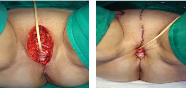
There were four radical vulvectomies, three simple vulvectomies (Figures 2A and 2B) and two wide resections (Table 1). In vulvar reconstructions we performed 11 fasciocutaneous flaps: four flap transpositions from the posterior thigh; two flaps from the medial thigh (Figures 3A and 3B); two VY advancement flaps (Figures 4A and 4B); two with simple advancing flaps; and one rotation of the
Figure 1 Figure 1 Figure 1 Figure 1
-Figure 1 - NIV usual type in patient in use of imunossupressant for renal transplant.
Table 1 Table 1 Table 1 Table 1
-Table 1 - Types of oncologic resections.
Type of Surgery Type of Surgery Type of Surgery Type of Surgery
Type of Surgery N (%)N (%)N (%)N (%)N (%)
Wide resections 2 (22%)
Simple Vulvectomy 3 (33%)
posterior thigh region (Figures 5A and 5B). In two patients, requiring extensive resection, bilateral flaps were used (Table 2).
In all patients we used suction drains in donor, recipient and lymphadenectomy regions, removing them when the flow rate in 24 hours was less than 50ml. In addition, a bladder catheter was introduced and stayed for 14 to 21 days.
The minimum postoperative follow-up was three months and the average was eight months.
There were no major losses of the flaps made. In 22% (2/9) of patients there was partial dehiscence of the inguinal wound, healed by second intention, and without
aesthetic or functional compromise. There was also a perivaginal suture dehiscence in an immunosuppressed patient, which was handled with dressing and healing occurred by second intention.
DISCUSSION
DISCUSSION
DISCUSSION
DISCUSSION
DISCUSSION
The realization of vulvectomy is not common in our hospitals, possibly because the diseases that lead to this behavior are not common, determining the referal of patients to the few institutions that have experience with the procedure. This enhances the knowledge and skill of
Figure 2 -Figure 2 -Figure 2 Figure 2
-Figure 2 - A) Raw area after vulvectomy; B) The dissection in the subfascial plan, made possible the direct approximation of the edges of the wound.
Figure 3 -Figure 3 -Figure 3 Figure 3
these professionals working in such facilities. The partnership between Gynecology and Plastic Surgery services brings even greater benefits. In addition to providing adequate treatment of the tumor, it offers tranquility and hope to patients who will have the possibility of immediate reconstruction, with the best possible functional and aesthetic results.
The liabilities are greater for Plastic Surgeon. The balance, positive or negative, of the reconstruction is usually
attributed to his/her conduct and surgical option. Patients want to be healed, but also desire, above all, to have their shapes and functions preserved. The interdisciplinary treatment increases confidence and creates partnership and complicity on the part of patients, facilitating postoperative follow-up.
In 1985, Kaplan8 pointed to the need to preserve local characteristics in vulvar reconstructions. However, unlike what we currently advocate, he used skin grafts in smaller injuries and flaps in bigger ones. He emphasized that local, fasciocutaneous-type flaps should not be used because they were not trusted or had satisfactory results. On this occasion, the knowledge of the blood supply of the flaps was not the same as today, which explains his insecurity.
Mayer and Rodriguez9 reported a case of use of abdominal flap based on the superficial external pudendal artery. They argued that the scar would be better disguised when compared to thigh flaps. Nevertheless, they presented a case where the cosmetic result does not seem acceptable
Figure 4 Figure 4 Figure 4 Figure 4
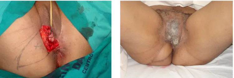
-Figure 4 - A) Extensive raw area after resection of the lesion; B) Two months after reconstruction with bilateral fasciocutaneous flaps of posterior thigh.
Figure 5 Figure 5 Figure 5 Figure 5
-Figure 5 - A) Partial Vulvectomy, leaving perineal raw area; B) Aspect four months after dermoadipose flap with V-Y advance.
Table 2 Table 2 Table 2 Table 2
-Table 2 - Types of fasciocutaneous flaps.
Type of Surgery Type of Surgery Type of Surgery Type of Surgery
Type of Surgery N (%)N (%)N (%)N (%)N (%)
Posterior thigh flap 4 (36.3%)
Medial thigh flap 2 (18.2%)
V-Y advance 2 (18.2%)
Simple advance 2 (18.2%)
and left a narrow skin island between the donor and recipient flap areas, favoring complications. We believe that the reconstructions are usually best used when the tissues have some functional and/or aesthetics interaction to the area to be repaired, the thigh and perineal surroundings being our regions of choice.
In 1995 Franco et al.10 stressed the importance of a joint work between specialties and showed the various applications of fasciocutaneous flaps from the thigh for reconstruction of the vulva. At the time, the open wound left by vulvectomies used to be larger and often in continuity with the inguinal dissection, which conferred greater risk.
Other publications reinforce our premise of using flaps to preserve the characteristics of natural sites, leave well hidden scars, keep the sensory innervation and benefit from local blood supply 4,11-13.
Salgarello et al.14, in an interesting article, created an algorithm for the reconstruction of the vulva and systematized it, taking into consideration the size and location of raw areas left after vulvectomies. They also indicate systematically the fasciocutaneous flaps, using various donor sites, including thighs. They recommend not to advance the flaps beyond the midline to avoid too much tension on its extremity and possible necrosis or dehiscence. Muneuchi et al.15, on they turn, use as the first choice for reconstruction of the vulva, the abdominal dermoadipose flap based on perforating branches of the deep inferior epigastric artery (DIEP flap). They recognize that the current preference lies with the fasciocutaneous flaps from the thigh, but they believe that the abdominal flap has a more reliable vascularization.
Lee et al.16 recommend flaps with preserved sensory innervation and of thickness adequate to the site. To do this they use an insulated VY advancement flap from the gluteal sulcus. In practice, we observed that this type of flaps is a good indication when there is preservation of the anterior half of the vulva and the raw area is not very extensive. This facilitates the advancement of the flap and the good location of the scars. Otherwise, in larger lesions, the need for large displacements and mobilization of the flaps may compromise their viability and make an array of scars that do not resemble the natural appearance of the vulva.
Staiano et al.17 perform many types of flaps, of which approximately half are fasciocutaneous from the thigh. As they treated many cases of advanced or recurrent tumors, they also used longer myocutaneous flaps and had higher rates of complications (53%). They stressed that these complications, the most common being wound
dehiscence, were influenced by multiple surgeries and radiotherapy.
In our patients, fasciocutaneous flaps from the thigh have been presented as good options for reconstruction of the vulva. The age of the patients usually determine laxity of the local tissue, which allows the availability of sufficient tissue to cover the wound, leaving little sequel in the donor area. The vascular axes of the thigh on its medial and posterior faces allow the construction of extended flaps with good viability, as well as preserving sensitivity.
Early diagnosis allows smaller resections and simpler reconstruction procedures. In some cases, after detachment from the surrounding area, the direct approximation of medium sized wounds is possible (Figure 1). In contrast, larger resections demand more complex flaps and, when combined with lymph node dissection, bring a greater chance of complications (Figure 2 and 3). The lymphorrhea observed in the inguinal region may take more than three weeks to regress and, in this period, it is advisable to maintain suction drains to accelerate its resolution. The only patient who had the drains removed inadvertently in the early postoperative period presented with inguinal dehiscence, which resolved after periodic aspiration punctures and healing by secondary intention.
For perineal and/or isolated perianal wounds, depending on the length, we used rotation, VY advance, dermoadipose or fasciocutaneous flaps from the gluteal region (Figure 4).
In the past, en block resections were common, with continuity between the genital and inguinal wounds. This allowed the accumulation of lymph beneath the flaps used for vulvar reconstruction and increased the chances of dehiscence.
Another important point to be emphasized is the postoperative maintenance of urinary catheter. Catheterization should be maintained until there is good healing of the wound around the urethra and vaginal canal, which usually happens after two to three weeks. Early withdrawal of the bladder catheter can cause urine collection beneath the flap and consequent complications.
In our sample, there was no tumor recurrence and the results allowed patients to return to normal daily life.
R E S U M O R E S U M O R E S U M O R E S U M O R E S U M O
Objetivo Objetivo Objetivo Objetivo
Objetivo: Analisar o emprego de técnicas de reconstrução imediata de vulva, pós-ressecção cirúrgica, com retalhos fasciocutâneos das faces medial e/ou posterior da coxa. MétodosMétodosMétodosMétodos: Estudo de coorte transversal, retrospectivo, para análise do resultado daMétodos reconstrução cirúrgica imediata, com retalhos fasciocutâneos em nove pacientes submetidas à vulvectomia, no período de maio de 2009 a agosto de 2010. ResultadosResultadosResultadosResultadosResultados: A média de idade foi 61 anos (variação 36 a 82 anos). Em 56% dos casos, o diagnóstico foi neoplasia intraepitelial vulvar (NIV) tipo usual. A vulvectomia radical foi realizada em 45% das pacientes, a vulvectomia simples em 33% e as ressecções amplas, em 22%. Foram confeccionados 11 retalhos fasciocutâneos, sendo 36,3% de transposições de retalho posterior de coxa, 18,2% de retalhos mediais de coxa, 18,2% de retalhos em avanço em V-Y, 18,2% de retalhos em avanço simples e 9,1% de rotação de retalho de região posterior de coxa. Não houve casos de perdas importantes dos retalhos confeccionados. Conclusão
Conclusão Conclusão Conclusão
Conclusão: Os retalhos fasciocutâneos de coxa são, atualmente, boas opções para a reconstrução imediata da vulva pós-ressecção oncológica devido à preservação da sensibilidade e da disponibilidade tecidual nas áreas doadoras. A associação do Cirurgião Plástico com o Ginecologista oferece tranquilidade às pacientes e determina bons resultados pós-operatórios.
Descritores Descritores Descritores Descritores
Descritores: Vulva. Vulva/cirurgia. Doenças da vulva. Doenças da vulva/reabilitação. Neoplasias da vulva.
REFERENCES
REFERENCES
REFERENCES
REFERENCES
REFERENCES
1. Sankaranarayanan R, Ferlay J. Worldwide burden of gynaecological cancer: the size of the problem. Best Pract Res Clin Obstet Gynaecol. 2006;20(2):207-25.
2. Judson PL, Habermann EB, Baxter NN, Durham SB, Virnig BA. Trends in the incidence of invasive and in situ vulvar carcinoma. Obstet Gynecol. 2006;107(5):1018-22.
3. van Seters M, van Beurden M, de Craen AJ. Is the assumed natural history of vulvar intraepithelial neoplasia III based on enough evidence ? A systematic review of 3322 published patients. Gynecol Oncol. 2005;97(2):645-51.
4. Höckel M, Dornhöfer N. Vulvovaginal reconstruction for neoplastic disease. Lancet Oncol. 2008;9(6):559-68.
5. McCraw JB, Dibbell DG, Carraway JH. Clinical definition of independent myocutaneous vascular territories. Plast Reconstr Surg. 1977;60(3):341-52.
6. Pecorelli S. Revised FIGO staging for carcinoma of the vulva, cervix, and endometrium. Int J Gynaecol Obstet. 2009;105(2):103-4. Erratum in: Int J Gynaecol Obstet. 2010;108(2):176.
7. Sideri M, Jones RW, Wilkinson EJ, Preti M, Heller DS, Scurry J, et al. Squamous vulvar intraepithelial neoplasia: 2004 modified terminology, ISSVD Vulvar Oncology Subcommittee. J Reprod Med. 2005;50(11):807-10.
8. Kaplan AL. Vulvar reconstruction. Clin Obstet Gynecol. 1985;28(1):211-9.
9. Mayer AR, Rodriguez RL. Vulvar reconstruction using a pedicle flap based on the superficial external pudendal artery. Obstet Gynecol. 1991;78(5 Pt 2):964-8.
10. Franco T, Conceição JCJ, Franco D, Gonçalves LFF, Silva CSC, Silva RO. Reconstrução de vulva pós-vulvectomia. Rev bras ginec obstet. 1995;17(8):803-10.
11. Spear SL, Pellegrino CJ, Attinger CE, Potkul RK. Vulvar reconstruction using a mons pubis flap. Ann Plast Surg. 1994;32(6):602-5.
12. Moschella F, Cordova A. Innervated island flaps in morphofunctional vulvar reconstruction. Plast Reconstr Surg. 2000;105(5):1649-57. 13. Hashimoto I, Nakanishi H, Nagae H, Harada H, Sedo H. The gluteal-fold flap for vulvar and buttock reconstruction: anatomic study and adjustment of flap volume. Plast Reconstr Surg. 2001;108(7):1998-2005.
14. Salgarello M, Farallo E, Barone-Adesi L, Cervelli D, Scambia G, Salerno G, et al. Flap algorithm in vulvar reconstruction after radical, extensive vulvectomy. Ann Plast Surg. 2005;54(2):184-90. 15. Muneuchi G, Ohno M, Shiota A, Hata T, Igawa HH. Deep inferior epigastric perforator (DIEP) flap for vulvar reconstruction after radical vulvectomy: a less invasive and simple procedure utilizing an abdominal incision wound. Ann Plast Surg. 2005;55(4):427-9. 16. Lee PK, Choi MS, Ahn ST, Oh DY, Rhie JW, Han KT. Gluteal fold V-Y advancement flap for vulvar and vaginal reconstruction: a new flap. Plast Reconstr Surg. 2006;118(2):401-6.
17. Staiano JJ, Wong L, Butler J, Searle AE, Barton DP, Harris PA. Flap reconstruction following gynaecological tumour resection for advanced and recurrent disease—a 12 year experience. J Plast Reconstr Aesthet Surg. 2009;62(3):346-51.
Received on 06/05/2011
Accepted for publication 01/07/2011 Conflict of interest: none
Source of funding: none
How to cite this article: How to cite this article: How to cite this article: How to cite this article: How to cite this article:
Franco D, Almeida G, Arnaut Júnior M, Arbex G, Furtado Y, Franco T. Analysis of the use of fasciocutaneous flaps for immediate vulvar reconstruction. Rev Col Bras Cir. [periódico na Internet] 2012; 39(1). Disponível em URL: http://www.scielo.br/rcbc
Mailling address: Mailling address: Mailling address: Mailling address: Mailling address: Diogo Franco


