Predictive factors for short gastric vessels division during
Predictive factors for short gastric vessels division during
Predictive factors for short gastric vessels division during
Predictive factors for short gastric vessels division during
Predictive factors for short gastric vessels division during
laparoscopic total fundoplication
laparoscopic total fundoplication
laparoscopic total fundoplication
laparoscopic total fundoplication
laparoscopic total fundoplication
Fatores preditivos da necessidade de secção dos vasos gástricos curtos nas
Fatores preditivos da necessidade de secção dos vasos gástricos curtos nas
Fatores preditivos da necessidade de secção dos vasos gástricos curtos nas
Fatores preditivos da necessidade de secção dos vasos gástricos curtos nas
Fatores preditivos da necessidade de secção dos vasos gástricos curtos nas
fundoplicaturas totais videolaparoscópicas
fundoplicaturas totais videolaparoscópicas
fundoplicaturas totais videolaparoscópicas
fundoplicaturas totais videolaparoscópicas
fundoplicaturas totais videolaparoscópicas
A
LEXANDREC
HARTUNIP
EREIRAT
EIXEIRA1; F
ERNANDOA
UGUSTOM
ARDIROSH
ERBELLA1; A
DORÍSIOB
ONADIMAN2; J
OSÉF
RANCISCODEM
ATTOSF
ARAH, ACBC-SP
2; J
OSÉC
ARLOSD
ELG
RANDE, TCBC-SP
1A B S T R A C T
A B S T R A C T
A B S T R A C T
A B S T R A C T
A B S T R A C T
Objective: Objective: Objective: Objective:
Objective: to determine clinical variables that can predict the need for division of the short gastric vessels (SGV), based on the gastric fundus tension, assessing postoperative outcomes in patients submitted or not to section of these vessels. MethodsMethodsMethodsMethodsMethods: we analyzed data from 399 consecutive patients undergoing laparoscopic fundoplication for gastroesophageal reflux disease (GERD). The section of the SGV was performed according to the surgeon evaluation, based on the fundus tension. Patients were divided into two groups: not requiring SGV section (group A) or requiring SGV section (group B). ResultsResultsResultsResultsResults: the section was not necessary in 364 (91%) patients (Group A) and required in 35 (9%) patients (Group B). Group B had proportionally more male patients and higher average height. The endoscopic parameters were worse for Group B, with larger hiatal hernias, greater hernias proportion with more than four centimeters, more intense esophagitis, higher proportion of Barrett’s esophagus and long Barrett’s esophagus. Male gender and grade IV-V esophagitis were considered independent predictors in the multivariate analysis. Transient dysphagia and GERD symptoms were more common in Group B. ConclusionConclusionConclusionConclusionConclusion: the division of the short gastric vessels is not required routinely, but male gender and grade IV-V esophagitis are independent predictors of the need for section of these vessels.
Key words: Key words: Key words: Key words:
Key words: Fundoplication. Video-Assisted Surgery. Gastroesophageal Reflux. Gastric Fundus.
1. Departamento de Cirurgia, Escola Paulista de Medicina, Universidade Federal de São Paulo - SP - Brazil; 2. Departamento de Cirurgia Geral e Oncológica, Hospital do Servidor Público Estadual de São Paulo - SP - Brazil.
INTRODUCTION
INTRODUCTION
INTRODUCTION
INTRODUCTION
INTRODUCTION
L
aparoscopic total fundoplication is an effective procedure
for the treatment of gastroesophageal reflux disease
(GERD)
1. However, some technical points are still
controversial, especially the need for short gastric vessels
(SGV) division
2. While most authors believe this step brings
better results
3-5, others showed similar outcomes whether
SGV are divided or not or even complications attributed to
SGV division
2,6-9.
This study aims to determine: (a) clinical variables
that may predict the need of SGV division based on gastric
fundus tension and (b) the outcomes in patients with or
without SGV division.
METHODS
METHODS
METHODS
METHODS
METHODS
We retrospectively studied 399 consecutive
patients (50% male, mean age 49 years) recorded in a
prospectively kept database that underwent laparoscopic
total fundoplication for the surgical treatment of GERD. This
study was approved by the local institutional review board.
(CEP 0742/11).
Patients were questioned before the operation
regarding the presence of symptoms. These were grouped
into esophageal symptoms (heartburn and regurgitation),
extra-esophageal symptoms (thoracic pain, respiratory
symptoms, such as cough and asthma or ear, nose and
throat symptoms) or dysphagia. Anthropometric variables
were also recorded. Individuals with partial fundoplication,
paraesophageal hernia, previous foregut operation or
conversion to conventional laparotomy were excluded from
the analysis.
All patients were submitted to an upper
endoscopy to evaluate the presence of hiatal hernia (HH),
esophagitis and Barrett´s esophagus. HH was classified
according to size in <4cm or
>
4cm. Modified Savary-Miller
endoscopic classification
10was used for grading esophagitis.
patients without esophagitis or with atypical symptoms.
pHmonitoring results were available for review in 62 (15%).
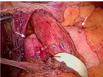
Surgical technique has been previously described
2.
In summary, an extensive mobilization of the posterior wall
of the gastric fundus followed the dissection of the distal
esophagus and diaphragmatic crus in all patients. SGV
division was done at the discretion of the surgeon based on
tension of the gastric fundus after performing a specific
maneuver (“drop-test” - Figure 1). A short-floppy total
fundoplication was performed without the aid of a bougie.
All procedures were performed by or under the supervision
of a single experienced surgeon. Patients were grouped
according to the necessity for SGV division (Group A – no
division; Group B – SGV division).
Follow-up visits were scheduled for 15, 30, 90,
180 and 360 days after the surgery and then annually,
irrespective of the presence of symptoms. Upper endoscopy
was performed annually or earlier if the patient had any
complaints related to the postoperative period. All selected
patients had at least a 6-month postoperative follow-up
period.
Chi-square, Student’s t test and logistic regression
were used when necessary. A value of p was considered
significant at the 0.05 level. Variables are expressed as
mean ± standard deviation.
RESULTS
RESULTS
RESULTS
RESULTS
RESULTS
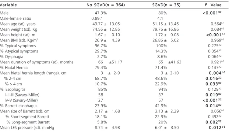
SGV division was deemed not necessary in 364
(91%) patients (Group A) but required in 35 (9%) patients
(Group B). Demographic data, symptoms distribution,
endoscopic and manometric data are depicted in table 1.
Group B had more males and a higher height. Endoscopic
parameters were worse for group B, with larger hiatal
hernias, higher proportion of hiatal hernias >4cm, more
severe esophagitis (Grade IV-V), higher proportion of Barrett
esophagus, and higher rate of long-segment Barrett
esophagus. Manometric parameters also disfavored group
B with decreased lower esophageal sphincter basal pressure.
Only male gender and grade IV-V esophagitis stood as
independent predictive factors for the need of SGV division
at the multivariate analysis (Table 2).
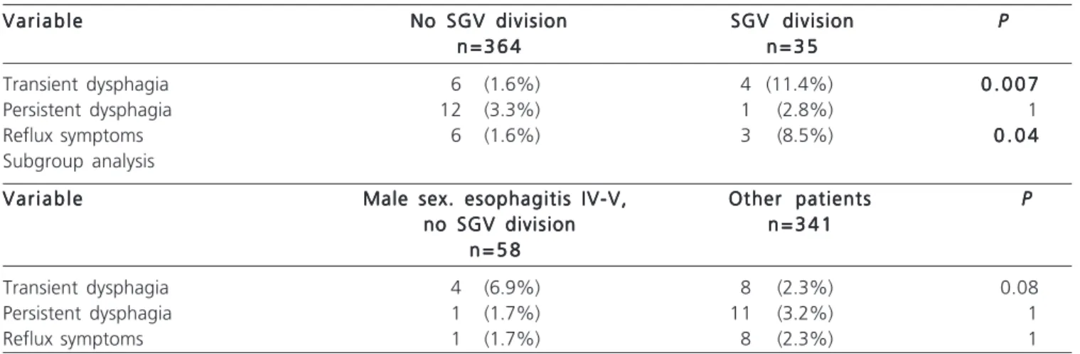
Average follow-up was 13.8 months. Outcomes
at last follow-up are depicted in table 3. Transient dysphagia
and GERD symptoms were more common in Group B.
If patients at higher risk for gastric fundus tension
(namely males with severe esophagitis) that did not
underwent SGV division are compared to the remaining
patients no difference in symptoms were noticed.
DISCUSSION
DISCUSSION
DISCUSSION
DISCUSSION
DISCUSSION
Our results show that: (a) male gender and severe
esophagitis are independent predictors for the need to SGV
division and (b) SGV division may lead to more transitory
dysphagia and GERD symptoms.
The effect of SGV division on the outcomes of
laparoscopic fundoplication have been evaluated in five
prospective randomized studies
2,6-9and their meta-analysis
11-14. These studies showed a longer operative time and
intraoperative bleeding
2,6,8,9,11-14, higher incidence of transient
dysphagia
2and gas-bloating syndrome
7when SGV are
divided. Our results also showed more postoperative GERD
symptoms. No benefit has been attributed to SGV division
15.
Very interestingly, however, authors that do not routinely
divide SGV find this step necessary at the time of the
fundoplication due to tension on the gastric fundus in up to
33% of patients
16even after extensive gastric fundus
mobilization by lysis of adhesions between the stomach
and diaphragm
2,6.
In this study, 8.8% (35/399) of the patients
needed SGVD for the completion of a short floppy
fundoplication. The likely cause of this low need of SGVD
is the extensive gastric fundus mobilization employed in all
patients, as advocated by Farah et al
2and Chrysos et al
6.
We believe that when this surgical step is used, one makes
a larger length of the gastric fundus available for the
construction of a floppy fundoplication around the
esophagus. So, even in those cases in which there is an
enlarged cardia, this maneuver lessens the likelihood of
the need of SGVD.
Some previous studies attempted to identify
anatomic parameters to predict gastric fundus tension and
consequent need to SGV division. Szor et al.
16found some
sort of tension in half of the cases during fundoplications in
cadavers, but no anatomic parameter predicted this tension.
Huntington et al.
17deemed necessary to divided SGV in
some patients based on the gastric fundus length and the
esophageal circumference. Severe esophagitis as a predictor
for fundus tension in our study may be linked to esophageal
circumference as progressive dilatation of the esophageal
Figure 1 Figure 1 Figure 1 Figure 1
-Figure 1 - Gastric fundus in place without the need of traction
diameter is observed as esophagitis severity increases
18,19.
Male gender may bring a higher chance of fundus tension
probably due to more exuberant visceral fat compared to
females. To the best of our knowledge, no other series
studied clinical parameters to predict gastric fundus
tension.
The current study studied a large number of
patients; however, it has the limitations of a retrospective
case series. As such, some parameters that could help
understand the results of the study were not evaluated,
such as the amount of visceral fat. Also, the time of
follow-up is short for a stronger conclusion that SGV division does
not affect long-term outcomes. More importantly, even
though a single surgeon operated all cases, gastric fundus
tension was based on subjective parameters.
We conclude that SGV division is not necessary
routinely but male sex and grade IV-V esophagitis are
independent predictors of the need of SGV division.
However, not all patients in these conditions need SGV
division as a subanalysis of these population that did not
underwent this step did not show worse outcomes compared
to other patients. Gastric fundus tension must still be
evaluated based on subjective parameters by experienced
surgeons.
Table 1 -Table 1 -Table 1 Table 1
-Table 1 - Demographic data, symptoms distribution, endoscopic and manometric data.
V a r i a b l e V a r i a b l eV a r i a b l e V a r i a b l e
V a r i a b l e No SGVD(n = 364)No SGVD(n = 364)No SGVD(n = 364)No SGVD(n = 364)No SGVD(n = 364) SGVD(n = 35)SGVD(n = 35)SGVD(n = 35)SGVD(n = 35)SGVD(n = 35) PPPPP Value Value Value Value Value Male 47.3% 80% < 0 . 0 0 1< 0 . 0 0 1< 0 . 0 0 1< 0 . 0 0 1< 0 . 0 0 1X 2X 2X 2X 2X 2
Male-female ratio 0.89:1 4:1
Mean age (sd). years 49.77 ± 13.05 51.15 ± 13.46 0.564t-S
Mean weight (sd). Kg 74.56 ± 12.85 79.76 ± 16.86 0.084t-S
Mean height (sd). m 1.67 ± 0.10 1.72 ± 0.08 < 0 . 0 0 1< 0 . 0 0 1< 0 . 0 0 1< 0 . 0 0 1< 0 . 0 0 1t - St - St - St - St - S
Mean BMI (sd). Kg/m2 26.9 ± 4.39 26.86 ± 5.02 0.969t-S
% Typical symptoms 96.7% 100% 0.275X2
% Atypical symptoms 29.7% 14.3% 0.054X2
% Dysphagia 2.7% 8.6% 0.064X2
Mean duration of symptoms (sd). months 66 ±51.17 65 ±41.63 0.921t-S
% Hiatal Hernia 79.4% 71.4% 0.137X2
Mean hiatal hernia length (range). cm 3 ± 2-9 3 ± 2-10 0 . 0 0 40 . 0 0 40 . 0 0 40 . 0 0 40 . 0 0 4t - St - St - St - St - S
% 2-4 cm 68.7% 48.6% 0 . 0 1 60 . 0 1 60 . 0 1 60 . 0 1 60 . 0 1 6X 2X 2X 2X 2X 2
% > 4 cm 10.7% 22.9% 0 . 0 3 30 . 0 3 30 . 0 3 30 . 0 3 30 . 0 3 3X 2X 2X 2X 2X 2
% Esophagitis 85% 94% 0.129X2
I-II-III (Savary-Miller) 58 37 0 . 0 1 90 . 0 1 90 . 0 1 90 . 0 1 90 . 0 1 9X 2X 2X 2X 2X 2
IV-V (Savary-Miller) 27 57 < 0 . 0 0 1< 0 . 0 0 1< 0 . 0 0 1< 0 . 0 0 1< 0 . 0 0 1X 2X 2X 2X 2X 2
% Barrett esophagus 23.9% 42.9% 0 . 0 1 40 . 0 1 40 . 0 1 40 . 0 1 40 . 0 1 4X 2X 2X 2X 2X 2
Mean size of Barrett (sd). cm 2.17 ± 1.68 3.13 ± 2.29 0.056t-S
% Short-segment Barrett 18.1% 22.9% 0.492X2
% Long-segment Barrett 5.8% 20% 0 . 0 0 20 . 0 0 20 . 0 0 20 . 0 0 20 . 0 0 2X 2X 2X 2X 2X 2
Mean LES pressure (sd). mmHg 8.74 ± 4.98 6.01 ± 3.50 0 . 0 1 20 . 0 1 20 . 0 1 20 . 0 1 20 . 0 1 2t - St - St - St - St - S
SGVD: short gastric vessels division; n: number; sd: standard deviation; y: years; m: meters; mo: months; cm: centimeters; LES: lower esophageal sphincter; X2: chi-square; t-S: Student’s t test.
Table 2 -Table 2 -Table 2 Table 2
-Table 2 - Multivariate analysis for the need to short gastric vessels division.
V a r i a b l e V a r i a b l eV a r i a b l e V a r i a b l e
V a r i a b l e Odds RatioOdds RatioOdds Ratio(95% CI)Odds RatioOdds Ratio(95% CI)(95% CI)(95% CI)(95% CI) PPPPP Value Value Value Value Value
Male gender 28.3 (2.25-355.22) 0 . 0 1 00 . 0 1 00 . 0 1 00 . 0 1 00 . 0 1 0
Weight 1.03 (0.98-1.09) 0.216
Hiatal hernia length 1.64 (0.75-3.59) 0.213
Esophagitis grade IV-V 19.5 (2.08-182.26) 0 . 0 0 90 . 0 0 90 . 0 0 90 . 0 0 90 . 0 0 9
Barrett esophagus and extension 0.11 (0.01-1.06) 0.056
LES pressure 0.79 (0.62-1.01) 0.063
R E S U M O
R E S U M O
R E S U M O
R E S U M O
R E S U M O
Objetivo: Objetivo: Objetivo: Objetivo:
Objetivo: determinar variáveis clínicas que possam predizer a necessidade de secção dos vasos gástricos curtos (VGC), baseado na tensão do fundo gástrico, avaliando os resultados pós-operatórios em pacientes submetidos ou não à secção destes vasos. Métodos:
Métodos: Métodos: Métodos:
Métodos: foram analisados os dados de 399 pacientes consecutivos submetidos à fundoplicatura total laparoscópica para a doença do refluxo gastroesofágico (DRGE). A secção dos VGC foi realizada de acordo com a avaliação do cirurgião, baseado na tensão do fundo gástrico. Os pacientes foram distribuídos em dois grupos: sem necessidade de secção dos VGC (grupo A) ou com necessidade de secção dos VGC (grupo B). Resultados:Resultados:Resultados:Resultados:Resultados: A secção não foi necessária em 364 (91%) pacientes (Grupo A) e necessária em 35 (9%) pacientes (Grupo B). O Grupo B tinha proporcionalmente mais pacientes do sexo masculino e maior estatura média. Os parâmetros endoscópicos foram piores para o Grupo B, com maiores hérnias hiatais, maior proporção de hérnias com mais de quatro centímetros, esofagite mais intensa, maior proporção de esôfago de Barrett e esôfago de Barrett longo. O sexo masculino e as esofagites graus IV-V foram considerados fatores preditivos independentes na análise multivariada. A disfagia transitória e os sintomas de DRGE foram mais comuns no Grupo B. Conclusão:Conclusão:Conclusão:Conclusão:Conclusão: A secção dos vasos gástricos curtos não é necessária rotineiramente, porém o sexo masculino e as esofagites graus IV-V são fatores preditivos independentes da necessidade da secção destes vasos.
Descritores: Descritores: Descritores: Descritores:
Descritores: Fundoplicatura. Cirurgia Vídeoassistida. Refluxo Gastroesofágico. Fundo Gástrico.
REFERENCES
REFERENCES
REFERENCES
REFERENCES
REFERENCES
1. Dallemagne B, Weerts J, Markiewicz S, Dewandre JM, Wahlen C, Monami B, et al. Clinical results of laparoscopic fundoplication at ten years after surgery. SurgEndosc. 2006;20(1):159-65. 2. Farah JFM, Grande JCD, Goldenberg A, Martinez JC, Lupinacci
RA, Matone J. Randomized trial of total fundoplication and fundal mobilization with or without division of short gastric vessels: a short-term clinical evaluation. Acta Cir Bras. 2007;22(6):422-9.
3. Soper NJ, Dunnegan D. Anatomic fundoplication failure after laparoscopic antireflux surgery. Ann Surg. 1999;229(5):669-76; discussion 676-7.
4. Wu JS, Dunnegan DL, Luttmann DR, Soper NJ. The influence of surgical technique on clinical outcome of laparoscopic Nissen fundoplication. Surg Endosc. 1996;10(12):1164-69; discussion 1169-70.
5. Hunter JG, Swanstrom L, Waring JP. Dysphagia after laparoscopic antireûux surgery. The impact of operative technique. Ann Surg. 1996;224(1):51-7.
6. Chrysos E, Tzortzinis A, Tsiaoussis J, Athanasakis H, Vasssilakis J, Xynos E. Prospective randomized trial comparing Nissen to Nissen-Rossetti technique for laparoscopic fundoplication. Am J Surg. 2001;182(3):215-21.
7. O‘Boyle CJ, Watson DI, Jamieson GG, Myers JC, Game PA, Devitt PG. Division of short gastric vessels at laparoscopic Nissen fundoplication: a prospective double-blind randomized trial with 5-year follow-up. Ann Surg. 2002;235(2):165-70.
8. Blomqvist A, Dalenbäck J, Hagedorn C, Lönroth H, Hyltander A, Lundell L. Impact of complete gastric fundus mobilization on outcome after laparoscopic total fundoplication. J Gastrointest Surg. 2000;4(5):493-500.
9. Watson DI, Pike GK, Baigrie RJ, Mathew G, Devitt PG, Britten-Jones R, et al. Prospective double-blind randomized trial of laparoscopic Nissen fundoplication with division and without division of short gastric vessels. Ann Surg. 1997;226(5):642-52.
10. Savary M, Miller G. The esophagus. Handbook and atlas of endoscopy. Solothurn, Switzerland: Grassman; 1978. p.135-42 11. Engström C, Jamieson GG, Devitt PG, Watson DI. Meta-analysis of
two randomized controlled trials to identify long-term symptoms after division of the short gastric vessels during Nissen fundoplication. Br J Surg. 2011;98(8):1063-7.
Table 3 Table 3 Table 3 Table 3
-Table 3 - Clinical follow-up.
V a r i a b l e V a r i a b l e V a r i a b l e V a r i a b l e
V a r i a b l e No SGV divisionNo SGV divisionNo SGV divisionNo SGV divisionNo SGV division SGV divisionSGV divisionSGV divisionSGV divisionSGV division PPPPP n = 3 6 4
n = 3 6 4 n = 3 6 4 n = 3 6 4
n = 3 6 4 n = 3 5n = 3 5n = 3 5n = 3 5n = 3 5
Transient dysphagia 6 (1.6%) 4 (11.4%) 0 . 0 0 70 . 0 0 70 . 0 0 70 . 0 0 70 . 0 0 7
Persistent dysphagia 12 (3.3%) 1 (2.8%) 1
Reflux symptoms 6 (1.6%) 3 (8.5%) 0 . 0 40 . 0 40 . 0 40 . 0 40 . 0 4
Subgroup analysis V a r i a b l e
V a r i a b l e V a r i a b l e V a r i a b l e
V a r i a b l e Male sex. esophagitis IV-V,Male sex. esophagitis IV-V,Male sex. esophagitis IV-V,Male sex. esophagitis IV-V,Male sex. esophagitis IV-V, Other patientsOther patientsOther patientsOther patientsOther patients PPPPP no SGV division
no SGV division no SGV division
no SGV division no SGV division n = 3 4 1n = 3 4 1n = 3 4 1n = 3 4 1n = 3 4 1 n = 5 8
n = 5 8n = 5 8 n = 5 8n = 5 8
Transient dysphagia 4 (6.9%) 8 (2.3%) 0.08
Persistent dysphagia 1 (1.7%) 11 (3.2%) 1
Reflux symptoms 1 (1.7%) 8 (2.3%) 1
12. Markar SR, Karthikesalingam AP, Wagner OJ, Jackson D, Hewes JC, Vyas S, et al. Systematic review and meta-analysis of laparoscopic Nissen fundoplication with or without division of the short gastric vessels. Br J Surg. 2011;98(8):1056-62.
13. Khatri K, Sajid MS, Brodrick R, Baig MK, Sayegh M, Singh KK. Laparoscopic Nissen fundoplication with or without short gastric vessel division: a meta-analysis. Surg Endosc. 2012;26(4):970-8. 14. Kösek V, Wykypiel H, Weiss H, Höller E, Wetscher G, Margreiter R,
et al. Division of the short gastric vessels during laparoscopic Nissen fundoplication: clinical and functional outcome during long-term follow-up in a prospectively randomized trial. SurgEndosc. 2009;23(10):2208-13.
15. Bonadiman A, Teixeira AC, Goldenberg A, Farah JF. Dysphagia after laparoscopic total fundoplication: anterior or posterior gastric wall fundoplication?.Arq Gastroenterol. 2014;51(2):113-7. 16. Szor DJ, Herbella FA, Bonini AL, Moreno DG, Del Grande JC. Gastric
fundus tension before and after division of the short gastric vessels in a cadaveric model of fundoplication. Dis Esophagus. 2009;22(6):539-42.
17. Huntington TR, Danielson L. Variation in fundic dimensions with respect to short gastric vessel division in laparoscopic fundoplication. SurgEndosc. 2001;15(1):76-9.
18. Csendes A, Miranda M, Espinoza M, Velasco N, Henríquez A. Perimeter and location of the muscular gastroesophageal junction or ‘cardia’ in control subjects and in patients with reflux esophagitis or achalasia. Scand J Gastroenterol. 1981;16(7):951-6.
19. Korn O, Csendes A, Burdiles P, Braghetto I, Stein HJ. Anatomic dilatation of the cardia and competence of the lower esophageal sphincter: a clinical and experimental study. J Gastrointest Surg. 2000;4(4):398-406.
Received on 28/09/2014
Accepted for publication 20/10/2014 Conflict of interest: none.
Source of funding: CAPES (Coordenação de Aperfeiçoamento de Pessoal de Nível Superior).