Citophotometric expression of Caspase-3 in papillary thyroid
Citophotometric expression of Caspase-3 in papillary thyroid
Citophotometric expression of Caspase-3 in papillary thyroid
Citophotometric expression of Caspase-3 in papillary thyroid
Citophotometric expression of Caspase-3 in papillary thyroid
carcinoma in nodular goiter colloid
carcinoma in nodular goiter colloid
carcinoma in nodular goiter colloid
carcinoma in nodular goiter colloid
carcinoma in nodular goiter colloid
Expressão citofotométrica da Caspase-3 no carcinoma papilífero da tireóide e
Expressão citofotométrica da Caspase-3 no carcinoma papilífero da tireóide e
Expressão citofotométrica da Caspase-3 no carcinoma papilífero da tireóide e
Expressão citofotométrica da Caspase-3 no carcinoma papilífero da tireóide e
Expressão citofotométrica da Caspase-3 no carcinoma papilífero da tireóide e
no bócio colóide
no bócio colóide
no bócio colóide
no bócio colóide
no bócio colóide
LUCIANA RODRIGUES QUEIROZDE SOUZA1; JURANDIR MARCONDES RIBAS-FILHO, TCBC-PR2; OSVALDO MALAFAIA, ECBC-PR2; NICOLAU GREGORI
CZECZKO, TCBC-PR2; CARMEN AUSTRALIA PAREDES MARCONDES RIBAS2; GLEIN DIASDE SOUZA1; CARLOS HESPANHA MARINHO-JUNIOR1
A B S T R A C T A B S T R A C T A B S T R A C T A B S T R A C T A B S T R A C T
Objective: Objective: Objective: Objective:
Objective: To quantitatively describe the cytophotometric expression of caspase-3 labeling in colloid nodular goiter and papillary carcinoma of the thyroid and compare its immunoexpression between these diseases. MethodsMethodsMethodsMethodsMethods: We performed caspase-3 protein immunohistochemical studies in 17 paraffin blocks from papillary thyroid carcinomas and 20 from thyroid colloid goiters through the SAMBA 4000 system - (Automatic Search Microscopic Analysis System), aiming the assessment of two variables: labeling index and optical density. Results: Results: Results: Results: Results: There was significant difference in caspase-3 labeling index between colloid goiter and papillary carcinoma, higher in carcinoma, and there was no significant difference in optical density. For colloid goiter, the correlation coefficient between labeling index and optical density was 0.72, thus rejecting the null hypothesis (p<0.001), indicating that there is a significant positive association between the labeling index and the optical density of caspase-3. For papillary thyroid carcinoma, the correlation coefficient between labeling index and optical density was 0.34. The statistical tests’ results indicated that it cannot be stated that there is an association between these parameters. ConclusionConclusionConclusionConclusionConclusion: For colloid goiter there is a positive and significant association between the variables labeling index and optical density of caspase-3, while for papillary carcinoma there is no such association. The comparative study between the quantitative analysis of caspase-3 showed that apoptosis is more evident in papillary carcinoma than in colloid goiter.
Key words Key words Key words Key words
Key words: Papillary carcinoma. Thyroid. Colloid goiter. Caspase-3. Immunohistochemistry.
Work done at the Medical Research Institute of the Curitiba Evangelical Hospital, Evangelical University of Paraná, Curitiba, Paraná, Brazil. 1. Master’s Degree Graduate, Post-Graduation in Principles of Surgery, Medical Research Institute of the Evangelical University of Paraná / Evangelical University Hospital in Curitiba, Curitiba, Paraná, Brazil; 2. PhD, Associate Professor, Post-Graduation in Principles of Surgery, Medical Research Institute of the Evangelical University of Paraná / Evangelical University Hospital in Curitiba, Curitiba, Paraná, Brazil.
INTRODUCTION
INTRODUCTION
INTRODUCTION
INTRODUCTION
INTRODUCTION
C
olloid (or nodular) goiter is an increase in volume of the thyroid gland caused by hyperplasia of the parenchyma, leading to a state of excessive follicular proliferation. The classification of goiter is described as diffuse and nodular, and it is uni or multinodular. According to the production of thyroid hormone, anatomo-clinical separation is made in toxic (or hyperactive) and nontoxic. The classification can also be in endemic or sporadic, given the endemic goiter that affects more than 10% of the population of a specific geographical area and sporadic goiter being caused by a range of factors – environmental, immunological and genetic – that interfere with hormone synthesis.Thyroid cancer is the most common malignancy of the endocrine system, predominating in females between 25 to 65 years old. As risk factors, exposure to radiation in
head and neck, personal history of goiter, thyroid nodule and family background of thyroid cancer are important1.
These tumors may histologically consist of two types of epithelial cells, parafollicular cells, which originate the medullary carcinomas and follicular cells, which give rise to adenomas and differentiated (papillary and follicular) or undifferentiated carcinomas (anaplastic)2.
of apoptosis. Caspases are cysteine proteases that have a cysteine at the active site and cleave their residues in a point further to the aspartic acid, resulting in functional inactivation or altering the activation of the substrate, either by cleavage of an inhibitory domain in the molecule itself or by inactivation of an inhibitory molecule6.
This paper aims to describe the quantitative cytophotometric expression of caspase-3 in colloid goiter and papillary carcinoma of the thyroid and compare the results of caspase-3 immunoexpression between them.
METHODS
METHODS
METHODS
METHODS
METHODS
The study material consisted of 37 paraffin blocks, 20 from colloid goiter and 17 from thyroid papillary carci-noma.
The blocks were cut on a rotation microtome with histological sections five microns thick. Two slides were prepared, one stained with H/E for the validation of the previously established diagnosis by two pathologists and the other stained by immunohistochemistry to study the caspase-3.
Preparation for analysis with Preparation for analysis withPreparation for analysis with Preparation for analysis withPreparation for analysis with immunohistochemistry
immunohistochemistry immunohistochemistry immunohistochemistry immunohistochemistry
Immunohistochemical analysis lasted 48 hours. The slides were placed in buckets for two sequential baths of xylene for 10 minutes, two of 100% alcohol for three minutes, one of 70% alcohol for three minutes and finally two baths with distilled water for one minute.
For antigen retrieval we used a buffer solution composed of sodium citrate (0.7 mmol/l), citric acid (0.6 mmol/l) and 450 ml of distilled water in the recipients containing the slides, taken to heat up to a temperature above 97°C for 20 minutes. Later they were left at room temperature for 30 minutes to cool and the recipients were rinsed three times in running water.
To block the endogenous peroxidase two baths of 2% hydrogen peroxide were performed for five minutes each. Then the slides were washed in water and placed in bottles with phosphate buffered saline (PBS) buffer consisted of 5.52 g monobasic sodium phosphate, 22.7 g of bibasic disodium phosphate and 1000 ml of distilled water.
For incubation of the slides with the antibody the recipient containing the first antibody (caspase-3) was initially shaken in the vortex machine to homogenization. The solution of this antibody was placed on slides and stored overnight in the refrigerator at 4°C.
After 12 hours, the slides were washed with distilled water, dried and placed individually in PBS for five minutes. Then they were dried and aligned in the incubation tray to hydrate the tissue.
In the sequence the second antibody (biotin) was pipetted, making the connection between the first
(caspase-3) and streptoavidin HRP, and between this and biotin, for 30 minutes of incubation. To finish the process, we put one drop of chromogen (DAB), amine-benzidine dioxide, which ligates with the streptoavidin.
I m m u n o h i s t o c h e m i s t r y q u a n t i t a t i v e I m m u n o h i s t o c h e m i s t r y q u a n t i t a t i v e I m m u n o h i s t o c h e m i s t r y q u a n t i t a t i v e I m m u n o h i s t o c h e m i s t r y q u a n t i t a t i v e I m m u n o h i s t o c h e m i s t r y q u a n t i t a t i v e analysis
analysis analysis analysis analysis
This process was evaluated by SAMBA 4000® System - automatic search microscopic analysis system. We used an Axioskop® microscope.
The luminous flux coming from the xenon bulb was controlled by a potentiometer capable of accurately assessing the amount of light. This beam passed through the condenser, the histological slide, the lens and then was separated in two parts: one beam for observation through the eyepiece of the microscope and the other for image capturing by the camcorder, which was sent to the monitor attached to the computer. The video camera used was a DXC-970M3CCD®, which standardizes the colors green, blue and red, so that the system works on the same level of the determined capture.
We used a Pentium® III computer with 16MB RAM and 12 gigabytes hard drive, which performed the functions of image importing from the microscope and video camera, execution of SAMBA 4000 system IMMUNO® software and data transfer for printing.
The analog images were captured with the camera and transformed into digital ones. The analysis aimed to transform the images stained by the marker in a numeric matrix; mathematical parameters were calculated that enabled the analysis of the microscopic images. Those images were processed using the SAMBA 4000 system and digitized in image points (pixels).
T h e l i g h t a b s o r b e d b y t h e t i s s u e w a s quantified and expressed in the range of variations of gray levels, ranging from 0 (black) to 255 (white). This process corresponds to the numbering of the image and involved two steps: generation of a matrix in gray levels and the transformation of that matrix in a binary numeric one.
Parameters analyzed by the Samba 4000 system The IMMUNO® program was used to analyze two variables that quantitatively characterized the histochemical staining reactions: labeling index and mean optical density.
The labeling index comprises the percentage of tissue area specifically marked by the immunohistochemistry test. The mean optical density reflects the intensity of the staining.
Statistical analysis Statistical analysis Statistical analysis Statistical analysis Statistical analysis
RESULTS
RESULTS
RESULTS
RESULTS
RESULTS
Colloid goiter Colloid goiter Colloid goiter Colloid goiter Colloid goiter
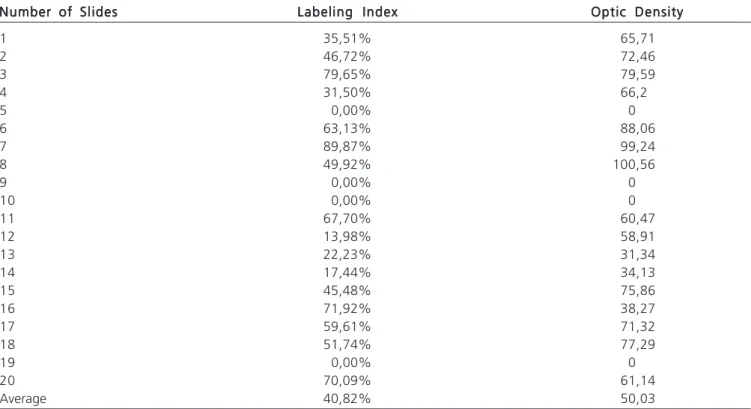
The average labeling index in the 20 colloid goiter slides was 40.82% and the optical density of 50.03 (Table 1).
The frequency of normality presented by the labeling index in colloid goiter predominated in the ranges from 25.1 to 50 (25%) and 50.1 to 75 (30%).
The frequency of normality displayed by the optical density in colloid goiter was more common in the ranges from 75.1 to 100 (25%) and 50.1 to 75 (35%).
Figure 1 illustrates the expression of caspase-3, staining in brown in colloid goiter slides.
Thyroid papillary carcinoma Thyroid papillary carcinoma Thyroid papillary carcinoma Thyroid papillary carcinoma Thyroid papillary carcinoma
The average labeling index in the 17 papillary carcinoma slides was 83.38% and the optical density of 64.57 (Table 2).
The frequency of normality presented by the labeling index in papillary carcinoma predominated in the ranges from 80.1 to 90 (17.65%) and 90.1 to 100 (58.82%). The frequency of normality displayed by the optical density was more common in the ranges from 50.1 to 60 (17.65%) and 60.1 to 70 (47.6%).
Figure 2 illustrates the expression of brownish staining caspase-3 in slides of thyroid papillary carcinoma. Correlation of the caspase-3 labeling index between colloid goiter and papillary thyroid carcinoma
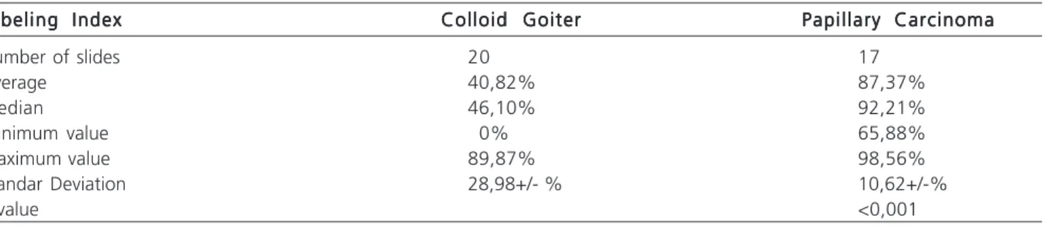
The correlation of the mean, median, minimum, maximum and standard deviation of the caspase-3 labeling index expression between colloid goiter and thyroid papillary carcinoma is shown in Table 3.
Correlation of caspase-3 optical density Correlation of caspase-3 optical density Correlation of caspase-3 optical density Correlation of caspase-3 optical density Correlation of caspase-3 optical density between colloid goiter and thyroid papillary carci-between colloid goiter and thyroid papillary carci-between colloid goiter and thyroid papillary between colloid goiter and thyroid papillary between colloid goiter and thyroid papillary carci-noma
nomanoma noma noma
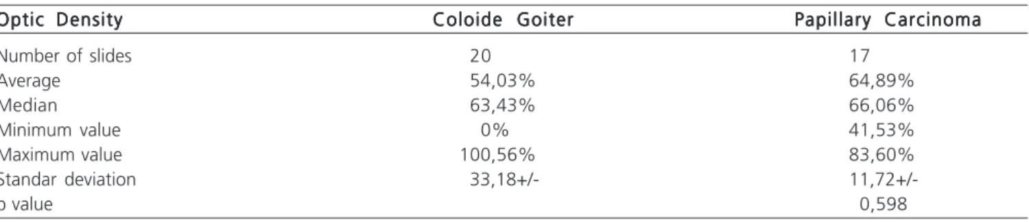
The correlation of the mean, median, minimum, maximum and standard deviations of the caspase-3 optical density expression between colloid goiter and papillary car-cinoma is shown in Table 4.
Table 1 Table 1 Table 1 Table 1
Table 1 - Expression of quantitative variables by caspase-3 for colloid goiter. Number of Slides
Number of Slides Number of Slides Number of Slides
Number of Slides Labeling IndexLabeling IndexLabeling IndexLabeling IndexLabeling Index Optic DensityOptic DensityOptic DensityOptic DensityOptic Density
1 35,51% 65,71
2 46,72% 72,46
3 79,65% 79,59
4 31,50% 66,2
5 0,00% 0
6 63,13% 88,06
7 89,87% 99,24
8 49,92% 100,56
9 0,00% 0
10 0,00% 0
11 67,70% 60,47
12 13,98% 58,91
13 22,23% 31,34
14 17,44% 34,13
15 45,48% 75,86
16 71,92% 38,27
17 59,61% 71,32
18 51,74% 77,29
19 0,00% 0
20 70,09% 61,14
Average 40,82% 50,03
F i g u r e 1 F i g u r e 1 F i g u r e 1 F i g u r e 1
Correlation of caspase-3 variables for each Correlation of caspase-3 variables for eachCorrelation of caspase-3 variables for each Correlation of caspase-3 variables for eachCorrelation of caspase-3 variables for each disease
disease disease disease disease
The correlation between optical density and labeling index for caspase-3 in colloid goiter is shown in Figure 3.
The correlation between optical density and labeling index for caspase-3 in papillary carcinoma is shown in Figure 4.
DISCUSSION
DISCUSSION
DISCUSSION
DISCUSSION
DISCUSSION
In analyzing the frequency of normality presented by the labeling index and the optical density in colloid goiter we observed that the pattern shown in both cases was abnormal, thereby demonstrating the variability of apoptotic responses. Thus, the pattern of abnormality presented in the two variables used demonstrates that the response to apoptosis occurs in a non-homogeneous pattern, with variability in apoptotic responses, as described by El May
Table 2 Table 2 Table 2 Table 2
-Table 2 - Quantitative Expression of variables by caspase-3 for papillary carcinoma. Number of Slides
Number of Slides Number of Slides Number of Slides
Number of Slides Labeling IndexLabeling IndexLabeling IndexLabeling IndexLabeling Index Optic DensityOptic DensityOptic DensityOptic DensityOptic Density
1 76,82% 82,06
2 95,50% 65,39
3 92,21% 67,22
4 69,81% 68,49
5 98,32% 55,49
6 81,85% 41,53
7 98,56% 83,6
8 91,88% 57,82
9 94,84% 65,65
10 97,21% 77,93
11 73,04% 45,04
12 65,88% 53,38
13 93,22% 67,74
14 93,62% 66,06
15 81,21% 61,48
16 85,91% 68,55
17 95,470% 75,71
Average 83,38% 64,57
Table 3 Table 3 Table 3 Table 3
Table 3 - Correlation of the caspase-3 labeling index between colloid goiter and papillary carcinoma. Labeling Index
Labeling Index Labeling Index Labeling Index
Labeling Index Colloid GoiterColloid GoiterColloid GoiterColloid GoiterColloid Goiter Papillary CarcinomaPapillary CarcinomaPapillary CarcinomaPapillary CarcinomaPapillary Carcinoma
Number of slides 20 17
Average 40,82% 87,37%
Median 46,10% 92,21%
Minimum value 0% 65,88%
Maximum value 89,87% 98,56%
Standar Deviation 28,98+/- % 10,62+/-%
p value <0,001
F i g u r e 2 - F i g u r e 2 - F i g u r e 2 - F i g u r e 2 -
et al.7. These authors refer apoptosis and necrosis as intrinsic processes in the development of goiter.
Brix et al.8 reported that the colloid goiter was characterized by a clinical, morphological and functional variation, with an unknown ratio of heterogeneity. Therefore, the increase in knowledge of molecular alterations can be used as an additional objective for the diagnosis and should provide the basis for new therapeutic strategies.
Table 4 Table 4 Table 4 Table 4
Table 4 - Correlation of caspases-3 optical density between colloid goiter and papillary carcinoma. Optic Density
Optic Density Optic Density Optic Density
Optic Density Coloide GoiterColoide GoiterColoide GoiterColoide GoiterColoide Goiter Papillary CarcinomaPapillary CarcinomaPapillary CarcinomaPapillary CarcinomaPapillary Carcinoma
Number of slides 20 17
Average 54,03% 64,89%
Median 63,43% 66,06%
Minimum value 0% 41,53%
Maximum value 100,56% 83,60%
Standar deviation 33,18+/-
11,72+/-p value 0,598
Following the analysis of statistical test, there was the rejection of the null hypothesis (p<0.001), i.e., it can be stated that for colloid goiter there is a significant positive association between caspase-3 labeling index and optical density.
Thomas et al.9 described colloid goiter as an enlarged thyroid gland not associated with hyperthyroidism or hypothyroidism and not resulting from inflammation or neoplasia. This corroborates the finding of the lower values
Figure 3 Figure 3 Figure 3 Figure 3
Figure 3 - Correlation of the parameters of caspase-3 in colloid goiter
Figure 4 Figure 4 Figure 4 Figure 4
Figure 4 - Correlation of the parameters of caspase-3 in papillary carcinoma.
Densidade
Índice de Marcagem
Densidade
for caspase-3 labeling when compared to that of papillary carcinoma.
Upon analysis of the frequency of normality displayed by the labeling index and the optical density in papillary carcinoma it was observed that the pattern displayed in both had normal behavior, which shows a homogeneous pattern in their response.
In this tumor the correlation coefficient between caspase-3 labeling index and optical density was 0.34, so the statistical analysis indicated no rejection of the null hypothesis (p=0.117). Therefore, we cannot say that for papillary carcinoma there is an association between the labeling index and the optical density of caspase-3.
There is a positive correlation between apoptosis and cell proliferation, associated with worse prognosis in several types of carcinomas. Mader et. al.10, for example, reported that the expression of apoptosis inducing proteins, such as caspase-3, had a positive correlation with the size of the tumor, metastasis and hence poor prognosis.
Rath et al.11 studied anticancer drugs, which exert a cytotoxic effect by inducing apoptosis through caspase-3 activity in thyroid carcinoma cells. Similarly, Spalletti-Cernia et al.12 reported RNases as inducers of apoptosis in thyroid tumors, associated with the activity of caspase-8 and caspase-9 and followed by activation of caspase-3, thus demonstrating the importance of the apoptotic process in thyroid cancer. In addition, Soung et al.13 observed mutations in carcinomas of various tissues, as in the CASP3 gene in human tumors, suggesting the importance of the caspase-3 encoding gene in cancer process.
The correlation between immunostaining for caspase-3 through the percentage of the labeling index and the optical density in colloid goiter and papillary carci-noma resulted in a statistically significant difference for the former (p<0.001) and no significant difference for the latter (p=0.598). Thus, one can say that there are significant differences between colloid goiter and papillary carcinoma as for the caspase-3 labeling index, which was higher in thyroid papillary carcinomas.
By correlating the statistical data with the caspase-3, the study remarks that both the colloid goiter and papillary thyroid carcinoma showed apoptosis. However, the expression in the carcinoma is higher and more homogeneous than in the goiter, featuring a higher degree of aggressiveness of carci-noma in relation to colloid goiter. This, in turn, showed a heterogeneous and poorly proliferative, less aggressive behavior.
CONCLUSION
CONCLUSION
CONCLUSION
CONCLUSION
CONCLUSION
With this study, we concluded that there is a significant positive association between the two variables of caspase-3 – labeling index and optical density – for colloid goiter, while there is no association between them for papillary carcinoma. The comparative study between the quantitative analysis of caspase-3 showed that apoptosis is more evident in papillary carcinoma than in colloid goiter.
R E S U M O R E S U M OR E S U M O R E S U M OR E S U M O
Objetivo: Objetivo:Objetivo:
Objetivo:Objetivo: Descrever a expressão citofotométrica quantitativa do marcador caspase-3 no bócio colóide e no carcinoma papilífero da tireóide e comparar a imunoexpessão entre as doenças. Métodos: Métodos: Métodos: Métodos: Realizou-se estudo imunoistoquímico da proteína caspase-Métodos: 3 em 17 blocos de parafina de carcinoma papilífero da tiróide e 20 de bócio colóide, através do sistema SAMBA 4000 – (Sistema de análise microscópica de busca automática), objetivando-se analisar duas variáveis: índice de marcagem e densidade óptica. Resultados:
Resultados:Resultados:
Resultados:Resultados: Houve diferença significativa quanto ao índice de marcagem da caspase-3, entre o bócio colóide e o carcinoma papilífero, sendo maior no carcinoma, e não foi encontrada diferença significativa quanto à densidade óptica. Para o bócio colóide, o coeficiente de correlação estimado entre o índice de marcagem e a densidade óptica foi igual a 0,72, indicando assim, a rejeição da hipótese nula (p <0,001), afirmando-se que existe associação positiva e significativa entre o índice de marcagem e a densidade óptica da caspase-3. Para o carcinoma papilífero da tiróide, o coeficiente de correlação estimado entre o índice de marcagem e a densidade óptica 3 foi de 0,34. O resultado do teste estatístico indicou que não se pode afirmar que existe associação entre esses parâmetros. Conclusão: Conclusão: Conclusão: Conclusão: Para o bócio colóide existe associação positiva e significativa entre as duasConclusão: variáveis, índice de marcagem e a densidade óptica da caspase-3, enquanto que para o carcinoma papilífero não existe essa associação. O estudo comparativo entre a análise quantitativa da caspase-3, demonstrou que a apoptose é mais evidente no carcinoma papilífero do que no bócio colóide.
Descritores: Descritores:Descritores:
Descritores:Descritores: Carcinoma papilífero. Tiróide. Bócio colóide. Caspase-3. Imunoistoquímica.
REFERENCES
REFERENCES
REFERENCES
REFERENCES
REFERENCES
1. Coeli CM, Brito AS, Barbosa FS, Ribeiro MG, Sieiro AP, Vaisman M. [Incidence and mortality from thyroid cancer in Brazil]. Arq Bras Endocrinol Metabol.,2005;49(4):503-9. Epub 2005 Oct 19.
2. Matsuo SE, Martins I, Leoni SG, Hajjar D, Ricarte-Filho JC, Ebina Kimura ET. Marcadores biológicos tireoideanos. Arq Bras Endocrinol Metab. 2004;48:115-25.
3. Bravo R, Frank R, Blundell PA, Macdonald-Bravo H. Cyclin/PCNA is the auxiliary protein of DNA polymerase-a. Nature.
4. Aebersold R, Anderson L, Caprioli R, Druker B, Hartwell L, Smith R. Perspective: a program to improve protein biomarker discovery for cancer. J Proteome Res. 2005;4(4):1104-9.
5. Canevari AR, Rogatto RS. Câncer de cabeça e pescoço. In: Ferreira CG, Rocha JC. Oncologia molecular. São Paulo: Atheneu; 2004. p. 198-200.
6. Kumar S. Mechanisms mediating caspase activation in cell death. Cell Death Differ. 1999;6(11):1060-6.
7. El May MV, Zekri S, Boubaker, S, Ladgham A, El May A. Chronic iodine overload and apoptosis in cold nodules from endemic multinodular goiters. Arch Inst Pasteur Tunis. 2005;82(1-4):69-74. 8. Brix TH, Kyvik KO, Hegedüs L. Major role of genes in the etiology of simple goiter in females: a population-based twin study. J Clin Endocrinol Metab. 1999;84(9):3071-5.
9. Thomas GA, Williams ED. A etiology of simple goiter. Baillieres Clin Endocrinol Metab. 1988;2(3):703-18.
10. Mader AMA, Patrício FRS, Rigueiro MP, Lourenço LG. Estudo clíni-co-patológico, da proliferação celular e da apoptose no adenocarcinoma gástrico da cárdia. Arq Gastroenterol. 2006;43(3): 184-90.
11. Rath GM, Schneider C, Dedieu S, Rothhut B, Soula-Rothhut M, Ghoneim C, Sid B, Morjani H, El Btaouri H, Martiny L. The C-terminal CD47/IAP-binding domain of thrombospondin-1 prevents camptothecin- and doxorubicin-induced apoptosis in human thyroid carcinoma cells. Biochim Biophys Acta. 2006;1763(10): 1125-34.
12. Spalletti-Cernia D, Sorrentino R, Di Gaetano S, Arciello A, Garbi C, Piccoli R, D’Alessio G, Vecchio G, Laccetti P, Santoro M. Antineoplastic ribonucleases selectively kill thyroid carcinoma cells via caspase-mediated induction of apoptosis. J Clin Endocrinol Metab. 2003;88(6): 2900-7.
13. Soung YH, Lee JW, Kim SY, Park WS, Nam SW, Lee JY, Yoo NJ, Lee SH. Somatic mutations of CASP3 gene in human cancers. Hum Genet. 2004;115:112-5.
Received in 15/09/2009
Accepted for publication in 17/11/2009 Conflict of interest: none
Source of funding: none
How to cite this article: How to cite this article:How to cite this article: How to cite this article: How to cite this article:
Souza LRQ, Ribas-Filho JM, Malafaia O, Czeczko NG, Ribas CAP, Sou-za GD, Marinho Júnior CH. Cytophotometry expression of caspase-3 in thyroid papillary carcinoma and colloid goiter. Rev Col Bras Cir. [periódico na Internet] 2010; 37(5). Disponível em URL: http:// www.scielo.br/rcbc