Rev Odontol UNESP. 2012 Sept-Oct; 41(5): 353-359 © 2012 - ISSN 1807-2577
ARTIGO ORIGINAL
Inluence of the rotary and/or oscillatory reciprocating
systems in the morphological changes of narrow and
curved molar root canals anatomy
Inluência dos sistemas rotatórios e/ou oscilatórios recíprocos nas alterações morfológicas
anatômicas de canais radiculares atresiados e curvos
Joedy Maria Costa Santa ROSA
a, Fábio Roberto DAMETTO
a, Cicero Romão GADÊ-NETO
a,
Rejane Andrade de CARVALHO
a, Diana Santana de ALBUQUERQUE
b, Cornelis PAMEIJER
c,
Mário TANOMARU-FILHO
d, Renato de Toledo LEONARDO
daPostgraduate Program, Department of Dentistry, UnP – Potiguar University,
LIU – Laureate International University, 59056-000 Natal - RN, Brazil
bDepartment of Dentistry, UFPE – Pernambuco University, 54753-220 Camaragibe - PE, Brazil cDentistry, Farmington, Connecticut, USA
dDepartment of Restorative Dentistry, Araraquara Dental School, UNESP – Univ Estadual Paulista,
14801-903 Araraquara - SP, Brasil
Resumo
Objetivo: Esse estudo avaliou quatro sistemas endodônticos mecanizados, ProTaper Universal, K3 Endo, Twisted file, (rotatórios) e Endo – Eze TiLOS(oscilatório) para verificar e medir as alterações na anatomia original do
canal radicular e desvios nos terços cervical,médio e apical. Material e método: Foram utilizadas 60 raízes
mésio-vestibulares de molares inferiores extraídos de humanos para coleta de medições de ângulos e classificação de Schneider. Os espécimes foram incluídos em resina de Éster vinil e montadas em muflas de Teflon, seccionadas transversalmente nos terços cervical,médio e apical para posterior fotografia usando-se câmera digital Cyber Shot DSC-TX10,acoplada à um microscópio 3101XY DFVasconcelos com 40× de aumento para se mensurar a àrea da secção anatômica transversal do canal radicular utilizando o programa AutoCAD 2008, comparando as àreas pré e pós instrumentação. Todos os espécimes ajustados na mufla foram radiografados de maneira padronizada para permitir a avaliação do ângulo de Schneider pré e pós-instrumentação.Uma vez coletados os dados,os mesmos foram
comparados estatisticamente usando-se o programa BioEstat 5.0. Resultado: A análise dos resultados mostrou
que no terço cervical os sistemas rotatórios foram mais eficazes que o sistema oscilatório Endo-Eze TiLOS,com diferenças estatisticamente significantes (p ≤ 0,05). As alterações anatômicas relativas ao desvio nos terços apical e médio foram similares, mas apicalmente o sistema ProTaper promoveu mais desvios quando se avaliou o ângulo
de Schneider pré e pós instrumentação (p ≤ 0,05). Conclusão: Utilizando-se a alteração do ângulo de Schneider e
a diferença entre área dentinária inicial (antes do preparo) e final (pós-preparo), concluiu-se que todos os sistemas causaram desvio na anatomia original do canal radicular.
Descritores: Tratamento de canal radicular; instrumentação; endodontia.
Abstract
354 Rosa, Dametto, Gadê-Neto et al. Rev Odontol UNESP. 2012; 41(5): 353-359
INTRODUCTION
Endodontics is science that embodies etiology, diagnosis, prevention, and treatment of apical periodontitis and its repercussion in the organism1. Apical Periodontitis is the disease
that endodontists spend most of their practicing lives treating. Understanding of the pathogenesis and microbiology of this disease must be a prerequisite to outcome2. Absence of root canal
infection ater root canal preparation and illing contributes with treatment success of teeth with apical periodontitis3.
Mechanical preparation plays a key role in this context by physically removing infected dentin and facilitating the access of irrigant solutions to the root canal system, mainly the apical critical curved area. he instrumentation of this critical area with instruments with larger sizes reduces infection more than apical instrumentation with small diameter instruments. he use of instruments with large diameters is diicult and requires the pre-enlargement of the root canal in the cervical and middle thirds, eliminating all the interferences, and may drive to procedural errors4, like the transportation of the root canal. In
order to minimize the risk of deformation of the root canal and transport of the apical portion during biomechanical preparation, a large number and options of techniques for instrumentation of root canals have been proposed.
Due to its high elasticity and lexibility, NiTi instruments can maintain a constant central position in the main canal, specially in the curved area, reducing apical transportation5. he kinematic
of use of these instruments is also an important aspect to be considered, once reciprocating movement promotes an extended cyclic fatigue life in comparison with conventional rotation, that constantly leads to the fracture of the ile6. herefore, the goal of
this study was to assess the occurrence and measurement of root canal area, before and ater instrumentation, in the 3 anatomical thirds, and apical transportation using Schneider7 method, using
as sample, the mesiobuccal root canals of lower molars, comparing 3 standard rotary systems (Protaper Universal, K3 Endo, Twisted File) and the oscillatory reciprocating TiLOS System.
MATERIAL AND METHODS
It was selected 60 human irst and second molars with separated root canals, extracted due advanced periodontal disease and/or tooth extensive decay, obtained through donations from dentists in the city of Maceió/AL Brazil. he teeth were stored in containers with 10% formalin until experiment started. hey were cleaned with periodontal curettes to remove the organic debris and calculus, washed and sterilized in an autoclave. he radiographic examination was performed with periapical ilm in ortho-position radial, using a positioner (HR
Indusbello-Plus – Indusbello Ind. e Com. Ltda, Londrina, Brasil) developed for this purpose. A wax #7 (Newwax – Technew Com Ind Ltda, Brazil) was used to stabilize the tooth on the platform, his way, all teeth were placed in the same distance from the cone and radiographic apparatus, in the same position in relation to the radiographic ilm. It was discarded all teeth with root calciication, root canal treatments, incomplete root formation, open apex and mesiobuccal root curvature higher than 20 degrees (according to Schneider classiication of 1971). he length of all evaluated teeth ranged from 18 to 24 mm. he 60 teeth were divided randomly in 4 experimental groups of 15 teeth each. he initial values of the angle of Schneider were recorded for later comparison. For the inclusion of the teeth in resin, a mule was confectioned in Telon with three grooves of 2 mm deep on its inner the face, on both sides of the mule. Aiming to guide the positioning of the resin block in the same position, the two parts of the mule have the aid of two guide pins and two screws. he teeth were fastened to the top of the mule with plastic glue SCOTCH FLEX (3M do BRASIL, Sumaré/SP) always taking care that the root apex touched the bottom base of the mule. Once centralized, the mule was illed with vinyl ester resin DERAKANE-470 (Dow Chemical Company-USA), resulting in a resin block. Ater the complete polymerization of the resin, the blocks were removed from the mule and brought to a cutting machine Expec. (Gerber Cuttinhg-EUA). A diamond disk 0.15 mm thick cut transversely at 3 and 8 mm counted from the bottom of the resin block. To adapt the resin block in the cutting machine, a device was mounted using silicone Cloning (DFL S.P. Brazil).
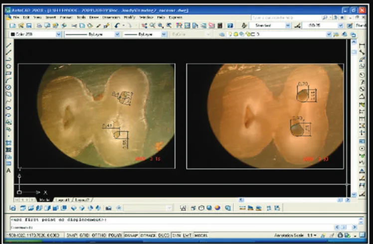
hree cross sections were then obtained and were denominated, apical, medium and cervical. Images of the sections were done using Sony Cyber Shot digital camera with a DF Vasconcelos operatory microscope, with 40× magniication. he obtained images were transferred to the sotware AutoCAD 2008, and the initial and inal mesial root canal areas were measured and statistically compared (Figure 1).
RESULT
1.
Angle of Schneider
It can be observed in Figure 2, representative of the average of degree before and ater the instrumentation, which all systems promote a rectiication in the root canal and consequently changes in the curvature degrees. In all groups was veriied also a decrease in the curvature degree.
he obtained values in degrees, before and ater the instrumentation, of each system were submitted to analysis of variance of repeated measures (ANOVA) with 5% of signiicance level (Table 1).
(p ≤ 0,05). Apical and middle third changes in anatomy were similar, but apically, the ProTaper system caused more deviations when comparing the angle of Schneider,and areas before and after instrumentation(p ≤ 0,05). Conclusion: It was concluded that all the systems caused alteration in the original anatomy of the root canal when parameters as angle of Schneider and areas before and after preparation were used.
Figure 1. Measuring of the mesial root canals areas using the sotware AutoCAD 2008.
Figure 2. Average of the initial and inal curvature degrees.
Table 1. Averages, standard deviation and value of p of the analyze of variance of the Protaper system
System Average and deviation ater PQM
Average and deviation before
PQM Test t - p
Protaper Universal 43.64 ± 13.52 31.47 ± 10.57 0.0137
K3 Endo 39.50 ± 14.78 33.14 ± 13.24 0.2399
Twisted File 36.00 ± 16.62 29.29 ± 13.97 0.2568
356 Rosa, Dametto, Gadê-Neto et al. Rev Odontol UNESP. 2012; 41(5): 353-359
It can be observed that among the tested systems that only the instrumentation with Protaper Universal promoted signiicant change in the curvature degree of the root canal, showing statistically signiicant diference (p ≤ 0,05) between the initial and inal values of the angle of Schneider.
he other systems maintained the original standards with minimum change in the root canal curvature, not presenting statistically signiicant diference (p ≥ 0.05).
2.
Root Canal Area
he evaluation of the efect of the used system, rotary or oscillating, over the canal area was perform by the coeicient between the inal areas ater the instrumentation and the initial area.
CERVICAL THIRD
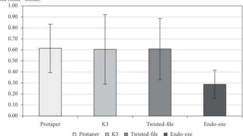
he Figure 3 represents the average of the obtained coeicient between the area before and ater the instrumentation in the cervical third. It can be observed that the three rotary systems provided the highest increase of the root canal area in the cervical third when comparing with oscillating system.
When performed the F test of the variance analysis, with 5% of signiicance level, it established that the G1, G2 and G3 groups statistically diferentiate of the Group 4 (p < 0,05), however, not presenting signiicant diference between itself (Table 2).
MEDIUM THIRD
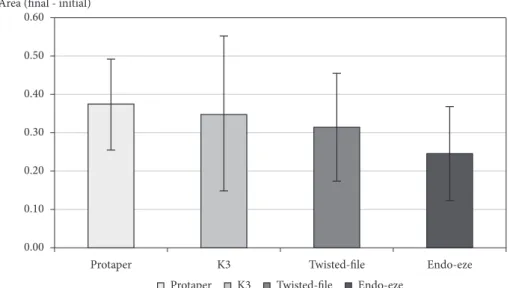
he Protaper system showed greater area of enlargement of the root canal, followed by the K3 Endo, Twisted File system and the EndoEze TiLOS. (Figure 4).
However, the F test of the variance analysis does not presented statistically signiicant diference (p > 0.05) between the groups as the average of the medium third area (Table 3).
APICAL THIRD
he K3 Endo system presented higher average of enlargement of the root canal, followed by the Protaper Universal system, Twisted File and EndoEze TiLOS (Figure 5).
he F test of the variance analysis, with 5% of level of signiicance does not showed statistically signiicant diference (p > 0.05) between the groups as the average of the apical third area (Table 4).
DISCUSSION
NiTi instruments are used in Endodontics since 1992. Since then, a lot of problems regarding fracture, deviation, excessive enlargement that weaken teeth, foraminal and apical transportation and iatrogenic changes in the original anatomy of the root canals started to be evident, specially narrow and curved canals. All these problems compromise the success of the endodontic therapy.
In this study it was evaluated mesial canals of lower molars, once these canals are representative of this kind of anatomy, are very diicult to prepare and became a huge challenge for mechanized instrumentation8-20.
he obtained results presented statistic diferences between the diferent kinematics(rotary or oscillating)in relation to the
Table 2. Averages and standard deviations of the area in cervical third
Groups Averages of the values in mm2
Protaper 0.61 ± 0.22 a
K3 Endo 0.60 ± 0.32 a
Twisted File 0.61 ± 0.28 a
Endo - Eze 0.29 ± 0.13 b
Averages accompany with diferent letters indicate statistically signiicant diference in the level of 5%.
Table 4. F test of variance analysis to comparison of the area averages between the groups
Variation sources Freedom degrees Medium square F p-value
Mistake treatments 3
46
0.068
0.030 2.25 0.0931
Table 3. F Testof the variance analysis to comparison of the area averages between the groups
Variation sources Freedom degrees Medium square F p-value
Mistake treatments 463 0.0370.023 1.65 0.188
Figure 4. Averages and standard deviation of the averages of the obtained quotient between the inal and initial medium third areas.
Figure 5. Averages and standard deviation of the averages of the obtained quotient between the inal and initial areas in the apical third.
root canal instrumentation area. he rotary systems presented more efective wears in the cervical third, when compared to the oscillatory system .Deplazes et al.21 (2001) founded, in the same
third, equivalent increase wears. In spite of the positive efect of this enlargement, once it makes an easier access to the apical third, it can result in weakening the tooth structure, and/or causing lateral root perforation in risk areas(the distal face of mesial canals in lower molars for exemple). Gluskin et al.22 (2001), comparing
the efects of the manual instrumentation associate to Gates-Glidden bur with the GT rotation system, founded signiicant diferences in the remaining thickness of dentin in risk areas. hey concluded that iatrogenic wears weaken the tooth a lot, in a tentative to create an appropriate root access.hus, it is preferable the use of lexible instruments with bigger taper and diameter to perform it. Similar indings were also veriied by Bergmans et al.23
358 Rosa, Dametto, Gadê-Neto et al. Rev Odontol UNESP. 2012; 41(5): 353-359
According to the results the EndoEze TiLOS oscillatory system promoted enlargement and wear of this third with a higher safety, because of its low wear, once it uses instruments with lower taper and metallic mass in the cervical third. In the medium and apical thirds the systems showed similar performance results, going against Heck24 (2005) studies that evaluated the efect of
three instrumentation techniques: hybrid, step back and rotary in the apical third of lower molars mesial canals. he author concluded that the compared techniques did not present a similar performance in the apical third, since the diameter values in the apical third weren’t similar. However, the results of this research conirm the studies of Veltry et al.25 (2004) and Ozgur
Uyanik et al.26 (2006), no statistically diference between diferent
techniques was found.
Bergmans et al.23 (2002) compared the inluence of the variable
taper of the Protaper Universal system with the constant taper inluence of the K3 Endo system in the preparation of the mesial canals of lower molars. hey observed that the Protaper system instruments promotes a centering apical prepare, respecting the root canal axis. However, these instruments promote the displacement of the preparation area, in cervical third, dangerous for the risk area, while the K3 Endo system instruments makes a displacement run into the safety area. However, in the cervical third wear area does not have evidences of diferences between the Protaper and K3 Endo systems, the present study found diferent results from the studies of Bergmans, because the Protaper system promoted more changes in the apical third, with higher incidence of deviation. Rasquin et al.27 (2007) compared Endo
Eze and Race systems, and found similar results to the ones we found with EndoEze, with more centered preparation, preserving
tooth structures. Zanin28 (2006) also found centered preparation
with minimally invasive wears. hese studies were opposite to founding from those by Camargo29 (2004) and Paque et al.30
(2005), that founded high levels of deviation.
he Twisted File rotary system, when compared to the Protaper and K3 Endo systems, presented excellent results as the maintenance of the canal anatomy and low levels of apical
deviation. Studies of Kariem, Elmallah31 (2011) demonstrated
that the Twisted File system present better results when compared the K3 Endo system. his fact was not conirmed in the present study, with all systems showing similar results. hey stated that Twisted File instruments transported the least root canal preparations, with a signiicant diference when compared with ProTaper group. hey justiied their results in accordance to the manufacturing of TF with twisting a Nickel Titanium wire, resulting in more lexibility than NiTi grounded instruments. Fayyad, Elgendy32 (2011) airmed that the non-ISO progressive
taper in the ProTaper system shows a higher cutting ability with selective areas of cutting.
CONCLUSION
Based in the obtained evidences in the present study it can established that the systems showed optimum results in the root canal preparation, maintaining the original root canal anatomy, with exception of the Protaper system, which showed higher transportations at apical third. However, it is important to consider that the scarce literature comparing the continuous and alternate systems, drives researchers towards new works using also diferent methodologies and protocols
REFERENCES
1. Leonardo MR, Leonardo RT. Endodontics - biological concepts and technological resources. São Paulo: Artes Médicas; 2011. 2. Morgan A, Alexander I. Endodontics - does the biology matter? Roots. 2010;6:36-8.
3. Trope M, Debelian G. BioRace NiTi complete endodontic system: achieving biologically desirable apical sizes-safely and eiciently. Endod Prac. 2011;4:31-6
4. Seltzer FC, Kwon TK, Karabucak B. Comparison of apical transportation between two rotary ile systems and two hybrid rotary instrumentation sequences. J Endod. 2010;36:1226-9. PMid:20630304. http://dx.doi.org/10.1016/j.joen.2010.03.011
5. Kunert GG, Fontanella VRC, Moura AAM, Barletta FB. Analysis of apical root transportation associated with Protaper Universal F3 and F4 instruments by using digital subtraction radiography. J Endod. 2010; 36: 1052-5. PMid:20478464. http://dx.doi.org/10.1016/j. joen.2010.02.004
6. De-Deus G, Moreira EJL, Lopes HP, Elias CN. Extended cyclic fatigue life of F2 ProTaper instruments used in reciprocating movement. Int Endod J. 2010;43:1063-8. http://dx.doi.org/10.1111/j.1365-2591.2010.01756.x
7. Schneider SW. A comparison of canal preparation in straight and curved root canal. Oral Surg. 1971;32:271–5 http://dx.doi. org/10.1016/0030-4220(71)90230-1
8. Chan AWK, Cheung GSP. A comparison of stainless steel and nickel-titanium K-iles in curved root canals. Int Endod J. 1996; 29:370-5. http://dx.doi.org/10.1111/j.1365-2591.1996.tb01400.x
9. Coleman CL, Svec TA. Analysis of NiTi versus stainless steel instrumentation in resin simulated canals. J Endod. 1997;23:232-5. http:// dx.doi.org/10.1016/S0099-2399(97)80053-2
10. Glosson CR, Haller RH, Dove SB, De Rio CE. A comparison of root canal preparations using Ni-Ti hand, Ni-Ti engine-driven, and K-lex endodontic instruments. J Endod. 1995;21:146-51. http://dx.doi.org/10.1016/S0099-2399(06)80441-3
12. Haller RH, Glosson CR, Dove SB, del Rio CE. Nickel-titanium hand and engine- driven root canal preparations: a comparison study [abstract PC12]. J Endod. 1994;20:209. http://dx.doi.org/10.1016/S0099-2399(06)80419-X
13. Harlan AL, Nicholls JI, Steiner JC. A comparison of curved canal instrumentation using nickel-titanium or stainless steel iles with the balanced-force technique.J Endod. 1996;22:410-3. http://dx.doi.org/10.1016/S0099-2399(96)80241-X
14. Kosa DA, Marshall G, Baumgartner JC. An analysis of canal centering using mechanical instrumentation techniques. J Endod. 1999;25:441-5. http://dx.doi.org/10.1016/S0099-2399(99)80275-1
15. Kuhn WG. Efect of tip design of nickel-titanium and stainless steel iles on root canal preparation. J Endod. 1977;23:735-8. http://dx.doi. org/10.1016/S0099-2399(97)80345-7
16. Rhodes JS, Pitt Ford TR, Lynch JA, Liepins PJ, Curtis RV. A comparison of two nickel-titanium instrumentation techniques in teeth using microcomputed tomography.Int Endod J. 2000; 33:279-85. http://dx.doi.org/10.1046/j.1365-2591.1999.00306.x
17. Royal JR, Donnely JC. A comparison of maintenance of canal curvature using balanced-force instrumentation with three diferent ile types. J Endod. 1995;21:300-4. http://dx.doi.org/10.1016/S0099-2399(06)81005-8
18. Serene TP, Adams JD, Saxena A. Nickel-titanium instruments: applications in endodontis. St. Louis: Ishiyaku Euroamericana; 1995. 19. Short JA, Morgan LA, Baumgartner JC.A comparison of canal centering ability of four instrumentation techniques.J Endod. 1997;23:503-7.
http://dx.doi.org/10.1016/S0099-2399(97)80310-X
20. Tucker DM, Wenckus CS, Bentkover SK. Canal wall planning by engine-driven nickel-titanium instruments, compared with stainless-steel hand instrumentation.J Endod. 1997;23:170-3. http://dx.doi.org/10.1016/S0099-2399(97)80269-5
21. Deplazes P, Peters C, Barbakow F. Comparing apical preparations of root canals shaped by titanium rotary instruments and nickel-titanium hand instruments. J Endod. 2001;27:196-202. PMid:11487151. http://dx.doi.org/10.1097/00004770-200103000-00015
22. Gluskin AH, Brown DC, Buchanan LSA. A reconstructed computerized tomographic comparison of Ni-Ti rotary GT iles versus traditional instruments in canal shaped by novice operators.Int Endod J. 2001;34:476-84. http://dx.doi.org/10.1046/j.1365-2591.2001.00422.x 23. Bergmans L, Van Cleynenbrenugel J, Beullens M, Wevers M, Van Meerbeek B, Lambrechts P. Smooth lexible versus active tapered shat
design using NiTi rotary instruments. Int Endod J. 2002;35:820-8. http://dx.doi.org/10.1046/j.1365-2591.2002.00574.x
24. Heck AR. Avaliação da alteração morfológica do canal radicular após o preparo com três técnicas de instrumentação e do tempo gasto para sua execução [tese de doutorado]. Piracicaba: Faculdade de Odontologia da UNICAMP; 2005.
25. Veltri M, Mollo A, Pini PP, Ghelli LF, Balleri P. In vitro comparison of shaping abilities of Protaper and GT rotary iles.J Endod. 2004;30:163-6. PMid:15055435. http://dx.doi.org/10.1097/00004770-200403000-00009
26. Ozgur Uyanik M, Cehreli ZC, Ozgen Mocan B, Tasman Dagli F. Comparative evaluation of thrre nickel-titanium instrumentation systems em human teeth using computer tomography.J Endod. 2006;32:668-71. PMid:16793477. http://dx.doi.org/10.1016/j.joen.2005.12.015 27. Rasquin LC, Carvalho BF, Lima RKP.In vitro evaluation of root canal preparation using oscillatory and rotary systems in lattened root
canals.J Appl Oral Sci. 2007;15:65-9. PMid:19089103. http://dx.doi.org/10.1590/S1678-77572007000100014
28. Zanin FP. Avaliação “in vitro” de três diferentes técnicas de instrumentação quanto ao deslocamento do canal radicular [dissertação mestrado]. Araraquara: Faculdade de Odontologia da UNESP; 2006.
29. Camargo JMP. Estudo comparativo do preparo do canal radicular de dentes artiiciais utilizando diferentes técnicas automatizadas de instrumentação [tese doutorado]. Araraquara: Faculdade de Odontologia da UNESP;2004.
30. Paque F, Barbakow F, Peters OA. Root canal preparation with Endo-Eze AET: changes in root canal shape assessed by micro-computed tomography. Int Endod J. 2005;38:456–64. PMid:15946266. http://dx.doi.org/10.1111/j.1365-2591.2005.00968.x
31. Kariem MEB, Elmallah, WE. Comparison of canal transportation and changes in canal curvature of two Nickel-Titanium rotary instruments. J Endod, 2011;37:1290-2. PMid:21846551. http://dx.doi.org/10.1016/j.joen.2011.05.024
32. Fayyad DM, Elgendy AAE. Cutting eiciency of Twisted versus machined Nickel-Titanium endodontic iles. J Endod. 2011;37:1143–6. PMid:21763910. http://dx.doi.org/10.1016/j.joen.2011.03.036
CONFLICTS OF INTERESTS
he authors declare no conlicts of interest.
CORRESPONDING AUTHOR
Mário Tanomaru Filho
Rua Humaitá, 1680, 3 andar, 14801-903 Araraquara - SP, Brasil e-mail: [email protected]