Endodontics
Cacio Moura-Netto(a) Renato Miotto Palo(b) Carlos Henrique Ribeiro Camargo(b)
Cornelis Hans Pameijer(c)
Marcia Regina Ramalho da Silva Bardauil(d)
(a)Department of Endodontics, School of
Dentistry, Univ Cruzeiro do Sul - UNICSUL, São Paulo, SP, Brazil.
(b)Department of Endodontics, School of
Dentistry, Univ Estadual Paulista - UNESP, São José dos Campos, SP, Brazil.
(c)Department of Endodontics, School of
Dentistry, Univ of Connecticut Health Center, Farmington, CT, USA.
(d)Department of Biochemistry, School of
Dentistry, Univ Paulista - UNIP, São Paulo, SP, Brazil.
Corresponding Author:
Cacio de Moura-Netto E-mail: caciomn@usp.br
Micro-CT assessment of two different
endodontic preparation systems
Abstract: The aim of this study was to compare two endodontic prepa-ration systems using micro-CT analysis. Twenty-four one-rooted man-dibular premolars were selected and randomly assigned to two groups. The samples (n = 12) of Group 1 were prepared using the ProTaper Uni-versal rotary system, while Group 2 (n = 12) was prepared using the En-doEZE AET system complemented by manual apical preparation with K-type hand iles up to #30. A 2.5% sodium hypochlorite solution was used in both groups for irrigating. Both groups were scanned by high-resolution microcomputed tomography before and after preparation (SkyScan 1172, SkyScan, Kontich, Belgium). The root canal volume and surface area was measured before and after preparation, and the differ-ences were calculated and analyzed for statistically signiicant differdiffer-ences using ANOVA complemented by the Tukey test (p < 0.05). The results showed no statistically signiicant differences between the mean volumes of dentin removal by the two systems. However, the EndoEZE AET sys-tem presented a signiicantly greater mean surface area compared to the ProTaper system (p < 0.05). The EndoEZE AET system enabled prepara-tion of a greater root canal surface area when compared to the ProTaper Universal system. There seemed to be no difference in dentin volume loss between the two systems used.
Descriptors: Dental Pulp Cavity; Root Canal Therapy; Endodontics.
Introduction
Root canal preparation is the most important phase of endodontic treatment and consists of cleaning and shaping canal space adequately.1
However, complete mechanical preparation of the root canal system is rarely achieved, because of the variations and complexity of the root ca-nal anatomy.2-4
In recent years, rotary instruments and oscillating systems have drawn considerable interest based on the eficacy, speed and safety of the preparation procedure.
The ProTaper Universal system (Dentisply Maillefer, Ballaigues, Swit-zerland) is known as having better cutting eficiency and lexibility, and especially indicated for curved canals. This NiTi alloy rotary system has six iles in its pack:
• 3 shaping iles for the coronal and middle third and
• 3 inishing iles for the apical third. Declaration of Interests: The authors
certify that they have no commercial or associative interest that represents a conflict of interest in connection with the manuscript.
Submitted: Jun 01, 2012
A study examining breakage and distortion of ProTaper, K3 Endo, and ProFile systems5
demon-strated no difference between the two groups with respect to breakage, but distortion was more pro-nounced for the ProFile system. The EndoEZE sys-tem (Ultradent Products, South Jordan, USA) was designed to promote a perimetric or circumferential preparation of coronal and middle thirds of root canals; however, when compared to FlexMaster, neither system was capable of completely preparing oval root canals.4 The latter system is composed of
4 stainless steel (SS) iles that have a similar tip di-ameter (0.10 to 0.13 mm) and a larger taper, start-ing with 0.02 of the irst ile to 0.06 of the last one. Because the iles are made of stainless steel, it is possible to apply a brushing motion in conjunction with the 30° oscillating movement of the handpiece. When using this system, preparation of the apical third must be complemented with hand iles. Theo-retically, oscillating systems may be more effective than rotary ones, since the latter may not reach all regions of the root canal, especially when oval-shaped canals are instrumented. This is because it is dificult to keep a rotating instrument in place, espe-cially in the middle part of the root canal. An oscil-lating ile, on the other hand, moves in all directions and has a short amplitude.4
Although microcomputed tomography (µCT) scanning is time consuming, it has major advantag-es6-8 in pre- and post-operative comparisons. µCT
has a high resolution and is imminently suitable for accurately evaluating the effect of root canal prep-arations according to Paqué et al.,6 Rhodes et al.,9
and Elayouti et al.10
Assuming that preparation systems with differ-ent kinematics could lead to distinctive patterns of inal preparation, these patterns may result in rela-tively untouched areas that probably retain bacteria and tissue remnants. The aim of this study was to compare the effect of volume change and surface area instrumentation of root canals using the rotary ProTaper Universal (PTU) system versus the oscil-lating EndoEZE AET (EEA) systems by means of microcomputed tomography (micro CT).
Methodology
Specimen selection and preparation
This study was approved by the Research Eth-ics Committee of the School of Dentistry, University of São Paulo (protocol no. 225 / 2008). A total of 24 single-rooted human mandibular premolars with fully developed roots and without any previous end-odontic treatment were selected for the study. The teeth were indicated for extraction for implant re-placement, periodontal or orthodontic reasons. The teeth were conserved in saline solution at 4°C. The root length of the sample ranged from 17 to 19 mm.
Acquisition and reconstruction of micro-CT images
All the teeth were scanned by high-resolution micro-computed tomography (SkyScan 1174, Sky-Scan, Kontich, Belgium). The x-ray tube operated at 80 kV, 10 W and 100 µA, with a 1 mm alumi-num ilter and a focal spot size of 5 µm. The speci-men was scanned with a 90°-vertical axis rotation step and a single rotation step of 0.9°. The small sample-holder device for µCT, SkyScan 1174 (Sky-Scan, Kontich, Belgium) was used to it the speci-men with the crown positioned downward and its long axis, perpendicular to the loor of the specimen holder and the x-ray source. The scanning time for each sample was approximately 25 min using a pix-el size of 14 µm. The angular projections acquired with NRecon volumetric reconstruction software (SkyScan, 1.4.0, Kontich, Belgium) produced two-dimensional cross-sectional slices through the root. The root canal volumes were measured in mm3 and
the root canal surface areas in mm2 using the
NRe-con software.
2D and 3D analyses
The 2D/3D analyses were performed using the CTan 1.7.0.0 version software (Skyscan, Kontich, Belgium). Select threshold levels were determined in the histogram from binary images. The white part of the binary images represented dentin (solid ob-ject), and the lower grey levels represented the root canal space for the subsequent quantitative 2D and 3D analyses: root canal surface areas (mm2) and
the program in a tabular comma-delineated text ile. Finally, CTvol 1.10.1.0 version software (Sky-scan, Kontich, Belgium) was used for 3D volumetric visualization. Dentin and root canal space models were developed separately and superimposed auto-matically, using different colors for pre- and post-instrumented canals in order to illustrate the meth-odology.
Experimental groups
The teeth were randomly assigned to two groups (each group n = 12) according to the endodontic preparation system. The procedures described be-low were conducted in all samples by the same op-erator (CMN), an endodontist with experience in both preparation systems. After coronal access, a size 15 K-ile (Dentsply Maillefer, Ballaigues, Swit-zerland) was inserted into the root canal until it was visible at the apical foramen, after which the work-ing length was established 1.0 mm short of the apex. A 2.5% sodium hypochlorite (NaOCl) solution was used as an irrigating solution in both groups. In Group 1, a ProTaper Universal rotary system (Gr. PTU) was used up to an F3 ile (0.30 mm tip di-ameter and 9% larger taper). Group 2 was prepared using the EndoEZE AET system (Gr. EEA), comple-mented by manual apical preparation with K-type hand iles up to a #30 ile. The last ile of this system was the S3, with a 0.13 mm tip diameter and taper of 0.06. The root canals were lushed with 10 mL of 2.5% NaOCl after using each ile. Afterwards, 10 mL of 17% ethylenediaminetetraacetic acid was used to remove the smear layer. Finally, the prepara-tion was completed by rinsing with 20 ml of sterile water.
All teeth were scanned again using the same pro-tocol as described above, and the volumes and sur-face areas were measured after preparation. It was possible to superimpose the initial and inal scan using the NRecon software, in order to illustrate the pattern of preparation for each system. All data were tabulated and the differences between initial and inal volumes and areas were calculated.
The normality of the data was conirmed by the Kolmogorov-Smirnov test, and the groups were sta-tistically compared using Analysis of Variance
com-plemented by Tukey’s test with a level of signiicance of 5% (p = 0.05).
Results
Table 1 shows the mean values and standard deviations of the differences between initial and i-nal root cai-nal volumes and surface area. ANOVA complemented by Tukey’s test showed no signiicant differences among the mean volume changes of the two systems. However, the EEA system presented a statistically signiicant higher mean surface area compared to the ProTaper system (p < 0.05).
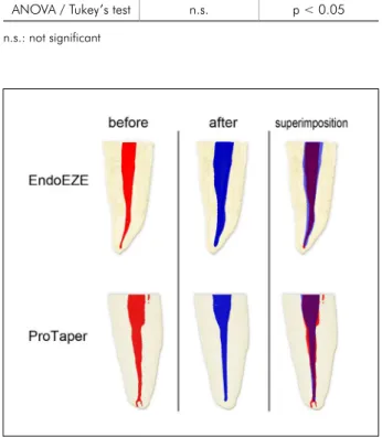
Figure 1 illustrates the difference between the PTU and EEA systems in regard to how each sys-tem acts on the walls of root canal. The anatomy of the canal before instrumentation is indicated in
Table 1 - Mean values and standard deviations (SD) of the differences between initial and final root canal volume and surface area.
Group ∆Volume (mm3) ∆Area (mm2)
ProTaper Universal 3.386 ± 0.828 6.052 ± 1.596 EndoEZE AET 2.816 ± 0.548 8.509 ± 1.951 ANOVA / Tukey’s test n.s. p < 0.05
n.s.: not significant
red, whereas blue denotes the post instrumentation coniguration. On the right, a superimposition of the pre- and the post-instrumentation scans demon-strates the complete root canal instrumentation for the EEA group. In contrast, the PTU example for superimposition shows areas in red, which are not instrumented.
Discussion
Unlike the PTU system, the EEA system offers a safe method for canal instrumentation without causing instrument fractures, enabled by the me-chanical action properties of its instruments. Some authors relate instrument fracture to fatigue caused by repetitive bending stresses on curved canals.11,12
Others indicate torsion as the primary mechanism responsible for fracture.13 Torsion can be generated
when the rotary ile is obstructed against the canal wall or exposed to excessive pressure by the clini-cian.13 Fractures caused by fatigue or high levels of
torsion may cause failure of the endodontic treat-ment.14
In the present study, a comparison was made be-tween a rotary and an oscillating system used for instrumenting root canals. The study data indicated that the EEA system prepared a larger root canal surface area in comparison to the PTU system. Stat-ed differently, the EEA system was in contact with a larger surface area of the canal during instrumenta-tion than the PTU system. This result means that the PTU system probably left more untouched areas than the EEA system. In our study, this difference was statistically signiicant. This may be attributed to a combination of alloy composition, design and mostly the movement applied by each system. The EndoEZE AET system is composed of 4 stainless steel iles (SS) with almost no difference in the tip diameter (0.10 to 0.13 mm). However, this system is composed of iles of increasing taper, starting with 0.02 of the irst ile to 0.06 of the last one. Because the iles are made of stainless steel, it is possible to apply a iling motion in conjunction with the 30° os-cillating movement of the handpiece. The combined action assures a more anatomical preparation, inso-far as the operator can determine what part of the root canal must be more or less prepared. Because
the purpose of this system is to perform the ana-tomical preparation, the apical enlargement must be completed with hand iles up to a 0.30 mm diam-eter.
In contrast, the ProTaper Universal is a rotary system that uses NiTi alloy iles that are used with an “in and out” movement combined with continu-ous rotation. This system prepares only the surfaces that are in contact with the ile. A iling or brushing motion is not recommended, since it increases the risk of ile fracture. The last ile of this system (F3) has a 0.030 mm tip diameter and a variable taper that increases 9% with the irst millimeter and then 5.5% up to the 14th millimeter. The inal
prepara-tion of this system is very similar to a taper of 0.06. If we compare these two systems, as they were used in this study, both had a 0.30 mm apical diameter and a 0.06 taper at the end.
Paqué et al.3 reported similar results for EEA.
However, the authors found poor results when the EEA system was used in curved canals. This is completely understandable if you imagine a 0.45 ta-per or a 0.6 tata-per stainless steel ile oscillating in a curved canal. In fact, we believe that although both systems have laws, they are highly indicated in different anatomic situations. While NiTi rotary systems are indicated for more rounded and curved anatomic situations, the EAA system is better used in lattened and isthmus areas, mostly present in the middle third of the roots. Using the ProTaper sys-tem to increase apical enlargement of curved canals did not result in complete apical preparation, but it did lead to unnecessary removal of dentin.10
Rüt-termann4 reported that neither rotary nor
oscillat-ing systems produced signiicantly different shifts of the canal centers in the middle part of the root. Furthermore, only a few of the preparations yielded excellent results, that is, having no uninstrumented canal walls left. These authors also observed that the oscillating and rotary systems had root canals prepared only in the buccal or lingual portion of the root, as well as roots having a circular bulge with unprepared lateral extensions.4
As reported by others8 and in agreement with
and surface areas. The instrumentation of root ca-nals respecting their anatomy and ensuring the least amount of dentin removal seems to be a challenging task, regardless of the system used.15
Conclusions
From the data gathered in the present study, it can be concluded that the EndoEZE system
pre-pared a signiicantly larger root canal surface area in comparison to the ProTaper Universal system. However, no difference in volume of dentin removal was observed between the two systems. Based on this, it can be inferred that the EndoEZE system is capable of performing a more anatomical prepara-tion, attaining areas that the Protaper Universal sys-tem was not capable of reaching.
References
1. Brkanic´ T, Stojšin I, Zivkovic´ S, Vukoje K. Canal wall thick-ness after preparation with NiTi rotary files. Microsc Res Tech. 2012 Mar;75(3):253-7.
2. De-Deus G, Barino B, Zamolyi RQ, Souza E, Fonseca Jr A, Fidel S, et al. Suboptimal debridement quality produced by the single file F2 ProTaper technique in oval-shaped canals. J Endod. 2010 Nov;36(11):1897-900.
3. Paqué F, Barbakow F, Peters OA. Root canal preparation with Endo-Eze AET: changes in root canal shape assessed by micro-computed tomography. Int Endod J. 2005 Jul;38(7):456-64. 4. Rüttermann S, Virtej A, Janda R, Raab WH. Preparation of
the coronal and middle third of oval root canals with a rotary or an oscillating system. Oral Surg Oral Med Oral Pathol Oral Radiol Endod. 2007 Dec;104(6):852-6.
5. Ankrum MT, Hartwell GR, Truitt JE. K3 Endo, ProTaper, and Profile systems: breakage and distortion in severely curved roots of molars. J Endod. 2004 Apr;30(4):234-7.
6. Paqué F, Ganahl D, Peters OA. Effects of root canal prepara-tion on apical geometry assessed by micro–computed tomog-raphy. J Endod. 2009 Jul;35(7):1056-9.
7. Bergmans L, Van Cleynenbreugel J, Wevers M, Lambrechts P. A methodology for quantitative evaluation of root canal instrumentation using microcomputed tomography. Int Endod J. 2001 Jul;34(5):390-8.
8. Yang G, Yuan G, Yun X, Zhou X, Liu B, Wu H. Effects of Two nickel-titanium instrument systems, Mtwo versus ProTaper universal, on root canal geometry assessed by micro–com-puted tomography. J Endod. 2011 Oct;37(10):1412-6.
9. Rhodes JS, Ford TR, Lynch JA, Liepins PJ, Curtis RV. Micro-computed tomography: a new tool for experimental endodon-tology. Int Endod J. 1999 May;32(3):165-70.
10. Elayouti A, Dima E, Judenhofer MS, Löst C, Pichler BJ. In-creased apical enlargement contributes to excessive dentin removal in curved root canals: a stepwise microcomputed tomography study. J Endod. 2011 Nov;37(11):1580-4. 11. Grande NM, Plotino G, Pecci R, Bedini R, Malagnino VA,
Somma F. Cyclic fatigue resistance and three dimensional analysis of instruments from two nickel-titanium rotary sys-tems. Int Endod J. 2006 Oct;39(10):755-63.
12. Zinelis S, Darabara M, Takase T, Ogane K, Papadimitriou GD. The effect of thermal treatment on the resistance of nickel-titanium rotary files in cyclic fatigue. Oral Surg Oral Med Oral Pathol Oral Radiol Endod. 2007 Jun;103(6):843-7. 13. Sattapan B, Palamara JE, Messer HH. Torque during canal
instrumentation using rotary nickel-titanium files. J Endod. 2000 Mar;26(3):156-60.
14. Cheung GSP. Instrument fracture: mechanisms, removal of fragments, and clinical outcomes. Endod Top. 2007 Mar;16(1):1-26.
