Functional Limitation and Intermittent Claudication: Impact of Blood
Pressure Measurements
Rita de Cassia Gengo e Silva
1, Cassiana Rosa Galvão Giribela
2, Nelson Wolosker
2, Fernanda Marciano
Consolim-Colombo
1Instituto do Coração do Hospital das Clínicas da Faculdade de Medicina da USP1; Instituto Central do Hospital das Clínicas da Faculdade de Medicina da USP2, São Paulo, SP, Brazil
Abstract
Background: Arterial hypertension is an important risk factor for Lower-Limb Occlusive Arterial Disease (LLOAD). However, the correlation between blood pressure and pulse pressure (PP) with LLOAD severity and functional impairment resulting from this disease is not well established in the Brazilian population.
Objective: To verify whether there is a correlation between blood pressure, PP, LLOAD severity and functional capacity in patients with symptomatic LLOAD.
Methods: A total of 65 patients (62.2 + 8.1 years, 56.9% males) were evaluated. They were divided into two groups: normal (A) and high (B) blood pressure. LLOAD severity was assessed using the ankle-brachial index (ABI) and functional capacity by the total and pain-free walking distance at the 6-minute walking test (6MWT).
Results: Group A consisted of 17 (26.1%) patients. The systolic (SBP), diastolic blood pressure (DBP), and PP were, respectively, 125.4 ±11.7, 74.5 ± 9.1 and 50.9 ± 10.0 mmHg in group A and 160.7 ± 19.6, 90.0 ± 12.2 and 70.7 ± 20.2 mmHg in group B. The ABI was significantly lower in group B (0.66 ± 0.12 vs. 0.57 ± 0.13, p <0.05). SBP and PP correlated with LLOAD severity and the distances walked at the 6MWT. Patients with PP > 40 mmHg walked shorter distances.
Conclusion: SBP and PP significantly correlated with the distances walked in the 6MWT, suggesting they are clinical markers of functional capacity impairment in patients with symptomatic LLOAD. (Arq Bras Cardiol 2012;98(2):161-166)
Keywords: Intermittent claudication; blood pressure; hypertension; peripheral arterial disease; walking.
Mailing Address: Rita de Cassia Gengo e Silva •
Rua José Maria Mamblona, 37 - Jd Maria Rosa - 06763-150 - Taboão da Serra, São Paulo, SP, Brasil
E-mail: [email protected], [email protected]
Manuscript received May 09, 2011; revised manuscript received August 03, 2011; accepted August 22, 2011.
reproducible and easy-to-perform method, which is useful in the detection of LLOAD with hemodynamic impact, as well as assessing patient functional capacity4,5.
Several risk factors contribute to the onset and progression of LLOAD, among which arterial hypertension (AH) is one of the most important factors1-7. Elevated systolic
blood pressure (SBP) and diastolic blood pressure (DBP) are accelerating atherosclerosis factors in the long-term8.
Some epidemiological studies have shown a strong association between arterial hypertension (AH) and LLOAD. There is evidence that AH affects up to 90% of individuals with LLOAD 7, and BP levels > 150/90 mmHg were observed
in about 25% of patients with intermittent claudication 8.
The Framingham study showed that hypertensive patients are up to four-fold more likely to develop intermittent claudication than normotensive individuals9.
More recently, emphasis has been given to the prognostic value of pulse pressure (PP) regarding cardiovascular events10. In this sense, there is evidence that PP is associated
with greater LLOAD severity in asymptomatic individuals11.
However, the association between AH, PP, LLOAD severity and functional capacity remains little understood, particularly in the Brazilian population.
Introduction
Lower-limb obstructive arterial disease (LLOAD) is an important manifestation of systemic atherosclerosis. LLOAD has high rates of morbidity and mortality and is associated with a higher incidence of cerebrovascular and cardiac events. Its most common clinical manifestation is intermittent claudication, characterized by the onset of pain in the lower limbs (LL) during walking, which rapidly disappears after cessation of the activity1. This symptom
has significant impact on quality of life of individuals with LLOAD, as it is strongly associated with impaired functional capacity2,3.
The objective of this study was to prospectively determine whether there is an association between BP, PP, disease severity and functional capacity in patients with symptomatic LLOAD.
Methods
The study assessed a total of 65 patients (62.2 + 8.1 years, 56.9% males) with symptomatic LLOAD (Rutherford grade I), stable intermittent claudication for at least six months; ABI < 0.90, and age > 40 years, who agreed to participate and signed the free and informed consent form. This study was approved by the Ethics Committee of the institution.
Anthropometric measurements
Weight and height measurements were obtained with a properly calibrated electronic scale (Personal, Filizola, SP, Brazil) and a stadiometer attached to the scale. Body mass index (BMI) was calculated by dividing the weight by the squared height and analyzed according to the recommendations of the World Health Organization (WHO)12.
Clinical and laboratory assessment
Medical history data were obtained from medical records, as well as laboratory test results.
The personal data were collected through interviews. Physical activity was defined as the practice of aerobic activity for at least three times a week for 30 minutes.
Blood pressure measurements
For blood pressure (BP) measurements, patients were placed in the supine position and remained at rest for 5 to 10 minutes in a quiet room with appropriate temperature and light. BP was measured through the oscillometric method, using validated and calibrated equipment (Omron HEM 741C, Omron Healthcare Inc., China). Two consecutive measurements were obtained with a one-minute interval between them. The second measurement obtained was always considered valid. SBP measurements were considered elevated when > 140 mmHg and DBP when > 90 mmHg 13. PP was calculated as the difference
between SBP and DBP.
Ankle-brachial index (ABI)
Blood pressure measurements to calculate the ABI were performed 5 minutes after the BP measurement described above. For this purpose, a portable vascular Doppler device (DV 610, Medmega, SP, Brazil) and a mercury sphygmomanometer with an appropriate cuff size for the patient’s arm circumference were used. SBP measurements were carried out in the following order: left brachial artery, left dorsalis pedis artery, left posterior tibial artery, right dorsalis pedis artery, right posterior tibial artery and right brachial artery. The ABI was calculated for each lower limb, establishing the ratio between the highest ankle pressure
(dorsalis pedis or posterior tibial) and highest pressure on the arm (right or left)1. The worst ABI value obtained was
used for the analysis of results (right or left ABI).
Six-minute walking test (6MWT)
The test was performed according to previously published guidelines14, in a 20-meter long corridor. All
patients were instructed to report symptoms that occurred during the test. Phrases of encouragement were spoken every two minutes. For the analysis, we used the total and pain-free walking distance.
The patients were divided into two groups: group A comprised 17 patients (26%) with normal blood pressure levels, and group B, 48 patients (74%) with elevated SBP and/or DBP levels.
The data were analyzed using descriptive statistics and are presented as mean ± standard deviation and absolute and relative frequencies. Inferential analysis was performed using Student’s t test for continuous variables, Chi-square test for categorical variables, and Pearson’s correlation for associations. P values < 0.05 were considered statistically significant.
Results
Initially, the sociodemographic and clinical characteristics of patients in this study were analyzed (Table 1).
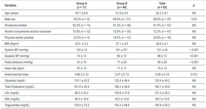
The mean age was 62.2 + 8.1 years and there was a predominance of male patients (56.9%). Although they were undergoing medical care, there was a persistence of cardiovascular risk factors, such as smoking and overweight. The mean ABI in group B was significantly lower than in group A (0.57 ± 0.13 versus 0.66 ± 0.12, p = 0.013), indicating greater LLOAD severity. However, mean serum levels of fasting glucose, total cholesterol and its fractions showed no significant difference between the groups.
The functional capacity assessment showed that groups A and B were similar (Table 2).
It can be observed that there was no difference between groups A and B regarding the total distance (275.8 ± 42.4
vs. 261.3 ± 86.5 meters, respectively, p > 0.05) and pain-free (200.0 ± 95.6 meters versus + 180.6 ± 100.5 meters, respectively, p > 0.05).
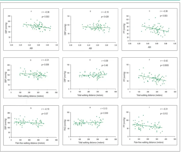
It is noteworthy the fact that patients with PP > 40 mmHg walked shorter distances at the 6MWT, both total (259.3 ± 77.2 vs. 334.7 ± 37.5 meters, p = 0.035) and pain-free (177.5 ± 95.4 vs. + 284.0 ± 95.3 meters, p = 0.019).
the 6MWT, both total (r = -0.42, p <0.001) and pain-free (r = -0.31, p = 0.012). The DBP did not correlate with the ABI, or with the walked distances.
Discussion
The main contribution of this study is the demonstration that high levels of SBP and PP correlated to LLOAD severity and to the functional impairment of patients with intermittent claudication. It is well established in the literature that hypertension is an important risk factor for the onset and progression of atherosclerosis and high BP has been linked to higher LLOAD prevalence7,8,15,16. The Framingham study showed that AH
increased by 2.5-fold the risk of LLOAD for men, and four-fold for women9.
The physiopathological mechanisms underlying the involvement of AH as a contributing factor to the development of vascular disease are complex. It should be emphasized that in spite of the existence of atherogenic cofactors interacting throughout the circulatory system, atherosclerosis develops in
segments with high pressure status, highlighting the central role of AH in this process17.
The relevance of other risk factors such as smoking, diabetes mellitus, obesity and hypercholesterolemia, however, has also been recognized and their association with a higher LLOAD prevalence has been demonstrated18-20. In
the present study, although patients were undergoing clinical treatment, it was observed that they maintained the habit of smoking, were overweight, and most were sedentary. It is known that the presence of risk factors determines vascular alterations in other vascular territories such as the carotid, coronary and cerebral arteries, worsening the cardiovascular prognosis for patients with LLOAD21-23.
Mehler et al24 demonstrated that intensive BP control in
diabetic patients with LLOAD results in marked reduction of cardiovascular events. Moreover, these authors showed that there is an inverse association between the ABI and the risk of cardiovascular events in these patients, which, however, was abolished in individuals who underwent intensive treatment to control BP. This protective effect conferred by BP control
Table 1 – Sociodemographic and clinical characteristics of the patients with symptomatic LLOAD
Variables Group A(n = 17) Group B(n = 48) (n = 65)Total p*
Age (years) 63.1 ± 6.9 61.9 ± 8.5 62.2 ± 8.1 NS
Male sex 35.3% (n = 6) 64.6% (n = 31) 56.9% (n = 37) 0.03
Smoker/ex-smoker 82.3% (n = 14) 81.2% (n = 39) 81.5% (n = 53) NS
Alcohol consumer/ex-alcohol consumer 70.6% (n = 12) 72.9% (n = 35) 72.3% (n = 47) NS
Physical activity practice 23.5% (n = 4) 18.5% (n = 12) 24.6% (n = 16) NS
BMI (Kg/m²) 25.0 ± 2.2 27.1 ± 4.5 26.6 ± 4.1 NS
Systolic BP (mmHg) 125 ± 12 161 ± 20 * 151 ± 24 < 0.001
Diastolic BP (mmHg) 74 ± 13 90 ± 12* 86 ± 13 <0.001
Pulse pressure (mmHg) 51 ± 10 71 ± 20 65 ± 20 < 0.001
Heart rate (bpm) 67 ± 13 71 ± 11 70 ± 12 NS
Ankle-brachial index 0.66 ± 0.12 0.57 ± 0.13 0.59 ± 0.14 0.013
Glycemia (mg/dL) 115.1 ± 42.5 122.4 ± 46.4 120.4 ± 45.2 NS
Total Cholesterol (mg/dL) 161.8 ± 28.4 186.2 ± 48.8 180.1 ± 45.6 NS
LDL (mg/dL) 88.2 ± 23.2 105.8 ± 37.6 101.3 ± 35.2 NS
HDL (mg/dL) 49.3 ± 16.8 49.3 ± 12.8 49.3 ± 13.8 NS
Triglycerides (mg/dL) 120.8 ± 72.2 154.3 ± 98.6 145.9 ± 93.3 NS
NS - non-signiicant; LLOAD - Lower-Limb Occlusive Arterial Disease; BMI - Body Mass Index; BP - Blood Pressure(*)descriptive levels for comparison between groups A and B.
Table 2 – Functional capacity of patients with symptomatic LLOAD, assessed by walking distances at the 6-minute walk test (6MWT)
Variables Group A
(n = 17)
Group B (n = 48)
Total
(n = 65) p
*
Total walking distance (meters) 275.8 ± 42.4 261.3 ± 86.5 265.1 ± 77.4 NS
Pain-free walking distance (meters) 200.0 ± 95.6 180.6 ± 100.5 185.7 ± 98.9 NS
Figure 1 – Correlation between blood pressure, pulse pressure, peripheral arterial disease severity and functional capacity (n = 65); A - Correlation between
systolic blood pressure (SBP) and peripheral arterial disease severity, measured by ankle-brachial index (ABI); B - Correlation between diastolic blood pressure (DBP) and peripheral arterial disease severity, measured by ABI; C - correlation between Pulse Pressure (PP) and peripheral arterial disease severity, measured by ABI; D - correlation between SBP and functional capacity, measured by total walking distance at the 6-minute walk test; E - correlation between DBP and functional capacity, measured by total walking distance at the 6-minute walk test; F - Correlation between PP and functional capacity, measured by total walking distance at the 6-minute walk test; G - Correlation between SBP and functional capacity, measured by pain-free walking distance in the 6-minute walk test; H - Correlation between DBP and functional capacity, measured by pain-free walking distance in the 6-minute walk test, I - Correlation between PP and functional capacity, measured by pain-free walking distance in the 6-minute walk test.
occurred independently from the classes of medications used in the treatment24.
Upon recognizing the significant impact of hypertension on LLOAD progression in the present study, we chose to divide the patients into two groups, according to the pressure level. It was observed that the risk factors were evenly distributed between the groups A (normal blood pressure) and B (high blood pressure), with arterial hypertension remaining as the only risk factor that differentiated them. Thus, we observed that SBP and PP showed an inversely proportional association with the ABI, demonstrating that patients with uncontrolled blood pressure and elevated PP had a higher LLOAD severity.
The increase in SBP is a process that accompanies aging. This increase is secondary to arterial wall stiffening and is also associated with an increase in cardiovascular event occurrence17. Possibly, the increased pressure load
increases the wall stress, resulting in vascular injury, caused by endothelial dysfunction and vascular smooth muscle remodeling17.
The SBP increase is not necessarily accompanied by a DBP increase of the same magnitude. This implies an increase in PP, of which prognostic value, regarding the occurrence of cardiovascular events, remains debatable10,25. The results of this
been shown that SBP and PP, but not DBP in patients with cardiovascular risk factors, but without clinically detected LLOAD, have an inversely proportional association with ABI11. Moreover, another study showed that PP has a
positive and significant correlation (r = 0.61, p <0.01) with blood flow in the calf, measured by pletismography26.
It is known that patients with intermittent claudication have impaired functional capacity27. The present study showed that
the pain-free walking distance accounted for 70% of the total walking distance, demonstrating that intermittent claudication was extremely limiting for these patients.
The total distance was significantly correlated with SBP and PP. On the other hand, the pain-free walking distance correlated only with PP. Thus, it is suggested that in patients with intermittent claudication, SBP and PP levels may be markers of functional limitation. These results corroborate the evidence already available in the literature. Selvin and Hirsch28 showed that the AH determines a greater chance
of walking limitation for individuals older than 60 years. In parallel, Safar et al26 demonstrated that there is a strong
correlation between the walked distance and the peripheral bed microvascular reserve in patients with LLOAD.
Notably, little is known about the associations between pressure variables (SBP, DBP and PP), severity of LLOAD (measured by the ABI) and functional capacity (measured by
walking distances at the 6MWT) in the Brazilian population. Although the study sample is small, the results contribute to the understanding of the correlations between these variables. It must be emphasized that other risk factors in addition to blood pressure were present in the studied sample, but differently from what was demonstrated for the pressure variables, the prevalence of smoking, blood glucose and total cholesterol, LDL and HDL were similar between groups A and B. Therefore, it can be concluded that SBP and PP correlated with the severity of LLOAD, and PP seems to be a marker of greater functional capacity limitation in patients with intermittent claudication.
Potential Conflict of Interest
No potential conflict of interest relevant to this article was reported.
Sources of Funding
There were no external funding sources for this study.
Study Association
This article is part of the thesis of doctoral submitted by Rita de Cássia Gengo e Silva, from Faculdade de Medicina da USP.
References
1. Hirsch AT, Haskal ZJ, Hertzer NR, Bakal CW, Creager MA, Halperin JL, et al. ACC/AHA Guidelines for the Management of Patients with Peripheral Arterial Disease (lower extremity, renal, mesenteric, and abdominal aortic): a collaborative report from the American Association for Vascular Surgery/Society for Vascular Surgery, Society for Cardiovascular Angiography and Interventions, Society of Interventional Radiology, Society for Vascular Medicine and Biology, and the American College of Cardiology/American Heart Association Task Force on Practice Guidelines (Writing Committee to Develop Guidelines for the Management of Patients With Peripheral Arteril Disease). J Am Coll Cardiol. 2006;47(6):1239-312.
2. Breek JC, Hamming JF, De Vries J, Aquarius AE, van Berge Henegouwen DP. Quality of life in patients with intermittent claudication using the World Health Organisation (WHO) Questionnaire. Eur J Vasc Endovasc Surg. 2001;21(2):118-22.
3. Ritti-Dias RM, Gobbo LA, Cucato GG, Wolosker N, Jacob Filho W, Santarém JM, et al. Translation and validation of the walking impairment questionnaire in Brazilian subjects with intermittent claudication. Arq Bras Cardiol. 2009;92(2):136-49.
4. Wolosker N, Rosoky RA, Nakano L, Basyches M, Puech-Leão P. Predictive value of the ankle-brachial index in the evaluation of intermittent claudication. Rev Hosp Clin Fac Med Sao Paulo. 2000;55(2):61-4.
5. Lyden SP, Joseph D. The clinical presentation of peripheral arterial disease and guidance for early recognition. Cleve Clin J Med. 2006;73(Suppl 4):S15-21.
6. Cournot M, Boccalon H, Cambou JP, Guilloux J, Taraszkiewicz D, Hanaire-Broutin H, et al. Accuracy of the screening physical examination to identify subclinical atherosclerosis and peripheral arterial disease in asymptomatic subjects. J Vasc Surg. 2007;46(6):1215-21.
7. Bartholomew JR, Olin JW. Pathophysiology of peripheral arterial disease and risk factos for its development. Cleve Clin J Med. 2006;73(Suppl 4):S8-14.
8. Piccinato CE, Cherri J, Moriya T. Hipertensão e doença arterial periférica. Rev Bras Hipertens. 2001;8(3):306-15.
9. Kannel WB, McGee DL. Update on some epidemiologic features of intermittent claudication: the Framingham Study. J Am Geriatr Soc. 1985;33(1):13-8.
10. Domanski MJ, Davis BR, Pfeffer MA, Kastantin M, Mitchell GF. Isolated systolic hypertension: prognostic information provided by pulse pressure. Hypertension. 1999;34(3):375-80.
11. Ostergren J, Sleight P, Dagenais G, Danisa K, Bosch J, Qilong Y, et al, (for the HOPE study investigators). Impact of ramipril in patients with evidence of clinical or subclinical peripheral arterial disease. Eur Heart J. 2004;25(1):17-24.
12. World Health Organization (WHO). Obesity: preventing and managing the global epidemic. Report of a WHO Consultation. Geneva; 2000. (WHO Technical Report Series, 894).
13. Sociedade Brasileira de Cardiologia / Sociedade Brasileira de Hipertensão / Sociedade Brasileira de Nefrologia. VI Diretrizes brasileiras de hipertensão. Arq Bras Cardiol. 2010;95(1 supl.1):1-51.
14. ATS Statement. Guidelines for the six minutes walk test. Am J Respir Crit Care Med. 2002;166(1):111-7.
15. Makdisse M, Ramos LR, Moreira F, Oliveira A, Berwanger O, Moscardi A, et al. Escore para rastrear idosos (> 75 anos) de alto risco para doença arterial periférica. Arq Bras Cardiol. 2007;88(6):630-6.
17. Simões MV, Schmidt A. Hipertensão arterial como fator de risco para doenças cardiovasculares. Medicina. Ribeirão Preto. 1996;29:214-9.
18. Passos VM, Barreto SM, Guerra HL, Firmo JO, Vidigal PG, Lima-Costa MF. The Bambuí Health and Aging Study (BHAS): prevalence of intermittent claudication in the aged population of the community of Bambuí and its associated factors. Arq Bras Cardiol. 2001;77(5):458-62.
19. Brevetti G, Oliva G, Silvestro A, Scopacasa F, Chiariello M. Peripheral Arteriopathy and Cardiovascular Events (PACE) Study Group. Prevalence, risk factors and cardiovascular comorbidity of symptomatic peripheral arterial disease in Italy. Atherosclerosis. 2004;175(1):131-8.
20. Makdisse M, Pereira AC, Brasil DP, Borges JL, Machado-Coelho GLL, Krieger JE, et al. Prevalência e fatores de risco associados à doença arterial periférica no projeto Corações do Brasil. Arq Bras Cardiol. 2008;91(6):370-82.
21. Leng GC, Lee AJ, Fowkes FG, Whiteman M, Dunbar J, Housley E, et al. Incidence, natural history and cardiovascular events in symptomatic and asymptomatic peripheral arterial disease in the general population. Int J Epidemiol. 1996;25(6):1172-81.
22. Meijer WT, Hoes AW, Rutgers D, Bots ML, Hofman A, Grobbee DE. Peripheral arterial disease in the elderly: the Rotterdam Study. Arterioscler Thromb Vasc Biol. 1998;18(2):185-92.
23. Huelmos A, Jiménez J, Guijarro C, Belinchón JC, Puras E, Sánchez C, et al. [Underrecognized peripheral arterial disease in patients with acute coronary syndrome: prevalence of traditional and emergent cardiovascular risk factors]. Rev Esp Cardiol. 2005;58(12):1403-10.
24. Mehler PS, Coll JR, Estacio R, Esler A, Schrier RW, Hiatt WR. Intensive blood pressure control reduces the risk of cardiovascular events in patients with peripheral arterial disease and type 2 diabetes. Circulation. 2003;107(5):753-6.
25. Mosley WJ, Greenland P, Garside DB, Lloyd-Jones DM. Predictive utility of pulse pressure and other blood pressure measures for cardiovascular outcomes. Hypertension. 2007;49(6):1256-64.
26. Safar ME, Totomoukouo JJ, Asmar RA, Laurent SM. Increased pulse pressure in patients with arteriosclerosis obliterans of the lower limbs. Arteriosclerosis. 1987;7(3):232-7.
27. Wolosker N, Nakano L, Rosoky RA, Puech-Leao P. Evaluation of walking capacity over time in 500 patients with intermittent claudication who underwent clinical treatment. Arch Intern Med. 2003;163(19):2296-300.