Clinical Evaluation of a Single-Tube Multiple
RT-PCR Assay for the Detection of 13
Common Virus Types/Subtypes Associated
with Acute Respiratory Infection
Dan Zhang1,2☯, Zhishan Feng3☯, Mengchuan Zhao2,3☯, Hao Wang2,4, Le Wang3, Shuo Yang3, Guixia Li3, Li Lu1*, Xuejun Ma2*
1Department of Pathophysiology, Guangzhou Medical University, Guangzhou city, Guangdong, China,
2Key Laboratory for Medical Virology, National Health and Family Planning Commission, National Institute for Viral Disease Control and Prevention, Chinese Center for Disease Control and Prevention, Changping district, Beijing, China,3Pediatric Research Institute, Children’s Hospital of Hebei Province, Hebei Medical University, Shijiazhuang, China,4Department of Infectious Diseases, Institute of Biomedicine, Sahlgrenska Academy, University of Gothenburg, Gothenburg, Sweden
☯These authors contributed equally to this work.
*[email protected](XM);[email protected](LL)
Abstract
Respiratory viruses are among the most important causes of human morbidity and mortality worldwide, especially for infants and young children. In the past years, a few commercial multiplex RT-PCR assays have been used to detect respiratory viruses in spite of the high cost. In the present study, an improved single-tube multiplex reverse transcription PCR assay for simultaneous detection of 13 respiratory viruses was evaluated and compared with a previously reported two-tube assay as the reference method using clinical nasopha-ryngeal aspirates samples. Of 310 prospectively tested respiratory specimens selected from children hospitalized with acute respiratory illness, 226 (72.90%, 226/310) and 214 (69.03%, 214/310) positive for one or more viruses were identified by the single-tube and the two-tube assays, respectively, with combined test results showing good concordance (Kappa value = 0.874). Individually, the single-tube assay for adenovirus (Adv), human metapneumovirus (HMPV), human rhinovirus (HRV), parainfluenza virus type 1 (PIV1), parainfluenza virus type 3 (PIV3) and parainfluenza virus type 4 (PIV4) showed the signifi-cantly superior sensitivities to those of the two-tube assay. No false positives were found. In conclusion, our results demonstrates the one-tube assay revealed significant improvements over the two-tube assay in terms of the better sensitivity, more accurate quality control, less nonspecific amplification, more cost-effective and shorter turn-around time and will be a valuable tool for routine surveillance of respiratory virus infection in China.
a11111
OPEN ACCESS
Citation:Zhang D, Feng Z, Zhao M, Wang H, Wang L, Yang S, et al. (2016) Clinical Evaluation of a Single-Tube Multiple RT-PCR Assay for the Detection of 13 Common Virus Types/Subtypes Associated with Acute Respiratory Infection. PLoS ONE 11(4): e0152702. doi:10.1371/journal.pone.0152702
Editor:Oliver Schildgen, Kliniken der Stadt Köln gGmbH, GERMANY
Received:December 30, 2015
Accepted:March 17, 2016
Published:April 4, 2016
Copyright:© 2016 Zhang et al. This is an open access article distributed under the terms of the
Creative Commons Attribution License, which permits unrestricted use, distribution, and reproduction in any medium, provided the original author and source are credited.
Data Availability Statement:All relevant data are within the paper.
Introduction
Respiratory viruses are among the most common causes of human morbidity and mortality worldwide, especially for infants and young children [1,2]. The rapid identification is impor-tant for both therapeutic and infection control purposes. Diagnoses of viral respiratory tract infection have been performed generally by non-molecular approaches such as direct immuno-fluorescence and viral culture. They are time-consuming, labor-intensive, and often lack sensi-tivity or specificity. In the last few years, multiplex RT-PCR assays have been developed to detect respiratory viruses, and many of them have been commercialized, such as xTAG RVP, RVP fast (Luminex Molecular Diagnostics, Toronto, Canada), Resplex II (Qiagen, Mississauga, Canada), FilmArray1Respiratory panel (Idaho Technology Inc., Salt Lake City, UT, USA), Anyplex™II RV16 and Seeplex RV assays (Seegene, seoul, Korea), AdvanSure™real-time RT-PCR (LG Life Science, Seoul, Korea) [3–7]. However, many of these assays are costly, or require specialized laboratory equipment [8,9].
In our previous study, the two–tube multiplex reverse transcription PCR assay (two-tube assay) to detect sixteen respiratory viruses based on the amplicon size differences using auto-mated electrophoresis system is described. The overall detection rate of the two-tube assay for each virus was comparable to those of the Luminex xTAG RVP Fast assay and Seeplex RV15 ACE assay, demonstrating the high sensitivity and specificity of the two-tube assay in the anal-ysis of clinical samples [10]. However, two-tube assay has a few drawbacks, such as the size dif-ference of some amplicons are not big enough leading to the difficulty in judging the results by untrained staff, the hands-on two-tube assay is till cumbersome and the internal control is not steadily detected, which limits its wide use in the routine screening in provincial and local Cen-ter for Diseases Control and Prevention (CDC) in China. In the present study, we adopted the two-tube assay as a reference, and have been progressively optimized and substantially improved the performance of simultaneous detection of thirteen respiratory viruses types/sub-types, the most frequently detected viral agents of respiratory tract infections documented by Beijing Monitoring Network for Pneumonia between 2012–2014 (unpublished data), in a sin-gle-tube assay while maintaining excellent sensitivity and specificity. The targeted 13 respira-tory viruses types/subtypes including influenza A virus (FluA), influenza B virus (FluB), seasonal influenza A virus subtypes H1N1 2009 pandemic (09H1N1), seasonal influenza A virus subtypes H3N2 (sH3N2), parainfluenza virus type 1 (PIV1), human rhinovirus (HRV), adenovirus (Adv), parainfluenza virus type 2 (PIV2), parainfluenza virus type 3 (PIV3), parain-fluenza virus type 4(PIV4), respiratory syncytial virus A (RSVA), respiratory syncytial virus B (RSVB) and human metapneumovirus (HMPV). The aim of this study is to provide a high throughput screening method for routine surveillance of respiratory virus infection in provin-cial and local Centers for Disease Control and Prevention, China.
Materials and Methods
Ethics Statement
All aspects of this study were performed in accordance with national ethics regulations and approved by the Institutional Review Boards of the Center for Disease Control and Prevention of Hebei. Children’s parents were apprised of the study’s purpose and of their right to keep information confidential. Written informed consent was obtained from parents or caregivers.
Clinical samples
Clinical specimens were obtained from 310 hospitalized patients who had acute respiratory infection(ARI) and admitted to the children's hospital of Hebei, China bet-ween May-October,
2015. Of those 110 (35.4%) were female and 200 (64.6%) were male. Ages were ranged from 1 months to 11 years old, and 279 (90% were under three years old. Trained staff collected naso-pharyngeal aspirates (NPA) by adding 3.5ml of transport medium and stored at−80°C.
Total RNA/DNA was extracted from195μL of clinical sample with addition of 5μL of
bacte-riophage MS2 as an internal control (105copies) prior to the extraction using the QIAamp Viral RNA Mini Kit (Qiagen, Hilden, Germany). The extracts were eluted into 50μL of
DNase-and RNase-free water DNase-and stored at−80°C.
Primers
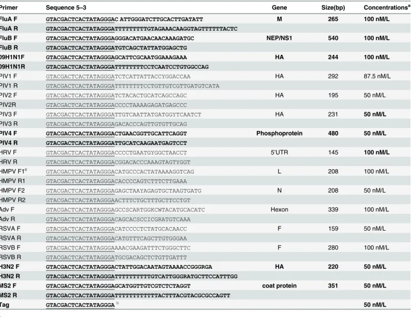
A total 14 pairs of chimeric primers were added to a single-tube to detect 13 respiratory viruses, and one pair of internal control primer (MS2 F, MS2 R) and one universal primer (Tag) was also added to the tube. The bacteriophage MS2 coat protein derived Virus-like particles (VLPs) was used as an internal control to monitor the extraction and detection process of each speci-mens [11,12]. The primers sequences, the target genes, the amplicon sizes, and primer working concentrations are listed inTable 1.
Two-tube Assay and Detection Method
Two-tube assay was performed as described [10]. Briefly, one-step RT-PCR Kit (Qiagen, Hil-den, Germany) was used for the amplification. A total of 25μL PCR mixture containing 2μL of
extracted RNA and varied primer concentrations was subjected to the following conditions: 50°C for 30min, 95°C for 15min, followed by 10 cycles of 95°C for 30s, 55°C for 30s, and 72°C 30 s; 10 cycles of 95°C for 30s, 65°C for 30s, 72°C for 30s; 25 cycles of 95°C for 30s, 48°C for 30s, and 72°C for 30s, and a final incubation at 72°C for 3min, and the products were analyzed on the QIAxcel automatic electrophoresis using QIAxcel DNA High-Resolution kit.
The Single-tube Assay and Detection Method
Single-tube assay was performed according to the protocols as described above in two-tube assay with a slight modification of the primer sequences and primer concentration (Table 1).
Analytical methods
The X2-test and Fisher’s exact test were performed to analyze the detection agreement between the single-tube assay and the two-tube assay and resolved discordant results.
Results
Performance evaluation using clinical samples
methods. All failed results and discordant results were resolved with repeated tests and direct sequencing using the same primers listed in theTable 1.
Comparison of the assay performance
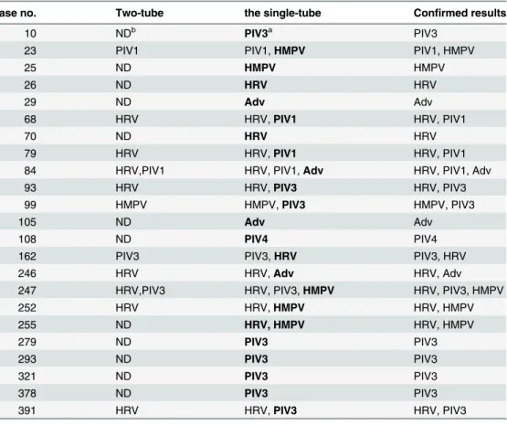
As shown inTable 2, there were 23 specimens with discordant results between the two-tube assay and the single-tube assay, all of them were confirmed by sequencing as true positives, A total of 24 additional viruses were detected only by the single-tube assay, including 5Adv, 4HMPV, 3HRV, 2PIV1, 9PIV3 and 1PIV4. The two-tube assay does not include the detection of PIV4. The sensitivity, specificity, negative prediction value (NPV), positive prediction value
Table 1. Primer information.
Primer Sequence 5–3 Gene Size(bp) Concentrationsa
FluA F GTACGACTCACTATAGGGAC ATTGGGATCTTGCACTTGATATT M 265 100 nM/L
FluA R GTACGACTCACTATAGGGATTTTTTTTTGTAGAAACAAGGTAGTTTTTTACTC
FluB F GTACGACTCACTATAGGGAGGGACATGAACAACAAAGATGC NEP/NS1 540 100 nM/L
FluB R GTACGACTCACTATAGGGATGTCAGCTATTATGGAGCTG
09H1N1F GTACGACTCACTATAGGGAGCATTCGCAATGGAAAGAAA HA 244 100 nM/L
09H1N1R GTACGACTCACTATAGGGATTTTTTTTCCTCAATCCTGTGGCCAG
PIV1 F GTACGACTCACTATAGGGATCTCATTATTACCYGGACCAA HA 292 87.5 nM/L
PIV1 R GTACGACTCACTATAGGGATTTTTTTTCCTGTTGTCGTTGATGTCATA
PIV2 F GTACGACTCACTATAGGGATCTACACTGCATCAGCCAGC HA 195 50 nM/L
PIV2R GTACGACTCACTATAGGGACCCCTAAAAGAGATGAGCCC
PIV3 F GTACGACTCACTATAGGGATTGTCAATTATGATGGYTCAATCT HA 231 50 nM/L
PIV3 R GTACGACTCACTATAGGGAGACACCCAGTTGTGTTGCAG
PIV4 F GTACGACTCACTATAGGGACTGAACGGTTGCATTCAGGT Phosphoprotein 480 50 nM/L
PIV4 R GTACGACTCACTATAGGGATTGCATCAAGAATGAGTCCT
HRV F GTACGACTCACTATAGGGACCCCTGAATGYGGCTAACCT 5’UTR 145 100 nM/L
HRV R GTACGACTCACTATAGGGACGGACACCCAAAGTAGTYGGT
HMPV F1c GTACGACTCACTATAGGGACATGCCCACTATAAAAGGTCAG L 208 100 nM/L
HMPV R1 GTACGACTCACTATAGGGACACCCCAGTCTTTCTTGAAA
HMPV F2 GTACGACTCACTATAGGGAGAGCTAAYAGAGTGCTAAGTGATG N 208 50 nM/L
HMPV R2 GTACGACTCACTATAGGGAACTTTCTGCTTTGCTTCCTGT
Adv F GTACGACTCACTATAGGGAGCCSCARTGGKCWTACATGCACATC Hexon 339 100 nM/L
Adv R GTACGACTCACTATAGGGACAGCACSCCICGRATGTCAAA
RSVA F GTACGACTCACTATAGGGACATCCCCTCTATGCACAACC F 159 50 nM/L
RSVA R GTACGACTCACTATAGGGACATGTTTCAGCTTGTGGGAA
RSVB F GTACGACTCACTATAGGGAAAACGAAGATTTCTGGGCTTC F 280 100 nM/L
RSVB R GTACGACTCACTATAGGGATGCGACAGCTCTGTTGATTT
H3N2 F GTACGACTCACTATAGGGACTATTGGACAATAGTAAAACCGGGRGA HA 220 50 nM/L
H3N2 R GTACGACTCACTATAGGGATTTTTTTTTTGTCATTGGGRATGCTTCCATTTGG
MS2 F GTACGACTCACTATAGGGAGCATGGTTGTCGTCTCTAGGT coat protein 351 50 nM/L
MS2 R GTACGACTCACTATAGGGATTTTTTTTTTTTACTTTACGTACGCGCCAGTT
Tag GTACGACTCACTATAGGGAb 50 nM/L
aPrimer sequences and primer concentrations varied from two-tube assay are marked in boldface. bThe underlined sequences are universal sequences.
cThe primers HMPV-1and HMPV-2 are designed to amplify the L gene and N gene, the amplicon sizes of both PCR products are exactly the same.
(PPV), the accordance rate, and the kappa value of each virus for the single-tube assay com-pared to two-tube assay are shown inTable 3.
Discussion
Early detection of respiratory virus infections allows clinicians to initiate immediate therapeu-tic interventions that can reduce complications, antibiotherapeu-tic use, and unnecessary laboratory test-ing. In this study, we compared the performance of a single-tube multiplex reverse
transcription PCR assay with the reference method, a two-tube assay, which has been con-firmed to be comparable to the commercial Luminex x TAG RVP Fast assay and Seeplex RV15 ACE detection kit in our previous study. The single-tube assay is also based on the QIAxcel capillary electrophoresis system, which is accessible in most of provincial Centers for Disease Control and Prevention in China. Of the 310 specimens from hospitalized children, a high prevalence of infection and co-infection with the common respiratory viral pathogens were revealed. HRV was found to be most frequently, followed by PIV3. The major discrepancies between the reference two-tube assay and the proposed single-tube assay were the detection rates of HRV, Adv, HMPV, PIV4, PIV1 and PIV3. The 24 viruses detected only by the single-tube assay (5Adv, 4HMPV, 3HRV, and 2PIV1, 9PIV3, 1PIV4) were confirmed by sequencing as true positives. Therefore, the single-tube assay is more sensitive than the two-tube assay for
Table 2. The confirmed results for specimens with discordant results between the single-tube assay and the two-tube assay.
Case no. Two-tube the single-tube Confirmed results
10 NDb PIV3a PIV3
23 PIV1 PIV1,HMPV PIV1, HMPV
25 ND HMPV HMPV
26 ND HRV HRV
29 ND Adv Adv
68 HRV HRV,PIV1 HRV, PIV1
70 ND HRV HRV
79 HRV HRV,PIV1 HRV, PIV1
84 HRV,PIV1 HRV, PIV1,Adv HRV, PIV1, Adv
93 HRV HRV,PIV3 HRV, PIV3
99 HMPV HMPV,PIV3 HMPV, PIV3
105 ND Adv Adv
108 ND PIV4 PIV4
162 PIV3 PIV3,HRV PIV3, HRV
246 HRV HRV,Adv HRV, Adv
247 HRV,PIV3 HRV, PIV3,HMPV HRV, PIV3, HMPV
252 HRV HRV,HMPV HRV, HMPV
255 ND HRV, HMPV HRV, HMPV
279 ND PIV3 PIV3
293 ND PIV3 PIV3
321 ND PIV3 PIV3
378 ND PIV3 PIV3
391 HRV HRV,PIV3 HRV, PIV3
aDiscordant results are highlighted in boldface. bND stands for not detected.
the detection of HRV, HMPV, Adv, PIV, PIV3 and PIV4. The overall detection rate of the sin-gle-tube assay for each virus was comparable to that of the two-tube assay (kappa>0.75) dem-onstrating the high sensitivity and specificity of the single-tube assay in the analysis of clinical samples.
Compared with the two-tube assay, the single-tube assay revealed better sensitivity, more accurate quality control, less nonspecific amplification, more cost-effective and shorter turn-around time. This improvement might be attributed to several aspects as follows. First, we have replaced human genome RNase P gene in the two-tube assay with bacteriophage MS2 as an internal control. The utility of MS2-based armored RNA as an assay internal control has been documented in many clinical applications [13,14]. In this study, the internal control MS2 was added to each specimen prior to extraction as an exogenous control to monitor the whole pro-cess from nucleic acid extraction to RT-PCR. Second, we inserted a few T nucleotides when appropriate between the specific and universal sequences in the primer design to ensure each amplicon can be easily distinguished by the QIAxcel machine. Third, homologous tail sequences was added to the 5' end of each chimeric primer [15]. The homo-tag assisted non-dimer (HAND) system introduced in this study was to prevent non-dimer formation and allow more, specific, sensitive, and stable detection of the viruses [16]. Fourth, for detection of multi-ple viral targets, simultaneous presence of multimulti-ple targets in one specimen may have led to competitive inhibition of amplification of less abundant targets and may explain some loss of assay sensitivity. In this study, we avoided this problem by optimizing temperature switch PCR parameters and further fine-tuning the concentration of primers. In addition, single-tube assay is more convenient and rapidly to apply to clinical specimens, saving the cost and turn-around time.
In our study, the most frequently detected pathogen was HRV, while in other study, the RSV was the most commonly detected viral pathogen [17,18]. This difference might be caused
Table 3. Performance of the single-tube assay for individual target compared with the two-tube assay.
No. of specimens: the single tube assay/ two-tube
Performance of the single tube assay compared with the two-tube assay
Viruses +/+ +/− −/+ −/− Sensitivity% Specificity% PPV% NPV% Accordance rate% Kappa value
FluA 6 0 0 304 100 100 100 100 100 1.00
FLuB 2 0 0 308 100 100 100 100 100 1.00
s09H1N1 0 0 0 310 NA 100 NA 100 100 NA
PIV1 7 2 0 301 100 99.34 77.78 100 99.35 0.87
PIV2 0 0 0 310 NA 100 NA 100 100 NA
PIV3 76 9 0 225 100 96.15 89.41 100 97.1 0.92
PIV4 0 1 0 309 NA 99.68 0 100 99.68 0
HRV 113 3 0 194 100 98.48 97.41 100 99.03 0.98
HMPV 31 4 0 275 100 98.57 88.57 100 98.71 0.93
Adv 11 5 0 294 100 98.33 68.75 100 98.39 0.81
RSVA 9 0 0 301 100 100 100 100 100 1.00
RSVB 9 0 0 301 100 100 100 100 100 1.00
H3N2 0 6 0 304 NA 98.06 0 100 98.06 0
Abbreviation: NA, not applicable.
This table shows the sensitivity, specificity, positive predictive value (PPV), negative predictive value (NPV), and the kappa values for each target using the confirmed results as the reference for comparison. All the accordance rate values were above 97.10%, except for NA, all the kappa values were above 0.75.
by the following two aspects. First, the detection of RSV increased during the winter, whereas HRV was detected year-round [19,20]. In this study, the samples collection time is not in win-ter (May to October). Second, HRV is a well-recognized cause of common colds and associated with upper respiratory tract complications. In this study, the patients were hospitalized for both the upper respiratory tract and lower respiratory tract infections.
The two-tube assay does not include PIV4 while single-tube assay does. For PIV4 and PIV2, only one PIV4 specimen was detected as positive and no PIV2 were detected by the single-tube assay with acute respiratory illnesses, which is consistent with previous reports that PIV2 and PIV4 were not identified as frequently as PIV1 and PIV 3 [21–23]. The different geographic location might also lead to the different seasonal distributions of PIVs. All of the FluA positives detected by the single-tube assay were subsequently sequenced to be H3N2 infection. 09H1N1 were not detected by either of two methods because H3N2 is the predominant seasonal FluA circulating in 2015 in China. Though PIV2 and 09H1N1 were not found in the samples, we detected a few stock clinical samples previously identified as 09 H1N1 or PIV2 using both sin-gle-tube and two-tube assays, and the results demonstrated both assays worked very well (data not shown). Moreover, no false positives were found by the single-tube assay, further suggest-ing the high specificity of the ssuggest-ingle-tube assay.
However, the limitations of our study are that only the main causes of respiratory infection were investigated. Other respiratory tract infection related viruses, such as coronaviruses 229E, HKU1, NL63, and OC43, human bocavirus (HBoV), enteroviruses (EV) [17,24,25], are not included in this study. Besides, the association of clinical outcome with the etiology informa-tion, particularly the role of co-infections in the clinical course and severity is not addressed. Given the high prevalence (18.38%, 57/310) and diversity of viral co-infections in our study, further research is also needed.
In summary, the improved single-tube assay in this study using automatic capillary electro-phoresis is a rapid, cost-effective and reliable method with high sensitivity and specificity for the simultaneous detection of thirteen respiratory virus infections, and will be a valuable tool for routine surveillance of respiratory virus infection in China.
Author Contributions
Conceived and designed the experiments: XM LL. Performed the experiments: DZ MZ HW. Analyzed the data: DZ MZ ZF SY LW. Contributed reagents/materials/analysis tools: DZ MZ ZF SY LW. Wrote the paper: XM LL GL.
References
1. Nair H, Nokes DJ, Gessner BD, Dherani M, Madhi SA, Singleton RJ, et al. Global burden of acute lower respiratory infections due to respiratory syncytial virus in young children: a systematic review and meta-analysis. Lancet (London, England). 2010; 375(9725):1545–55. Epub 2010/04/20. doi: 10.1016/s0140-6736(10)60206-1PMID:20399493; PubMed Central PMCID: PMCPMC2864404.
2. Thompson WW, Shay DK, Weintraub E, Brammer L, Cox N, Anderson LJ, et al. Mortality associated with influenza and respiratory syncytial virus in the United States. Jama. 2003; 289(2):179–86. Epub 2003/01/09. PMID:12517228.
3. Huh HJ, Park KS, Kim JY, Kwon HJ, Kim JW, Ki CS, et al. Comparison of the Anyplex(TM) II RV16 and Seeplex((R)) RV12 ACE assays for the detection of respiratory viruses. Diagnostic microbiology and infectious disease. 2014; 79(4):419–21. doi:10.1016/j.diagmicrobio.2014.01.025PMID:24985763.
4. Jung YJ, Kwon HJ, Huh HJ, Ki CS, Lee NY, Kim JW. Comparison of the AdvanSure real-time RT-PCR and Seeplex((R)) RV12 ACE assay for the detection of respiratory viruses. Journal of virological meth-ods. 2015; 224:42–6. Epub 2015/08/19. doi:10.1016/j.jviromet.2015.08.003PMID:26277911.
Diagnostic microbiology and infectious disease. 2014; 79(1):14–8. Epub 2014/03/04. doi:10.1016/j. diagmicrobio.2014.01.016PMID:24582583.
6. Bibby DF, McElarney I, Breuer J, Clark DA. Comparative evaluation of the Seegene Seeplex RV15 and real-time PCR for respiratory virus detection. Journal of medical virology. 2011; 83(8):1469–75. Epub 2011/06/17. doi:10.1002/jmv.22125PMID:21678451.
7. Renaud C, Crowley J, Jerome KR, Kuypers J. Comparison of FilmArray Respiratory Panel and labora-tory-developed real-time reverse transcription-polymerase chain reaction assays for respiratory virus detection. Diagnostic microbiology and infectious disease. 2012; 74(4):379–83. Epub 2012/09/18. doi:
10.1016/j.diagmicrobio.2012.08.003PMID:22981482.
8. Vallieres E, Renaud C. Clinical and economical impact of multiplex respiratory virus assays. Diagnostic microbiology and infectious disease. 2013; 76(3):255–61. Epub 2013/04/23. doi:10.1016/j.
diagmicrobio.2013.03.008PMID:23601453.
9. Sakthivel SK, Whitaker B, Lu X, Oliveira DB, Stockman LJ, Kamili S, et al. Comparison of fast-track diagnostics respiratory pathogens multiplex real-time RT-PCR assay with in-house singleplex assays for comprehensive detection of human respiratory viruses. Journal of virological methods. 2012; 185 (2):259–66. doi:10.1016/j.jviromet.2012.07.010PMID:22796035.
10. Li J, Qi S, Zhang C, Hu X, Shen H, Yang M, et al. A two-tube multiplex reverse transcription PCR assay for simultaneous detection of sixteen human respiratory virus types/subtypes. BioMed research inter-national. 2013; 2013:327620. Epub 2013/08/29. doi:10.1155/2013/327620PMID:23984344; PubMed Central PMCID: PMC3747601.
11. Zhan S, Li J, Xu R, Wang L, Zhang K, Zhang R. Armored long RNA controls or standards for branched DNA assay for detection of human immunodeficiency virus type 1. Journal of clinical microbiology. 2009; 47(8):2571–6. doi:10.1128/JCM.00232-09PMID:19494069; PubMed Central PMCID: PMC2725685.
12. Wei B, Wei Y, Zhang K, Yang C, Wang J, Xu R, et al. Construction of armored RNA containing long-size chimeric RNA by increasing the number and affinity of the pac site in exogenous rna and sequence coding coat protein of the MS2 bacteriophage. Intervirology. 2008; 51(2):144–50. doi:10.1159/ 000141707PMID:18594159.
13. Stevenson J, Hymas W, Hillyard D. The use of Armored RNA as a multi-purpose internal control for RT-PCR. Journal of virological methods. 2008; 150(1–2):73–6. Epub 2008/04/09. doi:10.1016/j.jviromet. 2008.02.007PMID:18395804.
14. Pasloske BL, Walkerpeach CR, Obermoeller RD, Winkler M, DuBois DB. Armored RNA technology for production of ribonuclease-resistant viral RNA controls and standards. Journal of clinical microbiology. 1998; 36(12):3590–4. Epub 1998/11/18. PMID:9817878; PubMed Central PMCID: PMCPMC105245.
15. Huang Q, Zheng L, Zhu Y, Zhang J, Wen H, Huang J, et al. Multicolor combinatorial probe coding for real-time PCR. PloS one. 2011; 6(1):e16033. Epub 2011/01/26. doi:10.1371/journal.pone.0016033
PMID:21264249; PubMed Central PMCID: PMCPMC3021529.
16. Brownie J, Shawcross S, Theaker J, Whitcombe D, Ferrie R, Newton C, et al. The elimination of primer-dimer accumulation in PCR. Nucleic Acids Res. 1997; 25(16):3235–41. Epub 1997/08/15. PMID:
9241236; PubMed Central PMCID: PMCPMC146890.
17. Liu C, Xiao Y, Zhang J, Ren L, Li J, Xie Z, et al. Adenovirus infection in children with acute lower respira-tory tract infections in Beijing, China, 2007 to 2012. BMC infectious diseases. 2015; 15:408. doi:10. 1186/s12879-015-1126-2PMID:26429778; PubMed Central PMCID: PMC4591558.
18. Jain S, Williams DJ, Arnold SR, Ampofo K, Bramley AM, Reed C, et al. Community-acquired pneumo-nia requiring hospitalization among U.S. children. The New England journal of medicine. 2015; 372 (9):835–45. Epub 2015/02/26. doi:10.1056/NEJMoa1405870PMID:25714161.
19. Cui B, Zhang D, Pan H, Zhang F, Farrar J, Law F, et al. Viral aetiology of acute respiratory infections among children and associated meteorological factors in southern China. BMC infectious diseases. 2015; 15:124. doi:10.1186/s12879-015-0863-6PMID:25884513; PubMed Central PMCID: PMC4365542.
20. Tran DN, Trinh QD, Pham NT, Pham TM, Ha MT, Nguyen TQ, et al. Human rhinovirus infections in hos-pitalized children: clinical, epidemiological and virological features. Epidemiology and infection. 2016; 144(2):346–54. Epub 2015/06/27. doi:10.1017/s0950268815000953PMID:26112743.
21. Liu WK, Liu Q, Chen DH, Liang HX, Chen XK, Huang WB, et al. Epidemiology and clinical presentation of the four human parainfluenza virus types. BMC infectious diseases. 2013; 13:28. Epub 2013/01/25. doi:10.1186/1471-2334-13-28PMID:23343342; PubMed Central PMCID: PMCPMC3560251.
23. Zhao LQ, Qian Y, Wang F, Zhu RN, Deng J. [Human parainfluenza virus infections in infants and young children with acute respiratory infections in Beijing]. Zhonghua er ke za zhi Chinese journal of pediat-rics. 2007; 45(2):91–5. Epub 2007/04/26. PMID:17456334.
24. Feng L, Li Z, Zhao S, Nair H, Lai S, Xu W, et al. Viral etiologies of hospitalized acute lower respiratory infection patients in China, 2009–2013. PloS one. 2014; 9(6):e99419. doi:10.1371/journal.pone. 0099419PMID:24945280; PubMed Central PMCID: PMC4063718.