Prevalence of oral hemangioma, vascular
malformation and varix in a Brazilian
population
Prevalência de hemangioma, malformação
vascular e variz de boca numa população
brasileira
Abstract: Hemangioma, vascular malformation and varix are benign vascular lesions, common in the head and neck regions. Studies about the prevalence of these lesions in the oral cavity are scarce. The aim of this study was to estimate the prevalence of and to obtain clinical data on oral hemangioma, vascular malformation and varix in a Brazilian population. Clinical data on those lesions were retrieved from the clinical forms from the iles of the Oral Diagnosis Service, School of Dentistry, Federal University of Minas Gerais, Brazil, from 1992 to 2002. Descriptive analysis was performed. A total of 2,419 clinical forms in the 10-year period were evaluated, of which 154 (6.4%) cases were cat-egorized as oral hemangioma, oral vascular malformation or oral varix. Oral varix was the most frequent lesion (65.6%). Females had more oral hemangioma and oral varix than males. Oral vascular malformation and oral varix were more prevalent in the 7th and 6th decades, respectively. Oral hemangioma and oral varix were more prevalent in the ventral surface of the tongue and oral vascular malformation, in the lips. Oral hemangioma was treated with sclerotherapy (54.5%), and vascular malformation was managed with sclero-therapy and surgery (19.4% each). The data of this study suggests that benign vascular lesions are unusual alterations on the oral mucosa and jaws.
Descriptors: Hemangioma; Peripheral vascular diseases, epidemiology; Blood vessels, abnormalities; Mouth mucosa.
Resumo: Hemangioma, malformação vascular e variz são lesões vasculares benignas co-muns na região de cabeça e pescoço. Estudos sobre a prevalência dessas lesões em boca são escassos. O objetivo deste estudo foi estimar a prevalência e realizar um levantamento de dados clínicos de hemangioma, malformação vascular e variz de boca. Dados clínicos dessas lesões foram consultados em ichas clínicas do arquivo do Serviço de Diagnóstico Oral da Universidade Federal de Minas Gerais, Brasil, no período de 1992 a 2002. Uma análise descritiva foi realizada. Foram obtidas neste período de 10 anos 2.419 ichas clí-nicas. Cento e cinqüenta e quatro (6,4%) casos foram categorizados como hemangioma, malformação vascular e variz de boca. Variz de boca foi a lesão mais freqüente (65,6%). Mulheres foram mais acometidas pelos hemangiomas e varizes de boca. Malformação vascular e variz de boca foram mais prevalentes na 7a e 6a décadas de vida, respectivamen-te. A localização prevalente do hemangioma e da variz de boca foi a superfície ventral da língua, e da malformação vascular, os lábios. Hemangioma de boca foi tratado com escle-roterapia em 54,5% dos casos e a malformação vascular com escleescle-roterapia e cirurgia em 19,4% cada. Este estudo sugere que lesões vasculares benignas são alterações incomuns na mucosa bucal e nos maxilares.
Descritores: Hemangioma; Doenças vasculares periféricas, epidemiologia; Vasos sangüí-neos, anormalidades; Mucosa bucal.
Priscila Henriques Corrêa(a)
Lara Cristina Caldeira Nunes(a)
Aline Cristina Batista Rodrigues Johann(b)
Maria Cássia Ferreira de Aguiar(c)
Ricardo Santiago Gomez(c)
Ricardo Alves Mesquita(c)
(a)DDSs; (b)MS; (c)PhDs, Professors –
Department of Oral Surgery, Oral Medicine and Oral Pathology, School of Dentistry, Federal University of Minas Gerais.
Corresponding author:
Ricardo Alves Mesquita
Faculdade de Odontologia da UFMG Disciplina de Patologia Bucal, sala 3204 Av. Antônio Carlos, 6627, Pampulha Belo Horizonte - MG - Brazil CEP: 31270-901
E-mail: ramesquita@ufmg.br
Introduction
Benign vascular lesions are a consequence of blood vessel abnormalities or endothelial cell prolif-eration.13 The International Society for the Study of
Vascular Anomalies (ISSVA), in 1996, approved a classiication system modiied from the one proposed by Mulliken, Glowacki20 (1982). The diseases were
subdivided into (a) tumors: hemangioma (HEM), pyogenic granuloma, rapidly involuting congenital hemangioma, noninvoluting congenital hemangio-ma, hemangiopericytohemangio-ma, tufted angioma and ka-posiform hemangioendothelioma; and (b) vascular malformation (VM).8
HEM is a benign proliferation of endothelial cells. It is the most common neoplasm of the in-fancy. HEM frequently is not present at birth and develops in three phases: proliferating, involution, and involuted.8 It presents as a red macula, papule
or nodule, depending on the congestion degree and on how deep it is in the tissue. Although HEM is a benign lesion, in some cases, it may lead to com-pression of surrounding structures, formation of issures, ulcers or hemorrhages, and functional and aesthetic problems.7,13 Oral HEM can be found in
the lips, tongue or buccal mucosa. It is more com-mon in white female, in twins and in premature infants.7,11,20
VM is considered an abnormality of the embry-onic development, a structural anomaly. VM may be composed by arteries, veins and/or capillaries. Clinically, they are similar to HEM; however, they are always present at birth and grow as the patient physically develops.20 It does not spontaneously
re-gress, remaining stable throughout life. Frequently, bone involvement is present as a radiolucent, mul-tilocular and well-circumscribed image.7 The few
studies on benign oral vascular lesions frequently do not distinguish between oral HEM and VM, or re-gard oral VM as a histological type of HEM, known as arteriovenous hemangioma.2
The treatments for benign vascular lesions are sclerotherapy, systemic corticosteroids, interferon α, laser, embolization, cryotherapy, and surgery. Whether they should be followed-up or treated de-pends on the patient’s age, and on lesion site and size.2,7,15,26
Varix (VAR) is an acquired benign vascular le-sion, generally asymptomatic, and does not require treatment.27 Age is a predisposing factor, as well
as tissue loosening and increased venous pressure. Oral VAR is rare in infants, but very common in old adults.16 Oral VAR is characterized as a red to
purple papule or nodule, commonly found on the tongue, lip or cheek, mainly in the seventh decade of life. Also, there are few studies on epidemiological data on oral VAR.16,27
The aim of this study was to estimate the preva-lence of and to report clinical data on benign oral vascular lesions (HEM, VM and VAR) in a Brazil-ian population, and also to relate these data with that found in the literature.
Material and Methods
The protocol of this study was approved by the Committee of Ethics in Research, Federal University of Minas Gerais (UFMG, number 190/02). Clini-cal forms from 1992 to 2002 were retrieved from the iles of the Oral Diagnosis Service, School of Dentistry, Federal University of Minas Gerais (FO-UFMG; Belo Horizonte, MG, Brazil) and evaluated. The Oral Diagnosis Service, FO-UFMG, uses criteria to distinguish between oral HEM and oral VM, as described by Mulliken, Glowacki20 (1982).
Oral HEM is a red, asymptomatic and well-circum-scribed lesion that develops during late fetal stages or in infancy, which grows quickly and generally spon-taneously regresses. Histologically, it presents hy-percellularity in the proliferating phase and ibrosis and diminished cellularity in the involuting phase. Oral VM is a red and asymptomatic lesion with undeined limits. It is present at birth and grows as the patient develops.20 Oral VM is characterized by
hypocellularity and vascular channels lined by lat mature endothelium. Oral VAR is characterized by a red to purple papule or nodule, commonly found in the tongue, lip or cheek, mainly in the seventh de-cade of life.16,27 Oral VAR is morphologically
com-posed by one to three extensive and tortuous blood vessels lined by lat mature endothelium.
Oral hemangioma Oral vascular malformation Oral varix
n. % n. % n. %
C
as
e
s
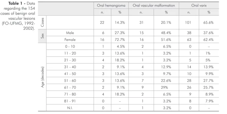
22 14.3% 31 20.1% 101 65.6%
Se
x Male 6 27.3% 15 48.4% 38 37.6%
Female 16 72.7% 16 51.6% 63 62.4%
A
g
e
(d
ec
a
d
es
)
0 - 10 1 4.5% 2 6.5% 0 –
11 - 20 3 13.6% 1 3.2% 1 1%
21 - 30 4 18.2% 1 3.2% 5 5%
31 - 40 2 9.1% 4 12.9% 14 13.9%
41 - 50 3 13.6% 3 9.7% 10 9.9%
51 - 60 3 13.6% 7 22.6% 28 27.7%
61 - 70 2 9.1% 9 29% 26 25.7%
71 - 80 4 18.2% 2 6.5% 9 8.9%
81 - 91 0 – 1 3.2% 8 7.9%
N.I. 0 – 1 3.2% 0 –
N.I.: No information.
Oral hemangioma Oral vascular malformation Oral varix
n. % n. % n. %
R
ace
White (Caucasian) 12 54.5% 13 41.9% 57 56.5%
Non-white 2 9.1% 3 9.7% 6 5.9%
N.I. 8 36.4% 15 48.4% 38 37.6%
Sy
m
pto
ms Asymptomatic 17 77.2% 22 71% 89 88.1%
Symptomatic 2 9.1% 4 12.9% 1 1%
N.I. 3 13.6% 5 16.1% 11 10.9%
Lo
caliz
atio
n
Upper lip 2 9.1% 8 25.8% 3 3%
Lower lip 5 22.7% 6 19.4% 3 3%
Floor of the mouth 0 – 0 – 9 8.9%
Ventral surface of the tongue 7 31.8% 4 12.9% 77 76.2%
Buccal mucosa 4 18.2% 7 22.6% 6 5.9%
Others 4 18.2% 6 19.4% 2 2%
N.I. 0 – 0 – 1 1%
N.I.: No information. Table 1 - Data
regarding the 154 cases of benign oral vascular lesions (FO-UFMG,
1992-2002).
Table 2 - Data regarding the 154 cases of benign oral vascular lesions (FO-UFMG,
1992-2002).
disease information (diagnosis, symptom, location, number, size and treatment) were obtained.
Results
A total of 2,419 clinical forms were evaluat-ed in the studievaluat-ed period (ten years). Of these, 154 (6.4%) were oral HEM, VM or VAR. Oral HEM
Discussion
The clinical parameters from the classiication of Mulliken, Glowacki20 (1982) were used to
distin-guish oral HEM from VM in our sample. Since the majority of studies on benign oral vascular lesions do not use this classiication, differences in epidemi-ological data on these diseases are common.9,17,21,26
However, since oral HEM is a neoplasm and oral VM is a disturbance of development, the distinc-tion between these two entities is important for the knowledge of the clinical behavior and manage-ment of these illnesses.7,20 The use of the Mulliken,
Glowacki20 (1982) classiication for benign oral
vas-cular lesions has also standardized the studies and epidemiological data on these lesions.
It was observed that benign oral vascular lesions represented 6.4% of all the diseases diagnosed in the Oral Diagnosis Service, FO-UFMG, and that oral VAR was the most prevalent lesion. Jainkittivong
et al.14 (2002) observed that oral VAR represented
59.6% of the oral mucosa conditions diagnosed in patients older than 60 years. Kovac-Kovacic, Skale-ric17 (2000) found a relative frequency of 16.2% for
oral VAR, with higher frequency (93%) in patients in their 7th and 8th decades of life. Few studies and
with variable relative frequency of oral HEM and VM are found in literature. In the study of Corbet
et al.6 (1994), oral HEM represented 2% of the
le-sions of the oral mucosa in patients with ages rang-ing from 65 to 74 years. Al-Khateeb et al.1 (2003), in
a study with infants, reported a relative frequency of 0.9% for HEM. Paltiel et al.23 (2000) reported on the
relative frequency of HEM and VM, and found that HEM was predominant in 53% and VM, in 56% of the cases. In our study, oral VM was more frequent than oral HEM. Unlike Paltiel et al.23 (2000), our
study was performed in a sample of jaw diseases. Oral HEM and VAR were more frequent in fe-males, as shown by the studies of Ettinger, Mander-son10 (1974), and Donnelly et al.7 (2000). Jackson
et al.13 (1993) observed a female : male ratio of 4:1
in patients with HEM. Jackson et al.13 (1993) and
Barrett, Speight2 (2000) observed that VM affects
equally females and males, as in our study.
Oral VAR was more frequently diagnosed in pa-tients in their 6th and 7th decades of life. Ettinger,
Manderson10 (1974) also observed a higher
occur-rence of oral VAR as age increased, stating that, with it, tissue loosening occurs, which is an impor-tant factor in the development of oral VAR. The in-cidence of this alteration in our study was 70.2% in patients older than 60 years, similar to that ob-served by Bean3,4 (1952; 1956), and Miles18 (1972).
Oral hemangioma Oral vascular malformation Oral varix
n. % n. % n. %
N
u
mbe
r
o
f
le
sio
ns Single 19 86.4% 25 80.6% 8 7.9%
Multiple 2 9.1% 4 12.9% 57 56.4%
N.I. 1 4.5% 2 6.5% 36 35.6%
Siz
e
(cm)
0.1 - 1 0 – 1 3.2% 0 –
1.1 - 2 1 4.5% 2 6.5% 0 –
2.1 - 3 0 – 1 3.2% 0 –
3.1 - 4 3 13.6% 5 16.1% 0 –
> 4 4 18.2% 1 3.2% 0 –
N.I. 14 63.6% 21 67.7% 101 100%
Tre
atme
nt
Sclerotherapy 12 54.5% 6 19.4% 3 3%
Surgery 3 13.6% 6 19.4% 2 2%
No treatment 0 – 6 19.4% 5 5%
N.I. 7 31.8% 13 41.9% 91 90%
N.I.: No information.
Otherwise, according to Kovac-Kovacic and Skale-ric17 (2000), oral VAR is more common in patients
between 51 and 60 years.
Jackson et al.13 (1993) observed that HEM is
the most common benign neoplasm in infancy, the majority arising between the 1st and the 4th week
of life. At ive and seven years of age, respectively 50% and 70% of the infants no longer have their lesions. This is conirmed by the studies of Mul-liken, Glowacki20 (1982), and Mulliken19 (1992).
VM is present at birth, grows together with the patient, and does not spontaneously regress.19,20
Hence, the clinical behavior of HEM and VM may explain the broad age range of these alterations found in our study, since our patients sought care in a later moment of their lives. Moreover, previ-ous studies13,19,20 analyzed HEM and VM located
in other anatomic regions, in which functional and/or aesthetic problems are more pronounced than in the jaws.
Previous studies did not refer to the race of the patients when analyzing benign vascular lesions7,10,13.
In our sample, all three lesions were more frequent in Caucasian patients. Parra et al.24 (2003) observed
that in the Brazilian population, approximately 39% of the population has European genes, 33% has Amerindian genes, and 28% has African genes. In addition, the Brazilian population census of 200012
showed that 54% of the population is Caucasian. Hence, the higher prevalence of benign oral vascu-lar lesions in Caucasian Brazilians is in accordance with the country’s race distribution.
Jackson et al.13 (1993) described HEM and VM
as asymptomatic lesions. In our analysis, both al-terations were also asymptomatic. Regarding oral VAR, studies in the literature do not describe symp-toms.10,27 However, we observed that oral VAR are
predominantly asymptomatic.
Oral VAR was located more frequently in the ventral surface of the tongue. Oral VM was more frequent in the upper lip, buccal mucosa and lower lip. Also, Barrett, Speight2 (2000) observed that
oral VM is more frequent in the lips and buccal mucosa. Oral HEM was more common in the
ven-tral surface of the tongue, followed by the lower lip and buccal mucosa. Nguyen et al.22 (2004)
ob-served that among HEM lesions of the head and neck, oral HEM was mostly located in the buccal mucosa.
As to the number of lesions, Takahashi et al.28
(1994) reported that oral HEM is a single lesion. Barrett, Speight2 (2000) stated that oral VM is
al-ways a single disease without systemic implication, which is also true in our study. In contrast, oral VAR presented predominantly as multiple lesions, as in the studies by Kleinman16 (1967) and Ettinger,
Manderson10 (1974).
Benign oral vascular lesions may be treated by sclerotherapy, systemic corticosteroids, interferon α, laser, embolization, cryotherapy, and surgery. Management and treatment decisions depend on the patient’s age, and on the lesion’s site and size.15,25
Sadeghi, Gingrass25 (1989) stated that surgery is
not indicated for bigger lesions due to post-surgi-cal hemorrhages. In such cases, arterial emboliza-tion is satisfactory.25 Satisfactory results are seen
with sclerotherapy in the treatment of small be-nign vascular lesions, including oral diseases.15 In
our sample, oral HEM and VM were treated with sclerotherapy or surgery. For oral VAR, treatment was carried out in ive cases, because lesions were single and with unusual localization (lips and buccal mucosa). Moreover, they were satisfactorily treated by sclerotherapy or surgery.
Conclusions
Benign oral vascular lesions are unusual altera-tions on the oral mucosa and jaws.
Acknowledgements
References
1. Al-Khateeb T, Al-Hadi Hamasha A, Almasri NM. Oral and maxillofacial tumours in north Jordanian children and ado-lescents: a retrospective analysis over 10 years. Int J Oral Maxillofac Surg. 2003;32(1):78-83.
2. Barrett AW, Speight PM. Superficial arteriovenous heman-gioma of the oral cavity. Oral Surg Oral Med Oral Pathol Oral Radiol Endod. 2000;90(6):731-8.
3. Bean WB. The caviar lesion under the tongue. Trans Am Clin Climatol Assoc. 1952;64:40-51.
4. Bean WB. The changing incidence of certain vascular lesions of the skin with aging. Geriatrics. 1956;11(3):97-102. 5. Centers for Disease Control and Prevention (CDC), Atlanta, in
collaboration with World Health Organization (WHO). EPI-INFO - A World-Processing, Database, and Statistics System for Epidemiology on Microcomputers. Geneva; 2003 [accessed 2004 Aug 12]. Available from: http://www.cdc.gov. 6. Corbet EF, Holmgren CJ, Phillipsen HP. Oral mucosal lesions
in 65-74-year-old Hong Kong Chinese. Community Dent Oral Epidemiol. 1994;22(5 Pt 2):392-5.
7. Donnelly LF, Adams DM, Bisset GS 3rd. Vascular malforma-tions and hemangiomas: a practical approach in a multidisci-plinary clinic. AJR Am J Roentgenol. 2000;174(3):597-608. 8. Enjolras O, Mulliken JB. Vascular tumors and vascular
mal-formations (new issues). Adv Dermatol. 1997;13:375-423. 9. Espinoza I, Rojas R, Arnanda W, Gamonal J. Prevalence of
oral mucosal lesions in elderly people in Santiago, Chile. J Oral Pathol Med. 2003;32(10):571-5.
10. Ettinger RL, Manderson RD. A clinical study of sublingual varices. Oral Surg Oral Med Oral Pathol. 1974;38(4):540-5. 11. Gampper TJ, Morgan RF. Vascular anomalies: hemangiomas.
Plast Reconstr Surg. 2002;110(2):572-88.
12. IBGE [página da internet; acesso12 aug 2004]. Disponível em: http://www.censo.ibge.gov.br.
13. Jackson IT, Carreno R, Potparic Z, Hussain K. Hemangiomas, vascular malformations, and lymphovenous malformations: classification and methods of treatment. Plast Reconstr Surg. 1993;91(7):1216-30.
14. Jainkittivong A, Aneksuk V, Langlais RP. Oral mucosal condi-tions in elderly dental patients. Oral Dis. 2002;8(4):218-23.
15. Johann AC, Aguiar MC, do Carmo MA, Gómez RS, Castro WH, Mesquita RA. Sclerotherapy of benign oral vascular le-sion with ethanolamine oleate: an open clinical trial with 30 lesions. Oral Surg Oral Med Oral Pathol Oral Radiol Endod. 2005;100(5):579-84.
16. Kleinman HZ. Lingual varicosities. Oral Surg Oral Med Oral Pathol. 1967;23(4):546-8.
17. Kovac-Kovacic M, Skaleric U. The prevalence of oral mucosal lesions in a population in Ljubljana, Slovenia. J Oral Pathol Med. 2000;29(7):331-5.
18. Miles AE. ‘Sans teeth’: changes in oral tissues with advancing age. Proc R Soc Med. 1972;65(9):801-6.
19. Mulliken JB. A biologic approach to cutaneous vascular anomalies. Pediatr Dermatol. 1992;9(4):356-7.
20. Mulliken JB, Glowacki J. Hemangiomas and vascular malforma-tions in infants and children: a classification based on endothelial characteristics. Plast Reconstr Surg. 1982;69(3):412-22. 21. Mumcu G, Cimilli H, Sur H, Hayran O, Atalay T. Prevalence
and distribution of oral lesions: a cross-sectional study in Turkey. Oral Dis. 2005;11(2):81-7.
22. Nguyen VA, Furhapter C, Romani N, Weber F, Sepp N. In-fantile hemangioma is a proliferation of beta 4-negative endo-thelial cells adjacent to HLA-DR-positive cells with dendritic cell morphology. Hum Pathol. 2004;35(6):739-44.
23. Paltiel HJ, Burrows PE, Kozakewich HP, Zurakowski D, Mulliken JB. Soft-tissue vascular anomalies: utility of US for diagnosis. Radiology. 2000;214(3):747-54.
24. Parra FC, Amado RC, Lambertucci JR, Rocha J, Antunes CM, Pena SD. Color and genomic ancestry in Brazilians. Proc Natl Acad Sci USA. 2003;100(1):177-82.
25. Sadeghi E, Gingrass D. Oral hemangioma treated with a scle-rosing agent. Report of a case. Int J Oral Maxillofac Surg. 1989;18(5):262-3.
26. Sato M, Tanaka N, Sato T. Oral and maxillofacial tumours in children: a review. Br J Oral Maxillofac Surg. 1997;35(2):92-5. 27. Southam JC, Ettinger RL. A histologic study of sublingual
vari-ces. Oral Surg Oral Med Oral Pathol. 1974;38(6):879-86. 28. Takahashi K, Mulliken JB, Kozakewich HP, Rogers RA,