Location and angulation of curvatures of
mesiobucal canals of mandibular molars
debrided by three endodontic techniques
Posição e angulação de curvaturas radiculares
em canais mesiobucais de molares inferiores
preparados por três técnicas endodônticas
Abstract: The aim of this study was to assess the correlation between the degree of angu-lation and the position of root curvatures and their inluence on the comparative results between the performances of the Progressive, Staged and Serial Preparation Techniques. The mesiobucal canals of 70 extracted mandibular molars were illed with a radiological contrast of 100% Barium sulphate and radiographed with a direct digital radiographic system, in an apparatus that guarantees that the samples remain in the same spatial posi-tion at all times. The images were then analyzed in the Coreldraw 10 program (MicroSafe, RJ, Brasil) in accordance with two criteria: the methods of Berbert, Nishiyama1 (1994) and Schneider11 (1971) to determine the position and the angle of the root curvatures, re-spectively. Initially, the possibility of correlation between these two variables was studied. The teeth were then selected according to angulation (greater than 25 degrees) and posi-tion of root curvatures (cervical, median and apical) in order to perform the endodontic techniques. After preparation, the samples were radiographed again and the images were superimposed in order to compare the pre- and post-operative areas. The difference be-tween them showed the percentage of widening for each technique. The results showed that there was no correlation between the angulations and the root curvature positions, and that the different positions did not interfere in the performance of the techniques. The Progressive Preparation technique produced the highest widening values for all the groups, irrespective of the root curvature position.
Descriptors: Root canal therapy; Root canal preparation; Anatomy; Abnormalities; Molar.
Resumo: O objetivo deste trabalho foi veriicar a correlação entre o grau de angulação e a posição das curvaturas radiculares, e a sua inluência nos resultados comparativos entre os desempenhos das técnicas do Preparo Progressivo, Escalonada e Seriada. Os canais mésio-vestibulares de 70 dentes molares inferiores extraídos foram preenchidos com um contraste radiológico de sulfato de Bário a 100% e radiografados em um sistema de ra-diograia digital direta, em um aparato que garantia que as amostras icassem sempre na mesma posição espacial. As imagens foram, então, analisadas no programa Coreldraw 10 (MicroSafe, RJ, Brasil) segundo dois critérios: os métodos de Berbert, Nishiyama1 (1994) e de Schneider11 (1971) para determinação da posição e do ângulo das curvaturas radicu-lares, respectivamente. Estudou-se inicialmente a possibilidade de correlação entre essas duas variáveis. Os dentes foram, então, selecionados segundo a angulação (superior a 25 graus) e posição das curvaturas radiculares (cervical, mediana e apical) para a realização das técnicas endodônticas. Após os preparos, as amostras foram novamente radiografa-das e as imagens sobrepostas para a comparação radiografa-das áreas pré e pós-operatórias. A dife-rença entre elas mostrou a porcentagem de alargamento para cada técnica. Os resultados mostraram não existir correlação entre as angulações e as posições das curvaturas radicu-lares, e que as diferentes posições não interferem no desempenho das técnicas. A técnica do Preparo Progressivo mostrou maiores valores de alargamento para todos os grupos, independentemente da posição da curvatura radicular.
Descritores: Tratamento do canal radicular; Preparo de canal radicular; Anatomia; Anormalidades; Molar.
Isa Geralda Teixeira Constante(a) Harry Davidowicz(b)
Fernando Branco Barletta(c) Abilio Albuquerque Maranhão de Moura(d)
(a)MSc in Endodontics; (b)Full Professor; (d)Full
Professor and Chairman – Endodontics Department, Paulista University.
(c)Professor, Graduate Program in
Endodontics, Lutheran University of Brasil, Canoas.
Corresponding author:
Abilio Albuquerque Maranhão de Moura Rua Barão do Triunfo, 1650, Ap. 22 Campo Belo
São Paulo - SP - Brazil CEP: 04602-006 E-mail: [email protected]
Introduction
Chemical-surgical preparation is the stage of endodontic treatment in which the root canal is cleaned and modeled. If this is well done, it should allow an enlargement of the original canal space without deforming it, enabling a three-dimensional obturation to be done, consequently promoting the repair of the periapical tissues.
However, the existence of alterations in the in-ternal anatomy of the root canal may make it dif-icult to carry out treatment. Different tooth wall thicknesses, various positions and angulations of root curvatures, isthmuses, among other alterations, involve speciic requirements for performing a good modeling. It is imperative for the professional who is going to work in Endodontics to have knowledge of the internal anatomy, to allow planning of the en-dodontic procedure, access to the pulp chamber and inal obturation.
Different degrees of root curvature lead to dif-ferent dificulties in canal preparation.11 Therefore,
curvatures have been classiied as slight (up to ive degrees), moderate (from ive to twenty degrees) and severe (greater than twenty-ive degrees).
The three-dimensional study of mesial root canal curvatures in mandibular molars in the buccolin-gual and mesiodistal directions2 showed that 100%
of the specimens presented with curvatures in both directions.
A method was developed for quantifying and locating the curvatures of root canals,1
determin-ing a quotient for establishdetermin-ing the position of the curvature along the root. Quotients lower than 0.5 represented curvatures that were concentrated in the apical third; quotients ranging from 0.5 to 2.0 represented curvatures concentrated in the middle third; and, inally, quotients above 2.0 represented curvatures in the cervical third. When applying this methodology to the radiographs of 50 extracted maxillary and mandibular molars, it was noted that the greatest concentration of curvatures was in the middle third.
Although the anatomy of each type of tooth pre-sented common characteristics,3,8,10 it also presented
very complex variations. Irregularities, such as acces-sory canals and apical deltas, were practically
inacces-sible to mechanical preparation; curvatures resulted in the asymmetrical removal of dentin during clean-ing, leading to apical transport by several degrees, and the real anatomy of the canal was much more complicated than it appeared to be radiographically, with curvatures in multiple positions and planes.
A series of endodontic techniques are recom-mended for the treatment of teeth with root curva-tures, each of them indicating suitable resources for overcoming the dificulties related to the intrinsic demands of the treatment of these teeth.
The Serial Technique was the irst technique introduced for the treatment of either straight or curved root canals.4 The Staged Technique13 was
recommended for the treatment of curved canals, which indicated the withdrawal of the larger caliber instruments that worked at shorter lengths than the working length. The “Progressive Preparation Tech-nique”7 combines the advantages of the crown-apex
principle with the optimization of manual instru-ments. It uses a different type of instrument (K-Flex or NiTi) depending on the degree of root curvature presented by the tooth. More accentuated curva-tures are worked on with more lexible instruments. The aim of this study was to assess the correlation between the degree of angulation and the position of root curvatures and their inluence on the compara-tive results between the performances of the Progres-sive, Staged and Serial Preparation Techniques.
Materials and Methods
The mesiobucal canals of 70 teeth were illed with a radiological contrast of 100% Barium sul-phate and direct digital radiographs were taken by an apparatus that guarantees that the radiographs are taken in the same spatial position. The images were then analyzed in the Coreldraw 10 program (MicroSafe, Rio de Janeiro, RJ, Brasil) in accor-dance with two criteria: The angulation and posi-tion of root curvatures.
Analysis of the position of curvatures was based on the Berbert and Nishiyama method1 that deines
segments were measured and the values divided. This division resulted in a quotient that indicated the location of the curvature: Cervical (greater than 2.0), median (from 0.5 to 2.0) and apical (smaller than 0.5) (Figure 1).
The angulations were analyzed on the basis of the Schneider method, which consists of tracing a straight line parallel to the long axis of the tooth and a second straight line starting from the apex un-til it meets with the irst straight line at the place where the canal begins to move away from the long axis.11 The acute angle formed is measured and is
denominated the root curvature angle. The roots are then classiied in accordance with the degree of the curvatures as straight (up to 5 degrees), moder-ate (from 5 to 20 degrees) and severe (over 25 de-grees) (Figure 2).
Forty-ive teeth, all of them with a curvature greater than 25 degrees, were selected and divided into three groups: One group for the Serial technique (GS), one for the Staged technique (GE), and one for the Progressive Preparation technique (GP). Each group was sub-divided into three sub-groups, one for each root curvature position: Sub-group A – cervical curvature, group B – median curvature, and sub-group C – apical curvature. The apical preparations of all the techniques were performed with #30 iles.
The Serial Technique was performed with the irst instrument working on the working length, with iling and widening movements, until it felt loose inside the canal. It was then replaced by the immediately higher number that would be used in the same kinematics and at the same length, up to instrument #30.
The Staged Technique was performed sequen-tially up to instrument #25, which was denominated the “memory instrument”. It was performed with a 1 mm reduction of the working length for each increase in instrument caliber, at all times reviewing the total working length with the memory instrument.
The Progressive Preparation Technique was per-formed by initially preparing the entry oriice of the canals with “Moura Manual Wideners”. These in-struments were confected by cutting 31 mm type K iles (#80, #70 and #60) with a Carborundum disk to a length of 20 mm and then wearing their tips. They were then used with half-turn maneuvers in a clockwise direction. After odontometry, the canal was debrided up to at least ile #25, denominated the “memory instrument”. This instrument was then put into the real working length passively, without acting actively against the tooth canal walls. The crown-apex preparation then began with a K type instru-ment, with a diameter 4 times greater than that of
Figure 1 - Analysis according to the Berbert and Nishiyama1
Figure 2 - Schneider11
(1971) Method.
the memory instrument, working in iling movements without apical pressure on the most cervical and mid-dle regions of the canal. The next instruments were
of successively smaller calibers and reached increas-ing apical depths of the root canal. They were inter-spersed with the memory instrument up to the real working length, from the greater to smaller caliber, until the memory instrument number was attained. This instrument was then replaced by a NiTi instru-ment of the same number, which now worked ac-tively in the apical region, followed by another NiTi instrument of an immediately higher diameter, #30, up to the working length, to perform apical prepara-tion. The apical preparations were performed with NiTi instruments, as all the teeth had curvatures greater than 25 degrees (severe curvatures).
The root canal preparations of all the techniques were performed with 1% NaOCl.
The teeth were radiographed again after the preparations, under the same conditions as those of the initial radiographs. The digital images were su-perimposed to compare the pre- and post-operative areas. The difference between them enabled the per-centage of widening to be calculated for each tech-nique, indicating how much each of them widened each group of teeth.
Results
Tables 1, 2 and 3 present the values resulting from the measurement of the angles (Å) and of the
Table 1 - Results of teeth with cervical curvature.
Tooth Å Q
8 28.24 2.211
14 37.24 2.402
15 25.46 2.104
19 33.6 2.047
32 33.2 2.059
34 25.34 2.644
36 39.4 2.044
38 29.51 2.183
41 44.9 4.362
45 28.02 2.307
50 26.8 2.253
52 34.19 2.520
53 29.37 2.125
54 41.24 2.222
57 38.9 2.800
59 27.1 2.706
69 25.2 2.294
Table 2 - Results of teeth with median curvature.
Tooth Å Q
2 30.9 1.766
3 26 1.551
4 29.3 1.064
5 22.37 1.705
6 22.9 1.021
7 22.65 1.284
11 43.24 1.547
16 32.3 0.909
17 30.9 0.935
18 28.6 0.881
21 37 0.721
22 29 1.291
23 29.22 0.897
24 29.5 0.991
25 37.92 0.659
26 25.4 1.145
27 26.37 0.836
29 26.5 1.048
30 25.2 1.101
33 27 1.168
35 24.7 1.027
37 36.5 0.786
39 25.6 1.462
47 26.39 1.247
49 21.49 1.644
58 20.1 1.744
62 25.86 0.740
63 28.7 1.739
65 20.1 1.844
66 21.2 1.829
70 23.6 1.000
51 20 0.789
74 33.4 0.982
75 28.4 1.068
40 21.3 0.744
Å = measurement of the angles (°); Q = root curvature position.
Table - Results of teeth with apical curvature.
Tooth Å Q
1 25.71 0.496
9 43.24 0.227
12 33.9 0.179
20 25.2 0.209
31 26.98 0.213
42 26 0.346
43 34.54 0.192
48 26.37 0.451
60 26.83 0.496
61 49.1 0.089
67 40.7 0.181
68 26.8 0.240
71 36.25 0.162
72 21.4 0.379
73 25.48 0.497
10 38.6 0.162
64 26.2 0.381
Å = measurement of the angles (°); Q = root curvature position.
root curvature positions (Q) in accordance with the two selection criteria in the CorelDraw 10 program.
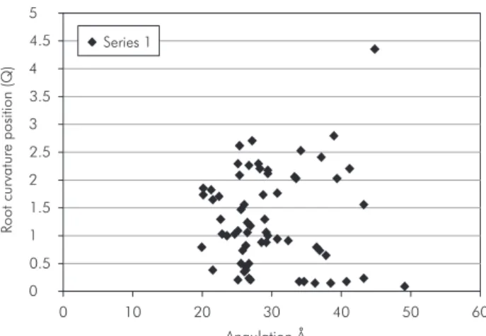
The Pearson Coeficient of Correlation was used to ind out whether there was any relation between
the variables Å and Q. The results indicated that r = 0, i.e., the two variables were independent; there was no correlation between them.
Graph 1 shows that the points were distributed in a dispersed manner, conirming that there was no relation between the variables.
It was observed that 50.72% of the samples pre-sented curvatures in the median region of the root, whereas the cervical and apical regions were in-volved in 24.64% and 24.64% of the samples, re-spectively.
From the initial group of 70 teeth, 45 teeth with root curvatures greater than 25 degrees in the dif-ferent positions of the above-mentioned sub-sections were then selected, in order to perform the tech-niques. The canal areas were measured before and after the preparations, and from the difference be-tween the inal and the initial areas, the percentage of widening for each technique was obtained (Table 4).
Table 4 - Comparison of the widening percentages among the techniques.
Groups GS GE GP
Sub-group A 51.25% 49.01% 52.73% Sub-group B 56.27% 53.23% 59.10% Sub-group C 43.59% 52.65% 54.89%
GS: serial; GE: staged; GP: progressive.
Table 5 - Result of the Kruskal-Wallis Test for analyzing wid-ening.
Curvature
Apical Median Cervical 0.867 0.177 0.792
p < 0.05.
GS (Serial Technique), GE (Staged Technique) and GP (Progressive Preparation Technique) with the re-spective sub-groups A – cervical curvature, B – me-dian curvature and C – apical curvature (Table 4). The results showed that the differences in the val-ues among the groups considered were not statisti-cally signiicant, i.e., the Technique and Curvature groups were similar.
Graph 2, however, shows that in all the sub-groups of teeth for each technique, the Progressive Preparation technique presented the best perfor-mance in terms of widening percentage.
Discussion
Tables 1, 2 and 3 show the tooth measurement values in relation to angulation and position of the curvature along the root. The lack of correlation be-tween these two variables (Graph 1) relects the ana-tomic diversity presented by the root canals in the studied group, indicating curvatures distributed in all regions of the root, in the most varying degrees. This result is in agreement with that of the studies of Cunningham, Senia2 (1992) and Berbert,
Nishi-yama1 (1994), who observed great variations in the
degrees of root curvature presented by the teeth in their studies. The larger percentage of curvatures in the median region also coincides with the indings of Berbert, Nishiyama1 (1994).
Fröner et al.3 (1999), Pécora et al.8 (2002) and
Peters10 (2004) observed that the apical anatomy of
mesial roots presented a morphological complexity related to the number and shape of the root canals, and that although the anatomy of each type of tooth was shown to have common characteristics, they also presented very complex variations. They also mentioned that the curvatures resulted in an asym-metric removal of dentin during cleaning, leading to apical transport by severous degrees.
The lack of statistically signiicant differences among the techniques with regard to the position of curvature (Table 5) showed that the root curvature position does not interfere directly in the perfor-mance of the studied techniques.
In addition, the studies of Schneider11 (1971),
Lim, Webber5 (1985), Pesce et al.9 (1997) and
Mi-gliau et al.6 (2004) observed the relation between
the degree of root curvature and deformations in preparation, as they found that a larger number of undesirable alterations in the original anatomy of
Graph 2 - Percentage of widening of the canal areas. 0 10 20 30 40 50 60 70 Sub-group C 51 .2 5 49 .0 1 52 .7 3 56 .2 7 53 .2 3 59 .1 0 43 .5 9 52 .6 5 54 .8 9
Sub-group B Sub-group A
Widening
Percentage
(%)
Serial Staged Progressive
Graph 1 - Correlation between the Angle and the root cur-vature Position. 0 0.5 1 1.5 2 2.5 3 3.5 4 4.5 5
0 10 20 30 40 50 60
References
1. Berbert A, Nishiyama CK. Curvaturas radiculares: uma nova metodologia para a mensuração e localização. Rev Gaúcha Odontol. 1994;42(6):356-8.
2. Cunningham CJ, Senia SA. A three-dimensional study of canal curvatures in the mesial roots of mandibular molars. J Endod. 1992;18(6):294-300.
3. Fröner IC, Imperador CA, Souza LG. Avaliação das alterações anatômicas do terço apical da raiz mesial de molares inferiores. Rev Odontol Univ São Paulo. 1999;13(2):149-52.
4. Heuer MA. The biomechanics of endodontic therapy. Dent Clin North Am. 1963;13(2):341-9.
5. Lim KC, Webber J. The effect of root canal preparation on the shape of the curved root canal. Int Endod J. 1985;18(4):233-9.
6. Migliau G, Turco S, Guida A, Gallottini I. Trattamento dei canali curvi dall’acciaio inossidabile al nichel-titanio. Minerva Stomatol. 2004;53(6):325-35.
7. Moura AAM, Moura Netto C, Carvalho CF. Técnica do preparo progressivo do canal radicular. ACDC em Ação. 2003;15(104):22.
8. Pécora JD, Sousa Neto MD, Silva RG. Revisão da anatomia interna dos dentes humanos. Medcenter [periódico online] 2002 [acesso em 13 nov 2006]. Disponível em URL: http://www. odontologia.com.br/artigos.asp?id= 54.
9. Pesce HF, Medeiros JMF, Moura AAM. Análise morfológica comparativa do preparo de canais radiculares curvos com dois tipos de instrumentos endodônticos. Rev Odontol Univ São Paulo. 1997;11(2):87-91.
10. Peters OA. Current challenges and concepts in the preparation of root canal systems: a review. J Endod. 2004;30(8):559-67.
11. Schneider SW. A comparison of canal preparations in straight and curved root canals. Oral Surg Oral Med Oral Pathol. 1971;32(2):271-5.
12. Sydney GB. Análise comparativa em dentes humanos extraí-dos, mediante o emprego de técnica escalonada e plataforma radiográfica, do índice e ângulo do desvio apical em função do tipo e número do instrumento memória e da curvatura original dos canais radiculares [Dissertação de Mestrado]. São Paulo: Faculdade de Odontologia da USP; 1993.
13. Weine FS. Endodontic therapy. Saint Louis: Mosby; 1972.
the canal would occur in canals with severe curva-ture. Components of force were created in the pres-ence of a curvature, which tended to displace the instrument in the opposite direction. However, Syd-ney12 (1993), while studying the occurrence of apical
transport in 6 groups of teeth with different angula-tions prepared with the Staged technique, observed that the inter-relation between the original degree of curvature and the presence of deviations was not constantly manifested.
The angulation factor was not studied in this work, as all the teeth selected for the application of the endodontic techniques presented angles greater than 25 degrees, i.e., they presented severe root curvature angulations, in accordance with the Sch-neider method.11 Thus, the dificulties related to the
treatment of teeth with accentuated curvatures were present for all the studied techniques.
Although the statistical analysis of the results of
the three techniques did not present statistically sig-niicant differences, the comparison among the per-centages of widening of the techniques did indicate that the Progressive Preparation technique presented the best results for all the groups of teeth (Table 4 and Graph 2), irrespective of the root curvature po-sition.
Conclusions
There was no correlation between the position and the degree of root curvature for the 70 teeth studied in this work.
There was no statistically signiicant difference among the techniques for the groups of different positions of the root curvature.
When comparing the percentage of widening, the Progressive Preparation Technique presented the best performance for all the groups of teeth, irrespective of the root curvature position.
1.
2.