Correspondence: Prof. Dr. Carlos Estrela, Centro de Ensino e Pesquisa Odontológica do Brasil (CEPOBRAS), Rua C-245, Quadra 546, Lote 9, Jardim América, 74290-200 Goiânia, GO, Brasil. Tel/Fax: +55-62-3945-7476. e-mail: [email protected]
Prevalence of Endodontically Treated Teeth
in a Brazilian Adult Population
Augusto César Braz HOLLANDA1
Ana Helena Gonçalves de ALENCAR1
Cyntia Rodrigues de Araújo ESTRELA1
Mike Reis BUENO2
Carlos ESTRELA1
1Dental School, Federal University of Goiás, Goiânia, GO, Brazil 2Dental School, University of Cuiabá, Cuiabá, MT, Brazil
This cross-sectional study examined the prevalence of endodontically treated teeth in 1,401 Brazilian adults. Panoramic radiographs were selected at the Radiological Center of Orofacial Images (CRIOF, Cuiabá, MT, Brazil) between August 2002 and September 2007. Three independent endodontists discussed interpretation criteria and classified specimens according to the following data: presence of root canal treatment, which was defined as partially or completely filled canal space, regardless of whether filling ended at the radiographic apex or not; presence of intracanal post; and associated apical periodontitis. Odds ratio, logistic regression and a chi-square test were used for statistical analyses. Significance level was set at p<0.05. Of 29,467 teeth evaluated, 6,313 (21.4%) were treated endodontically. Endodontic treatment was most frequent in maxillary premolars and molars, whereas mandibular incisors showed the lowest prevalence. Most endodontically treated teeth were found in people aged 46 to 60 years (47.6%, p<0.001) and the prevalence
increased with age in this age range. Females (61.9%, p<0.001) showed a higher prevalence of teeth with root fillings than males. The present study found a higher prevalence of endodontically treated teeth in a Brazilian adult population compared to the prevalence reported in epidemiological studies conducted in other countries.
Key Words: apical periodontitis, endodontic treatment, root canal filling, endodontic epidemiology, epidemiology.
INTRODUCTION
Several risk factors may affect the dental pulp. One of the most important injurious agents of the dental pulp is caries disease, caused by oral microorganisms. Pulp infection is an important caries sequel and the endodontic treatment should be planned according to disease prevalence. The study of endodontic epidemiol-ogy may contribute to increase this knowledge.
Previous studies have investigated the mecha-nisms of microbial aggression to the dental pulp, the development of apical periodontitis (AP), and treat-ments for pulp injuries (1,2). However, some factors may affect tooth survival, such as dental caries, peri-odontal disease and endodontic treatment. Dental caries causes some of the most severe injuries to the dental
pulp. A report on the oral health of the Brazilian popu-lation between 2002-2003, issued by the Brazilian Min-istry of Health (3), showed that 70% of the 12-year-old and 90% of the 15-19-year-old adolescents had at least one decayed tooth. Individuals aged 35 to 44 and 65 to 74 years show a substantial increase in the number of dental caries sequelae.
endodon-tic treatment and periapical radiolucencies in a Japanese adult population. Periapical status and length of root fillings of 672 adult patients seen at the Okayama University Hospital of Dentistry were evaluated using full-mouth intraoral radiographs. The total number of examined teeth was 16,232, and 3,320 teeth (20.5%) had been treated endodontically. The prevalence of root-filled teeth was higher in this Japanese population than in Europe or America (9-11,15,16,18). However, the ratio of teeth with apical radiolucency in root-filled teeth was within the range reported for other countries. Bueno and Estrela (19) examined the prevalence of endodontic treatment and AP in different populations using a systematic review. Their search yielded 262 studies and 63 met their inclusion criteria. The preva-lence of AP in association with endodontic treatment was high. There was a discrepancy between minimum and maximum prevalence rates. These indexes are important evaluation tools and show that continuous scientific updating is necessary.
Eriksen et al. (4) have pointed that the purposes of the few epidemiological studies in Endodontics pub-lished in the last decade were the adjustment of preven-tive and clinical procedures and the determination of endodontic treatment status.
According to the Brazilian Ministry of Health (3), data on the prevalence of dental caries in Brazil and the scarcity of epidemiological studies indicate that the prevalence of endodontic treatment should be
investi-gated. Therefore, the purpose of this study was to examine the prevalence of endodontically treated teeth in a Brazilian adult population.
MATERIAL AND METHODS
This cross-sectional study examined the preva-lence of endodontically treated teeth in 1,401 males and females with a mean age of 48 years. Panoramic radiographs were retrieved from randomly selected charts of patients seen at the Radiological Center of Orofacial Images (CRIOF, Cuiabá, MT, Brazil) be-tween August 2002 and September 2007. The sample consisted of 1,401 panoramic images captured with an Orthoralix 9200 AEC panoramic system (Gendex Den-tal Systems, Des Plaines, IL, USA) using 0.5-mm focal spot and Kodak dental film (T-MAT, 15X30, Manaus, AM, Brazil). The study protocol was independently reviewed and approved by the institutional Research Ethics Committee.
Three independent endodontists with over 5 years of clinical experience discussed interpretation criteria and then examined the radiographs. About 10% of all samples were initially examined by the observers for calibration and standardization of evaluation criteria. When a consensus was not reached after two observers examined the radiographs, the third observer made the final decision. The images were examined using an image-analysis software (Planimp software, CRIOF,
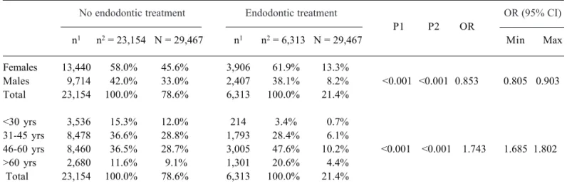
Table 1. Prevalence of endodontic treatment according to gender and age.
No endodontic treatment Endodontic treatment OR (95% CI)
P1 P2 OR
n1 n2 = 23,154 N = 29,467 n1 n2 = 6,313 N = 29,467 Min Max
Females 13,440 58.0% 45.6% 3,906 61.9% 13.3%
Males 9,714 42.0% 33.0% 2,407 38.1% 8.2% <0.001 <0.001 0.853 0.805 0.903 Total 23,154 100.0% 78.6% 6,313 100.0% 21.4%
<30 yrs 3,536 15.3% 12.0% 214 3.4% 0.7% 31-45 yrs 8,478 36.6% 28.8% 1,793 28.4% 6.1%
46-60 yrs 8,460 36.5% 28.7% 3,005 47.6% 10.2% <0.001 <0.001 1.743 1.685 1.802 >60 yrs 2,680 11.6% 9.1% 1,301 20.6% 4.4%
Total 23,154 100.0% 78.6% 6,313 100.0% 21.4%
n1=number of each tooth type; n2 = number of teeth without endodontic treatment; N= total number of teeth; P1 = 2; P2 = logistic
Cuiabá, MT, Brazil) in a PC workstation running Microsoft Windows XP professional SP-1 (Microsoft Corp., Redmond, WA, USA).
The criteria for radiographic detection of endo-dontic treatment of all teeth seen on the radiographs, except for third molars, were the presence of radio-paque material in the pulp chamber, in one or more root canals, or in a combination of these sites. The following conditions were recorded: poor or complete root canal filling, ending or not at the radiographic apex or not, presence of intracanal post and associated AP.
Odds ratio, logistic regression and a chi-square test were used for statistical analyses. Significance level was set at p<0.05. Interobserver agreement was as-sessed using kappa (k) values.
RESULTS
Of 29,467 evaluated teeth, 6,313 (21.4%) had been endodontically treated. Table 1 shows the distribu-tion of endodontically treated teeth according to age and gender. Table 2 shows the prevalence and distribution
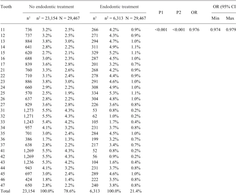
Table 2. Prevalence of endodontic treatment according to dental group.
Tooth No endodontic treatment Endodontic treatment OR (95% CI) P1 P2 OR
n1 n2 = 23,154 N = 29,467 n1 n2 = 6,313 N = 29,467 Min Max
11 736 3.2% 2.5% 266 4.2% 0.9% <0.001 <0.001 0.976 0.974 0.979 12 737 3.2% 2.5% 271 4.3% 0.9%
13 884 3.8% 3.0% 294 4.7% 1.0% 14 641 2.8% 2.2% 311 4.9% 1.1% 15 620 2.7% 2.1% 329 5.2% 1.1% 16 688 3.0% 2.3% 287 4.5% 1.0% 17 839 3.6% 2.8% 201 3.2% 0.7% 21 760 3.3% 2.6% 268 4.2% 0.9% 22 710 3.1% 2.4% 278 4.4% 0.9% 23 886 3.8% 3.0% 291 4.6% 1.0% 24 660 2.9% 2.2% 308 4.9% 1.0% 25 570 2.5% 1.9% 334 5.3% 1.1% 26 637 2.8% 2.2% 304 4.8% 1.0% 27 829 3.6% 2.8% 226 3.6% 0.8% 31 1,273 5.5% 4.3% 53 0.8% 0.2% 32 1,271 5.5% 4.3% 62 1.0% 0.2% 33 1,243 5.4% 4.2% 105 1.7% 0.4% 34 957 4.1% 3.2% 231 3.7% 0.8% 35 701 3.0% 2.4% 284 4.5% 1.0% 36 386 1.7% 1.3% 199 3.2% 0.7% 37 638 2.8% 2.2% 217 3.4% 0.7% 41 1,269 5.5% 4.3% 52 0.8% 0.2% 42 1,269 5.5% 4.3% 56 0.9% 0.2% 43 1,236 5.3% 4.2% 104 1.6% 0.4% 44 943 4.1% 3.2% 231 3.7% 0.8% 45 697 3.0% 2.4% 289 4.6% 1.0% 46 424 1.8% 1.4% 222 3.5% 0.8% 47 650 2.8% 2.2% 240 3.8% 0.8% Total 23,154 100.0% 78.6% 6,313 100.0% 21.4%
n1=number of each tooth type; n2 = number of teeth without endodontic treatment; N= total number of teeth; P1 = 2; P2 = logistic
of root canal treatment according to the tooth type. Maxillary premolars and molars were the teeth in which endodontic treatment was most frequent, whereas mandibular incisors showed the lowest prevalence. The largest number of endodontic treatments was found among individuals aged 46 to 60 years (47.6%, p<0.001). Females (61.9%, p<0.001) showed the greatest preva-lence (Table 1). Interobserver agreement was excellent, and kappa values were greater than 0.91.
DISCUSSION
The results of this cross-sectional evaluation are similar to those of previous studies (5,6,16), in which males had significantly fewer remaining natural teeth than females. The average number of root-filled teeth was also lower among males.
The most prevalent teeth were maxillary premolars followed by maxillary molars and mandibular premolars and molars (Table 1). This result is in accordance with those of Lupi-Pegurier et al. (6). Kirkevang et al. (5) found that significantly more molars had been endodon-tically treated (8.1%) than premolars (5.4%) or anterior teeth (2.5%). This difference may be explained by the number of root-filled teeth in each sample.
The analysis of prevalence of root canal treat-ment according to age revealed a higher prevalence in the 46-60-year-old range (n=8,460; 47.6%) and a de-crease in subjects older than 60 years (n=2,680; 20.6%). These values should be analyzed according to sample size and risk of caries disease, which were based on a Brazilian government’s study of the oral health of the Brazilian population (3). A substantial increase in the number of dental caries sequelae and lost teeth were found in the groups of individuals aged 35 to 44 and 65 to 74 years.
This cross-sectional study was designed ac-cording to the methodologies of previous studies (5-14). Images of a random sample from a database were examined to calculate the number of endodontically treated teeth. Panoramic radiographs are often used for such purpose in epidemiological studies (6,8-10). Lupi-Pegurier et al. (6) reported that the fact that all teeth can be seen using only one radiograph, the relatively low patient radiation dose, and the convenience and speed with which panoramic radiographs can be obtained are advantages over full-mouth periapical radiographs. Estrela et al. (13) evaluated the periapical status and
quality of root canal fillings, suggesting that epidemio-logical studies to assess the quality of periapical condi-tions using panoramic or periapical radiographs should be reviewed. Those authors found that CBCT images were more accurate than conventional methods.
In the present study, calibration was good and kappa was greater than 0.91, which indicates a high interobserver agreement. Studies that used similar meth-ods also found high kappa values (6,13,16).
The high rate of endodontic treatment, a critical sequela of dental caries, points to the negative impact of this important risk factor. The clinical impact of this study reinforces the need for permanent educational programs. The prevalence of endodontically treated teeth in the Brazilian adult population evaluated in the present study was higher than that observed in epide-miological studies conducted in other countries. Fe-males had more endodontically treated teeth than Fe-males. Endodontic treatment was most frequent in maxillary premolars and molars. Most endodontically treated teeth were found in 46-60-year-olds (47.6%) and the prevalence increased with age in this age range.
RESUMO
O objetivo do estudo transversal foi avaliar a prevalência de dentes tratados endodonticamente em uma população de Brasileiros adultos. Um total de 1.401 radiografias panorâmicas, oriundas do banco de imagens do Centro de Radiologia e Imagens Orofacial de Cuiabá (CRIOF, Cuiabá, MT, Brasil), entre agosto de 2002 e setembro de 2007 foi analisado. Três examinadores avaliaram todas as imagens, considerando-se a presença de tratamento endodôntico, indiferente à qualidade do tratamento (presença ou ausência de retentor intra-radicular ou periodontite apical). Os dados foram estatisticamente avaliados empregando-se razão de chances (odds ratio), regressão logística e teste Qui-quadrado. A partir de 29.467 dentes avaliados, 6.313 (21,4%) eram endodonticamente tratados. Os pré-molares e molares superiores foram os dentes com maior prevalência de tratamento, enquanto os incisivos inferiores representaram o grupo de menor prevalência. Indivíduos do gênero feminino (61,9%), e com idade entre 46 a 60 anos apresentaram maior prevalência de tratamento endodôntico. O presente estudo encontrou elevada prevalência de dentes tratados endodonticamente em adultos Brasileiros comparada com outros estudos epidemiológicos.
REFERENCES
1998;9:67-76.
2 . Estrela C, Holland R, Bernabé PFE, Souza V, Estrela CRA. Antimicrobial potential of medicaments used in healing pro-cess in dogs teeth with apical periodontitis. Braz Dent J 2004;15:181-185.
3 . Oral Health Project. Ministry of Health. Brazil 2003: oral health conditions of the Brazilian population 2002-2003. Brasília: National Coordination of Oral Health; 2004. 67 p (original document in Portuguese).
4 . Eriksen HM, Kirkevang LL, Petersson K. Endodontic epide-miology and treatment outcome: general considerations. Endod Topics 2002;2:1-9.
5 . Kirkevang LL, Hørsted-Bindslev P, Ørstavik D, Wenzel A. Frequency and distribution of endodontically treated teeth and apical periodontitis in an urban Danish population. Int Endod J 2001;34:198-205.
6 . Lupi-Pegurier L, Bertrand MF, Muller-Bolla M, Rocca JP, Bolla M. Periapical status, prevalence and quality of endo-dontic treatment in an adult French population. Int Endod J 2002;35:690-697.
7 . Tsuneishi M, Yamamoto T, Yamanaka R, Tamaki N, Sakamoto T, Tsuji K, et al.. Radiographic evaluation of periapical status and prevalence of endodontic treatment in an adult Japanese population. Oral Surg Oral Med Oral Pathol Oral Radiol Endod 2005;100:631-635.
8 . De Cleen MJH, Schuurs AHB, Wesselink PR, Wu M-K. Peri-apical status and prevalence of endodontic treatment in an adult Dutch population. Int Endod J 1993;26:112-119. 9 . Marques MD, Moreira B, Eriksen HM. Prevalence of. Apical
periodontitis and results of endodontic treatment in an adult, Portuguese population. Int Endod J 1998;31:161-165. 10. DeMoor RGJ, Hommez GMG, DeBoever JG, Delme KIM,
Martens GEI. Periapical health related to the quality of root canal treatment in a Belgian population. Int Endod J 2000;33:113-120.
11. Boltacz-Rzepkowska E, Laszkiewicz J. Endodontic treatment and periapical health in patients of the Institute of Dentistry in Lódz. Przegl Epidemiol 2005;59:107-115.
12. Estrela C, Leles CR, Hollanda ACB, Moura MS, Pécora JD. Prevalence and risk factors of apical periodontitis in endo-dontically treated teeth in a selected population of Brazilian adults. Braz Dent J 2008;19:34-39.
13. Estrela C, Bueno MR, Leles CR, Azevedo B, Azevedo JR. Accuracy of cone beam computed tomography and pan-oramic and periapical radiography for detection of apical periodontitis. J Endod 2008;34:273-279.
14. Estrela C, Bueno MR, Azevedo B, Azevedo JR, Pécora JD. A New Periapical Index Based on Cone Beam Computed To-mography. J Endod 2008;34:1325-1331.
15. Jiménez-Pinzón A, Segura-Egea JJ, Poyato-Ferrera M, Velasco-Ortega E, Rios-Santos JV. Prevalence of apical peri-odontitis and frequency of root filled teeth in an adult Spanish population. Int Endod J 2004;37:167-173.
16. Dugas NN, Lawrence HP, Teplitsky PE, Pharoah MJ, Fried-man S. Periapical health and treatment quality assessment of root-filled teeth in two Canadian populations. Intern Endod J 2003;36:181-192.
17. Kirkevang LL, Wenzel A. Risk indicators for apical peri-odontitis. Community Dent Oral Epidemiol 2003;31:59-67. 18. Buckley M, Spångberg LS. The prevalence and technical quality of endodontic treatment in an American subpopula-tion. Oral Surg Oral Med Oral Pathol Oral Radiol Endod 1995;79:92-100.
19. Bueno MR, Estrela C. Prevalence of endodontic treatment and apical periodontitis in several populations of world, de-tected by panoramic and periapical radiography and cone beam computed tomography. Rev Odontol Brasil Central 2008;17:79-90.
20. Amaral MA, Nakama L, Conrado CA, Matsuo T. Dental caries in young male adults: prevalence, severity and associ-ated factors. Braz Oral Res 2005;19:149-155.
Accepted October 24, 2008