20
Brandão AHF et al. Ophthalmic artery Doppler and endothelial function in preeclampsia
Radiol Bras. 2012 Jan/Fev;45(1):20–23
Dopplerfluxometry of ophthalmic arteries and assessment
of endothelial function in early and late preeclampsia
*
Dopplerfluxometria de artérias oftálmicas e avaliação da função endotelial nas formas precoce e tardia da pré-eclâmpsia
Augusto Henriques Fulgêncio Brandão1, Alexandre Simão Barbosa2, Ana Paula Brum Miranda Lopes2, Henrique Vitor Leite3, Antônio Carlos Vieira Cabral4
Objective: To identify possible differences between endothelial dysfunction evaluated by brachial artery flow-mediated dilation and central hyperperfusion evaluated by dopplerfluxometry of ophthalmic artery in women with early- and late-onset preeclampsia. Materials and Methods: Flow-mediated dilation testing and dopplerfluxometry of ophthalmic artery were performed in 81 patients (26 with early preeclampsia, 30 with late preeclampsia, and 25 normotensive pregnant women – control group). Results: As compared with the control group, patients with preeclampsia presented lower values of flow-mediated dilation, both in cases of early preeclampsia (7.62 ± 5.42% × 14.12 ± 6.14%; p = 0.02) and in cases of late preeclampsia (5.83 ± 4.12% × 14.12 ± 6.14%; p = 0.00). No statistically significant difference was observed between early- and late-onset preeclampsia (7.62 ± 5.42% × 5.83 ± 4.12%; p = 0.09). Values for dopplerfluxometry of ophthalmic artery were significant lower in patients with preeclampsia as compared with the control group, both in cases of early preeclampsia (0.631 ± 0.024 × 0.737 ± 0.032; p = 0.01) and in cases of late preeclampsia (0.653 ± 0.019 × 0.737 ± 0.032; p = 0.03). Again, no statistically significant difference was observed between early- and late-onset preeclampsia (0.631 ± 0.024 × 0.653 ± 0.019; p = 0.12). Basically, the results demonstrate a decrease in values for dopplerfluxometry of ophthalmic artery in patients with early and late presentations of preeclampsia as compared with the control group, although with no statistically significant difference between the two presentations of the disease. Conclusion: The present results indicate the presence of endothelial dysfunction and central hyperperfusion in patients with early- and late-onset preeclampsia.
Keywords: Preeclampsia; Flow-mediated dilation; Ophthalmic arteries.
Objetivo: Avaliar possíveis diferenças entre a disfunção endotelial, avaliada pela dilatação fluxo-mediada, e hiperper-fusão central, avaliada por dopplerfluxometria da artéria oftálmica, entre pacientes portadoras da forma precoce e tardia da pré-eclâmpsia. Materiais e Métodos: O teste de dilatação fluxo-mediada e a dopplerfluxometria da artéria oftálmica foram obtidos de 81 gestantes, sendo 56 portadoras de pré-eclâmpsia (26 na forma precoce e 30 na forma tardia) e 25 gestantes saudáveis (grupo controle). Resultados: Portadoras de pré-eclâmpsia apresentaram valores menores de dilatação fluxo-mediada quando comparadas ao grupo controle, tanto na forma precoce (7,62 ± 5,42% × 14,12 ± 6,14%; p = 0,02) como na forma tardia (5,83 ± 4,12% × 14,12 ± 6,14%; p = 0,00). Não houve dife-rença quando foram comparadas as duas formas (7,62 ± 5,42% × 5,83 ± 4,12%; p = 0,09). A dopplerfluxometria da artéria oftálmica apresentou-se significativamente menor nas pacientes portadoras de pré-eclâmpsia quando com-paradas ao grupo controle, tanto na forma precoce (0,631 ± 0,024 × 0,737 ± 0,032; p = 0,01) como na forma tardia (0,653 ± 0,019 × 0,737 ± 0,032; p = 0,03). Não houve diferença entre as duas formas de apresentação (0,631 ± 0,024 × 0,653 ± 0,019; p = 0,12). Os resultados basicamente demonstram redução nos valores de di-latação fluxo-mediada e dopplerfluxometria da artéria oftálmica nas formas tardia e precoce da pré-eclâmpsia quando comparadas ao grupo controle, sem, contudo, diferenças significativas entre as duas formas de apresentação da doença. Conclusão: Os resultados indicam a presença de disfunção endotelial e hiperperfusão central em gestantes com pré-eclâmpsia, tanto na forma precoce como na tardia.
Unitermos: Pré-eclâmpsia; Dilatação fluxo-mediada; Artérias oftálmicas. Abstract
Resumo
* Study developed at Universidade Federal de Minas Gerais (UFMG), Belo Horizonte, MG, Brazil.
1. MD, Fellow PhD degree, Scholar at Fundação de Amparo à Pesquisa de Minas Gerais (Fapemig), Belo Horizonte, MG, Bra-zil.
2. PhDs, MDs, Hospital das Clínicas da Universidade Federal
Brandão AHF, Barbosa AS, Lopes APBM, Leite HV, Cabral ACV. Dopplerfluxometry of ophthalmic arteries and assessment of endothe-lial function in early and late preeclampsia. Radiol Bras. 2012 Jan/Fev;45(1):20–23.
0100-3984 © Colégio Brasileiro de Radiologia e Diagnóstico por Imagem
ORIGINAL ARTICLE
de Minas Gerais (UFMG), Belo Horizonte, MG, Brazil. 3. PhD, Associate Professor, Universidade Federal de Minas Gerais (UFMG), Belo Horizonte, MG, Brazil.
4. PhD, Full Professor, Universidade Federal de Minas Gerais (UFMG), Belo Horizonte, MG, Brazil.
Mailing Address: Dr. Augusto Henriques Fulgêncio Brandão.
Rua Costa Rica, 333, ap. 701, Sion. Belo Horizonte, MG, Bra-zil, 30320-030. E-mail: [email protected]
21
Brandão AHF et al. Ophthalmic artery Doppler and endothelial function in preeclampsia
Radiol Bras. 2012 Jan/Fev;45(1):20–23 INTRODUCTION
Preeclampsia (PE) is a syndrome of multifactorial etiology globally responsible for the highest rate of maternal and fetal mortality(1). Endothelial dysfunction is
pointed out as the pathophysiological event behind the clinical manifestations and complications of such syndrome, from in-creased arterial pressure to hyperperfusion of the central nervous system(2,3).
The vascular endothelium is a paracrine structure capable of, among other func-tions, to control the arterial tone by the re-lease of vasoactive factors, particularly ni-tric oxide, that acts by promoting vasodi-latation of the muscular coat(4). Such
mecha-nism assumes a greater importance during gestation, since the potential for arterial dilatation is critical to accommodate the increase in maternal blood volume and to allow appropriate placental perfusion. Bra-chial artery flow-mediated dilation (FMD) is a sonographic test that allows the indi-rect evaluation of the endothelial function. The study is based on the arterial dilation capacity as a response to an induced tran-sient hypoxic stimulus(5,6).
Central hyperperfusion is a result from the loss of capacity of self-regulation of the arterial flow in the central nervous system. This condition progresses with develop-ment of cerebral edema that is a direct cause of the typical tonic-clonic seizures of eclampsia(7). The decrease in the
oph-thalmic artery resistive index (OARI) iden-tified at dopplerfluxometry of ophthalmic arteries indicates the involvement of cen-tral arteries that culminates in hyperper-fusion(8).
A classification of PE based on the pe-riod of symptoms onset has been proposed, creating two categories as follows: early PE – with onset before the 34th gestational week –, and late PE – occurring after the 34th gestational week(9). Such a
classifica-tion is compatible with the pathophysi-ological basis of PE as placental defi-ciency(10) and the maternal hemodynamic
condition(11) are taken into consideration in
the differentiation between forms of PE. Endothelial involvement and cerebral hyperperfusion may present distinct behav-iors in relation to early- and late onset PE. The present study was aimed at evaluating
the behavior of endothelial function and cerebral blood flow by means of FMD test and Doppler spectral analysis of oph-thalmic artery in women with early- and late-onset PE.
MATERIALS AND METHODS
Patients
The present cross-sectional study in-cluded 81 pregnant women divided into two groups as follows: 56 patients with PE and no other comorbidity, and 25 healthy pregnant women paired according to their ages and number of pregnancies. Among the 56 patients with PE, 30 presented late PE, and 26, early PE.
The diagnosis of PE was made in com-pliance with the criteria defined by the Na-tional High Blood Pressure Education Pro-gram Working Group on High Blood Pres-sure in Pregnancy, 2000. According to such classification, PE is defined as increase in arterial pressure after 20 weeks of gesta-tion (pressure levels ≥ 140 × 90 mmHg (in two measurements at a six-hour interval) associated with the presence of proteinuria (≥ 1+ measured either with a reagent strip test or 24 hour proteinuria > 0.3 g)(12).
Patients with comorbidities such as chronic arterial hypertension, renal disease, coronary disease and infectious diseases were excluded from the study. Twin preg-nancies, pregnancies with fetal malforma-tion or altered fetal growth were also ex-cluded as well as smoker patients, drug users, and patients taking nitrite-based drugs. Such situations are known to be as-sociated with endothelial injury.
The present study was approved by the Committee for Ethics in Research of Hos-pital das Clínicas – Universidade Federal de Minas Gerais (HC-UFMG). The se-lected patients received explanations and signed a term of free and informed con-sents. Subsequently, the patients underwent brachial artery FMD.
Brachial artery FMD
The technique to evaluate brachial ar-tery FMD was performed with a Medison Sonoace 8800 color Doppler ultrasonogra-phy apparatus with a 4–8 MHz linear trans-ducer. The patients were placed at rest in dorsal decubitus for 15 minutes. All the
patients had their arterial pressure mea-sured and their brachial artery was identi-fied medially in the antecubital fossa of the dominant upper limb. One image of the vessel was acquired at approximately 5 cm from the elbow of the upper limb, with a longitudinal section (B mode) at the mo-ment of lesser distention of the vessel cor-responding to cardiac diastole, and was obtained by means of image recovery on the cine loop display of the equipment. The image was frozen to get a mean of the three measurements of the vessel caliber (D1). After this first measurement, the sphygmo-manometer cuff positioned proximally to the site of the brachial artery measurement was inflated for five minutes up to a pres-sure > 250 mmHg, and later was slowly deflated. The mean of three further mea-surements of the vessel caliber was ob-tained with the already mentioned tech-nique one minute after the cuff deflation (D2). The FMD value was obtained by the following equation:
FMD (%) = [(D2 – D1)/D1] × 100
where: D1 = basal diameter; D2 = post-occlusion diameter.
All the studies were performed by a single investigator of the HC-UFMG, trained and certified in ultrasonography.
Dopplerfluxometry of ophthalmic arteries
22
Brandão AHF et al. Ophthalmic artery Doppler and endothelial function in preeclampsia
Radiol Bras. 2012 Jan/Fev;45(1):20–23 Statistical analysis
The normality for continuous variables was evaluated by means of the Shapiro-Wilk test. The Kruskal-Wallis test was uti-lized for comparison between groups of non-parametric variables, with the post-hoc Dunn procedure for comparison of pairs of groups. Analysis of variance (ANOVA) was utilized to compare parametric vari-ables. The results were expressed as me-dian ± interquartile range or mean ± stan-dard deviation for non-parametric and parametric variables, respectively. All the analyses were performed with the aid of the software Statistical Package for Social Sci-ences version 18 (SPSS; Chicago, IL, USA).
RESULTS
Table 1 shows the demographic charac-teristics of the three groups. The patients with late PE presented higher body mass index than the patients with early PE or those in the control group.
As regards FMD results, as compared with the control group, the patients with both early and late PE presented lower val-ues (7.62 ± 5.42% × 14.12 ± 6.14%; p =
0.02) and (5.83 ± 4.12% × 14.12 ± 6.14%;
p = 0.00), respectively. However, no
statis-tically significant difference was observed in the comparison between early- and late-onset PE (7.62 ± 5.42% × 5.83 ± 4.12%;
p = 0.09).
The OARI was significantly lower in the patients with early and late PE as com-pared with the control group (0.631 ± 0.024
× 0.737 ± 0.032; p = 0.01) and (0.653 ±
0.019 × 0.737 ± 0.032; p = 0.03),
respec-tively. Again, no statistically significant dif-ference was observed between early- and late-onset PE (0.631 ± 0.024 × 0.653 ± 0.019; p = 0.12).
The results for FMD and OARI are shown on Table 2.
DISCUSSION
Vascular endothelial injury, clinically characterized as endothelial dysfunction, was extensively demonstrated in patients with PE, by means of FMD(13,14). Lower
values of such test have already been dem-onstrated in patients who subsequently de-veloped PE, indicating that such test can be utilized to predict clinical manifestations of
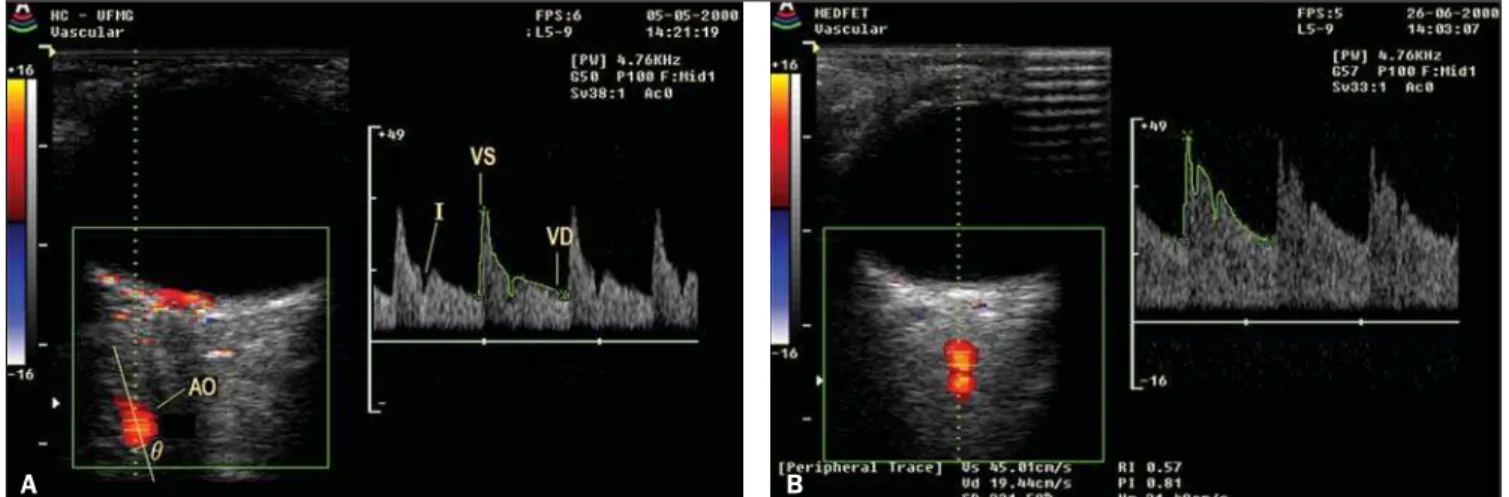
Figure 1. Dopplerfluxometry of ophthalmic artery. A: Doppler of ophthalmic artery of a normotense patient. B: Doppler of ophthalmic artery of a patient with preeclampsia demonstrating increased diastolic flow and consequential decrease in the resistive index.
A B
Table 1 Demographic characteristics of 81 pregnant women participating in the study.
Maternal age (years)
Gestational age (weeks)
Number of pregnancies
Body mass index (g/m2)
Early PE (n = 26)
29 ± 6.4
29 ± 3.4
1 ± 1.2
24.7 ± 8.5
Late PE (n = 30)
30 ± 4.2
36 ± 2.1
1 ± 1.8
29.2 ± 9.8
Control group (n = 25)
28 ± 7.1
30 ± 4.2
1 ± 2.1
23.6 ± 12.1
p value
0.18
0.02
0.43
0.04
Note: Body mass index calculated with basis on the pre-gestational weight of the patients.
Table 2 Results regarding brachial artery FMD and OARI.
Basal diameter of brachial artery (mm)
FMD (%)
OARI
Early PE
3.25 ± 6.27
7.62 ± 5.42
0.631 ± 0.024
Late PE
3.02 ± 7.22
5.83 ± 4.12
0.653 ± 0.019
Control group
3.12 ± 4.14
14.12 ± 6.14
0.737 ± 0.032
p value
0.23
0.003
0.017
FMD, flow-mediated dilation; OARI, ophthalmic artery resistive index.
PE(15,16). Apparently, endothelial
dysfunc-tion precedes clinical PE manifestadysfunc-tions and persists up to one year after delivery, which would also explain the higher inci-dence of cardiovascular complications in women with previous history of PE(17).
Hyperperfusion of the central nervous system demonstrated by lower OARI val-ues has also been observed in patients with PE(18). The utilization of such index plays
a relevant role in the differential diagnosis between PE and chronic arterial hyperten-sion. Patients with chronic arterial hyper-tension tend to present OARI results simi-lar to those of normotense pregnant women(19). Considering that the
dopplerfluxo-23
Brandão AHF et al. Ophthalmic artery Doppler and endothelial function in preeclampsia
Radiol Bras. 2012 Jan/Fev;45(1):20–23 metry of ophthalmic artery could play a relevant role nor only in the diagnosis but also in the definition of the approach to patients with high pressure levels during pregnancy.
The classification of PE into early- and late-onset PE has been extensively uti-lized(9). A study developed in the authors’
institution has demonstrated that early-on-set PE is associated with a high rate of maternal and fetal complications(20). Early
PE is responsible for 10% of cases of PE, but, besides prematurity, it is known that in such cases there is a higher rate of fetuses with intrauterine growth restriction(21).
Probably, the worst degree of placentation demonstrated by higher indices of uterine artery pulsatility, and that is also more en-hanced in cases of early PE, explains the placental insufficiency and the intrauterine growth restriction in these cases(22).
Patients with early PE probably would present lower values for FMD and OARI as compared with patients with late PE, explaining the higher rate of maternal com-plications. However, such results were not found in the present study. Explanations might be based on maternal constitutional factors of patients with late PE. In spite of the exclusion of comorbidities, such preg-nant women presented higher body mass indices and might have latent diseases that already run their course with endothelial dysfunction, among them plurimetabolic syndrome, which would lead to lower in-dices in the tests, particularly regarding FMD. Such lower values could explain the similarity in results as compared with the patients with early-onset PE who theoreti-cally would present a greater involvement
of the endothelial function solely resulting from PE.
Concluding, the present results indicate the presence of endothelial dysfunction and hyperperfusion of central nervous system in pregnant women with both early and late PE, but with no significant difference be-tween the clinical presentations of the syn-drome.
REFERENCES
1. World Health Organization. The World Health Report 2005 – make every mother and child count. Geneva: World Health Organization; 2005. 2. Roberts JM, Gammill HS. Preeclampsia: recent
insights. Hypertension. 2005;46:1243–9. 3. Cabral ACV, Cabral MA, Brandão A, et al.
Aspec-tos atuais da fisiopatologia da pré-eclâmpsia com repercussões na conduta. Femina. 2009;37:305– 8.
4. Lyall F, Greer IA. The vascular endothelium in normal pregnancy and pre-eclampsia. Rev Reprod. 1996;1:107–16.
5. Al-Qaisi M, Kharbanda RK, Mittal TK, et al. Measurement of endothelial function and its clini-cal utility for cardiovascular risk. Vasc Health Risk Manag. 2008;4:647–52.
6. Harris RA, Nishiyama SK, Wray DW, et al. Ul-trasound assessment of flow-mediated dilation. Hypertension. 2010;55:1075–85.
7. Young BC, Levine RJ, Karumanchi SA. Patho-genesis of preeclampsia. Annu Rev Pathol. 2010; 5:173–92.
8. Diniz AL, Moron AF, dos Santos MC, et al. Oph-thalmic artery Doppler as a measure of severe pre-eclampsia. Int J Gynaecol Obstet. 2008;100:216– 20.
9. von Dadelszen P, Magee LA, Roberts JM. Sub-classification of preeclampsia. Hypertens Preg-nancy. 2003;22:143–8.
10. Plasencia W, Maiz N, Poon L, et al. Uterine ar-tery Doppler at 11 + 0 to 13 + 6 weeks and 21 + 0 to 24 + 6 weeks in the prediction of pre-eclamp-sia. Ultrasound Obstet Gynecol. 2008;32:138–46. 11. Valensise H, Vasapollo B, Gagliardi G, et al. Early and late preeclampsia: two different maternal hemodynamic states in the latent phase of the disease. Hypertension. 2008;52:873–80.
12. [No authors listed]. Report of the National High Blood Pressure Education Program Working Group on High Blood Pressure in Pregnancy. Am J Obstet Gynecol. 2000;183:S1–S22. 13. Sierra-Laguado J, Garcia RG, López-Jaramillo P.
Flow-mediated dilatation of the brachial artery in pregnancy. Int J Gynaecol Obstet. 2006;93:60–1.
14. Brandão AHF, Lopes APBM, Salomão CMN, et al. Dilatação fluxo-mediada da artéria braquial como método de avaliação da função endotelial na pré-eclâmpsia e em gestantes normotensas. Rev Med Minas Gerais. 2011;21:9–13. 15. Takase B, Goto T, Hamabe A, et al.
Flow-medi-ated dilation in brachial artery in the second half of pregnancy and prediction of pre-eclampsia. J Hum Hypertens. 2003;17:697–704.
16. Savvidou MD, Noori M, Anderson JM, et al. Maternal endothelial function and serum concen-trations of placental growth factor and soluble endoglin in women with abnormal placentation. Ultrasound Obstet Gynecol. 2008;32:871–6.
17. Hamad RR, Eriksson MJ, Silveira A, et al. De-creased flow-mediated dilation is present 1 year after a pre-eclamptic pregnancy. J Hypertens. 2007;25:2301–7.
18. Barbosa AS, Pereira AK, Reis ZSN, et al. Oph-thalmic artery-resistive index and evidence of overperfusion-related encephalopathy in severe preeclampsia. Hypertension. 2010;55:189–93.
19. Hata T, Hata K, Moritake K. Maternal ophthalmic artery Doppler velocimetry in normotensive preg-nancies and pregpreg-nancies complicated by hyper-tensive disorders. Am J Obstet Gynecol. 1997; 177:174–8.
20. Reis ZSN, Lage EM, Teixeira PG, et al. Pré-eclâmpsia precoce e tardia: uma classificação mais adequada para o prognóstico materno e pe-rinatal? Rev Bras Ginecol Obstet. 2010;32:584– 90.
21. Crispi F, Domínguez C, Llurba E, et al. Placental angiogenic growth factors and uterine artery Doppler findings for characterization of different subsets in preeclampsia and in isolated intrauter-ine growth restriction. Am J Obstet Gynecol. 2006;195:201–7.