Potential Role in the Ctenophore,
Mnemiopsis leidyi
Kevin Pang1, Joseph F. Ryan2, Andreas D. Baxevanis2, Mark Q. Martindale1*
1Kewalo Marine Laboratory, Pacific Biosciences Research Center, University of Hawaii at Manoa, Honolulu, Hawaii, United States of America,2Genome Technology Branch, National Human Genome Research Institute, National Institutes of Health, Bethesda, Maryland, United States of America
Abstract
The TGF-b signaling pathway is a metazoan-specific intercellular signaling pathway known to be important in many developmental and cellular processes in a wide variety of animals. We investigated the complexity and possible functions of this pathway in a member of one of the earliest branching metazoan phyla, the ctenophoreMnemiopsis leidyi. A search of the recently sequenced Mnemiopsis genome revealed an inventory of genes encoding ligands and the rest of the components of the TGF-bsuperfamily signaling pathway. TheMnemiopsisgenome contains nine TGF-bligands, two TGF-b -like family members, two BMP--like family members, and five gene products that were unable to be classified with certainty. We also identified four TGF-breceptors: three Type I and a single Type II receptor. There are five genes encoding Smad proteins (Smad2, Smad4, Smad6, and two Smad1s). While we have identified many of the other components of this pathway, including Tolloid, SMURF, and Nomo, notably absent are SARA and all of the known antagonists belonging to the Chordin, Follistatin, Noggin, and CAN families. This pathway likely evolved early in metazoan evolution as nearly all components of this pathway have yet to be identified in any non-metazoan. The complement of TGF-bsignaling pathway components of ctenophores is more similar to that of the sponge, Amphimedon, than to cnidarians, Trichoplax, or bilaterians. The mRNA expression patterns of key genes revealed byin situhybridization suggests that TGF-bsignaling is not involved in ctenophore early axis specification. Four ligands are expressed during gastrulation in ectodermal micromeres along all three body axes, suggesting a role in transducing earlier maternal signals. Later expression patterns and experiments with the TGF-binhibitor SB432542 suggest roles in pharyngeal morphogenesis and comb row organization.
Citation:Pang K, Ryan JF, Baxevanis AD, Martindale MQ (2011) Evolution of the TGF-bSignaling Pathway and Its Potential Role in the Ctenophore,Mnemiopsis leidyi. PLoS ONE 6(9): e24152. doi:10.1371/journal.pone.0024152
Editor:Peter K. Dearden, University of Otago, New Zealand
ReceivedMay 23, 2011;AcceptedJuly 31, 2011;PublishedSeptember 8, 2011
Copyright:ß2011 Pang et al. This is an open-access article distributed under the terms of the Creative Commons Attribution License, which permits unrestricted use, distribution, and reproduction in any medium, provided the original author and source are credited.
Funding:KP was funded by a National Science Foundation (NSF) Graduate Research Fellowship. This work was also supported in part by the Intramural Research Program of the National Human Genome Research Institute, National Institutes of Health (NIH). The funders had no role in study design, data collection and analysis, decision to publish, or preparation of the manuscript.
Competing Interests:The authors have declared that no competing interests exist.
* E-mail: [email protected]
Introduction
The transforming growth factor-b (TGF-b) signaling pathway was first discovered about 30 years ago, a pathway in which certain secreted proteins had the capability of transforming cells and tissues. The first TGF-bgene was cloned in 1985 [1]. Since then, similar proteins were discovered in animals as diverse as flies, nematodes, and vertebrates, all of which had similar functions in tissue morphogenesis (reviewed in [2–5]). Through the use of cloning and sequencing technologies, it was soon discovered that the genes encoding for these proteins were all related and diversified from a common ancestral gene. There are roughly a dozen families belonging to the TGF-bsuperfamily, and these can be divided into two major classes: the TGF-b-like class and the bone morphoge-netic protein-like (BMP) class. The TGF-b-like class includes TGF-b sensu stricto, Lefty, Activin/Inhibin, and Myostatin/Gdf8. The BMP class includes Bmp2/4/Dpp, Bmp5–8, Bmp3, Gdf2, Gdf5–7, Vg1/Univin, ADMP, and Nodal. Besides being known for its roles in morphogenesis, TGF-bsignaling, especially via Bmp2/4/Dpp, is also known for its role in dorsal-ventral patterning in both protostomes and deuterostomes (reviewed in [6–7]).
The TGF-bprecursor protein has three distinct regions: (1) the signal peptide, which targets it to the endoplasmic reticulum and
and Myc, or co-activators, such as the Creb-binding protein (CBP) [13]. The MH1 domain is capable of interacting with DNA, while the MH2 domain interacts with Type I receptors and is involved with protein-protein interactions, such as R-Smad/Co-Smad binding.
Inhibition of TGF-b signaling can occur at multiple levels: extracellularly, cytoplasmically, and in the nucleus. Extracellularly, diffusible antagonists such as Chordin, Noggin, Follistatin and the CAN family (Cerberus/DAN/Gremlin) act as ligand traps, interfering with ligand binding to receptors [14]. In turn, the zinc metalloprotease Tolloid is capable of cleaving Chordin, thereby releasing BMPs to become active, showing that there are many levels of regulation involved with TGF-b signaling [15]. Besides cleaving Chordin, Tolloid also functions to cleave pro-collagens of the extracellular matrix [16], as well as other proteoglycans, some of which also are known to bind TGF-bligands [17].
Intracellularly, the pathway can be inhibited at many levels. At the level of the receptors, FKBP12 can block Type I receptor phosphorylation by binding to the GS domain [18]. BAMBI, a pseudoreceptor, can prevent the Type I and Type II receptors from forming a receptor complex [19]. Pathway modulation can also occur via inhibitor-Smads (I-Smad, Smad6/7), which have an MH2 domain (like other Smads) and can bind to Type I receptors, interfering with R-Smad binding and phosphorylation [20]. I-Smads can also compete with R-Smad in binding with Co-I-Smads. Another intracellular regulator of TGF-b signaling is the Smad ubiquitin regulatory factor (SMURF), an E3 ubiquitin ligase that targets R-Smads for degradation [21]. SMURF can also be recruited by I-Smads to degrade Type I receptors at the membrane. TGF-bsignaling is also regulated within the nucleus by the binding of co-repressors Ski/Sno [22]. These proteins recruit other repressors to block the activation of TGF-btarget genes.
All levels of the TGF-bsignaling pathway are highly conserved in metazoans, with pathway members present in all animals
studied to date [23,24]. Outside the metazoa, no TGF-breceptor or ligand has been discovered, so this pathway most likely evolved early in animal evolution. In the choanoflagellate, Monosiga brevicollis, an MH2 domain is present; however, it is unlike all known Smad proteins in that it is accompanied by a zinc finger domain [25]. Amongst the non-bilaterians (cnidarians, poriferans, the placozoan, and ctenophores), most of our knowledge regarding this pathway is gleaned from cnidarians [26–33]. Interestingly, this pathway has been implicated in axial patterning in cnidarians, similar to its role in dorsal-ventral patterning in bilaterians. Work in the sponge,Amphimedon queenslandica, has also shown that TGF-b signaling may be involved in axial patterning [34]. To date, there is nothing known about this pathway in the final group of non-bilaterians, the ctenophores. To better understand the evolution of this pathway, we need to be able to compare all the non-bilaterian taxa.
The ctenophore body plan and body axes are specified early in development. Developmental potential is segregated to different lineages; however, the exact molecules involved are unknown. Analysis of the genomic sequence of the lobate ctenophore,
Mnemiopsis leidyi, allowed us to identify a near-complete TGF-b signaling pathway composed of nine ligands, four receptors, and five Smads, revealing that the core components are present in all metazoans studied to date. Notably absent are extracellular diffusible antagonists, including Chordin, Follistatin, Noggin, and CAN family members. We looked at the expression of these genes during ctenophore development and found expression of ligands to be differentially expressed along all three body axes (oral-aboral, tentacular, and sagittal). While we do not believe this pathway is necessarily specifying these axes, since they are expressed after the axes are already specified, we do believe they are involved with transducing earlier signals.
Results
Ligand diversity
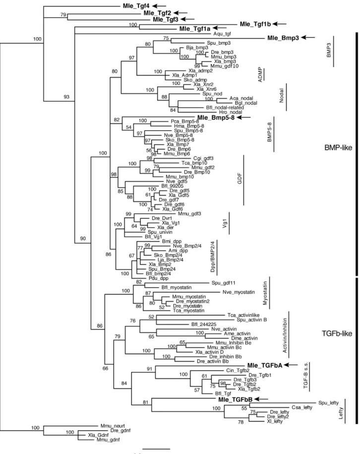
Similar to the situation previously seen for the Wnt/b-catenin pathway, searches of theMnemiopsisgenome have revealed a near complete TGF-b signaling pathway (Table 1). We were able to identify and isolate nine putative TGF-b ligands, four receptors, and five Smads. The nine ligands include members of both the TGF-b-like and the BMP-like clades. Due to the relatively high divergence of the ctenophore sequences, only four could be placed in supported families by phylogenetic analyses: MlTGFbA and
MlTGFbB, which are most closely related to TGF-b-like families TGF-bsensu strictoand Lefty (hence capitalized ‘‘TGF’’), as well as
MlBmp3 and MlBmp5–8 (Figure 2). However the posterior probability support is rather low (less than 95%), suggesting that there is a lack of phylogenetic signal in just the peptide domain sequence. When further analyses were run on the TGF-b-like clade using both the propeptide domain and the peptide domain,
MlTGFbAandMlTGFbBend up as sister to the Activin+Myostatin grouping (data not shown); therefore, we do not think these genes are actually TGF-bsensu strictoor Lefty orthologs per se, but rather divergent members of the TGF-b-like clade. The other five ligands (MlTgf1a,MlTgf1b,MlTgf2,MlTgf3, andMlTgf4) group as sister to the other families (hence lower case ‘‘Tgf’’). MlTGFbA and
MlTGFbBboth have eight cysteine residues, which are conserved in gene families of the TGF-brelated clade (Figure 3A).MlTgf1a,
MlTgf3, and MlBmp5–8 have seven conserved cysteines, while
MlTgf1b,MlTg4, andMlBmp3have only six.MlTgf1bis missing the first cysteine, whileMlTgf4 and MlBmp3 are missing the fourth cysteine at position 113 in the alignment. Two of the genes appear to be relatively recent tandem duplications (MlTgf1a and MlTgf1b)
Figure 1. Basic overview of TGF-bsignaling pathway.Binding of a ligand to a Type II receptor initiates signaling. The sequestering of a Type I receptor results in the activation of a Receptor-Smad (Smad1/5, Smad2/3). Together with the Co-Smad (Smad4), this complex enters the nucleus and activates the transcription of target genes. The pathway can be inhibited by extracelluar antagonists, or intracellularly via Inhibitor-Smad (Smad6/7) or the ubiquitin ligase SMURF.
doi:10.1371/journal.pone.0024152.g001
since they group closely together and are located adjacent to each other on the same scaffold. It is likely thatMlTgf1bis the result of a retroposition due to the fact that it is so closely linked toMlTgf1a
and it does not contain any introns. The seven remaining genes are on separate contigs.
Homology searches using SMART [35] predicted signal peptides, TGF-bpropeptides, and TGF-bpeptides forMlTGFbA,
MlTgf2, MlTGFbB, MlBmp5–8, and MlTgf1a (Figure 3B). For
MlBmp3 and MlTgf3, a signal peptide and TGF-b peptide are predicted, but the TGF-bpropeptide is not. In the case ofMlTgf4
and MlTgf1b, only the TGF-bpeptide is predicted. In the latter cases, the propeptides are missing or they are highly divergent and not detected by homology searches. The mature peptide cleavage site of RXXR is clearly present for all ligands, with the exceptions
of possible modification forMlBmp3(KSAR), MlTgf2(RAAVR), andMlTgf3(RQSKR).
Pathway members
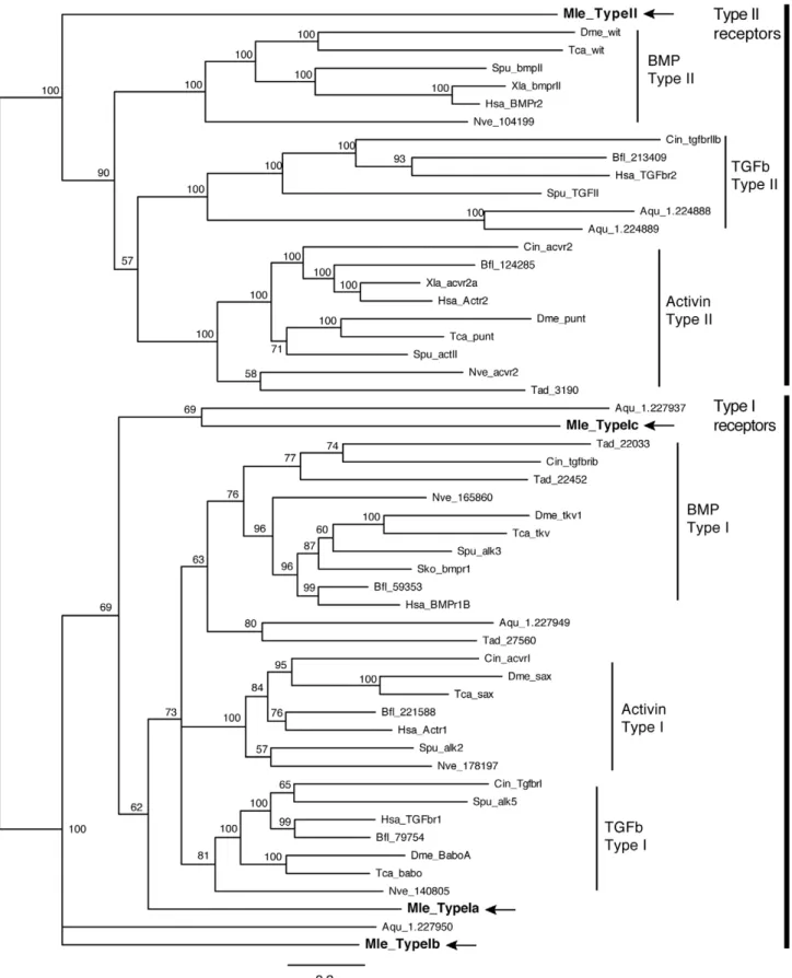
There is a single Type II receptor (MlTgfRII) and three Type I receptors (MlTgfRIa, MlTgfRIb, and MlTgfRIc). All contain the extracellular receptor domain, the single pass transmembrane domain, and the intracellular serine-threonine kinase. Additionally, all three Type I receptors possess the glycine-serine repeat (GS region) adjacent to the kinase domain, an arrangement that is characteristic of Type I receptors. Phylogenetic analyses that included sequences of TGF-b receptors from representative metazoans show that, while there is strong support for the different subclasses (Type II: BmpRII/wit, TGF-bRII, ActivinRII/punt;
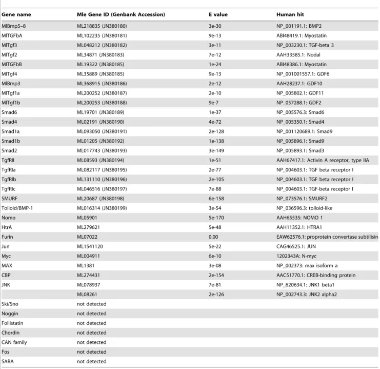
Table 1.TGF-bpathway members inMnemiopsisgenome.
Gene name Mle Gene ID (Genbank Accession) E value Human hit
MlBmp5–8 ML218835 (JN380180) 3e-30 NP_001191.1: BMP2
MlTGFbA ML102235 (JN380181) 9e-13 ABI48419.1: Myostatin
MlTgf3 ML048212 (JN380182) 3e-11 NP_003230.1: TGF-beta 3
MlTgf2 ML34871 (JN380183) 7e-12 AAH33585.1: Nodal
MlTGFbB ML19322 (JN380185) 1e-24 ABI48386.1: Myostatin
MlTgf4 ML35889 (JN380185) 9e-13 NP_001001557.1: GDF6
MlBmp3 ML368915 (JN380186) 2e-12 AAH28237.1: GDF10
MlTgf1a ML200252 (JN380187) 2e-10 NP_005802.1: GDF11
MlTgf1b ML200253 (JN380188) 9e-7 NP_057288.1: GDF2
Smad6 ML19701 (JN380189) 1e-37 NP_005576.3: Smad6
Smad4 ML02191 (JN380190) 4e-72 NP_005350.1: Smad4
Smad1a ML093050 (JN380191) 2e-128 NP_001120689.1: Smad9
Smad1b ML01205 (JN380192) 1e-138 NP_005896.1: Smad9
Smad2 ML017743 (JN380193) 3e-149 NP_005893.1: Smad3
TgfRII ML08593 (JN380194) 1e-51 AAH67417.1: Activin A receptor, type IIA
TgfRIa ML082117 (JN380195) 2e-77 NP_004603.1: TGF beta receptor I
TgfRIb ML131110 (JN380196) 2e-105 NP_004603.1: TGF beta receptor I
TgfRIc ML046516 (JN380197) 7e-88 NP_004603.1: TGF-beta receptor I
SMURF ML20687 (JN380198) 6e-158 NP_073576.1: SMURF2
Tolloid/BMP-1 ML016314 (JN380199) 3e-54 NP_036596.3: tolloid-like
Nomo ML05901 5e-170 AAH65535: NOMO 1
HtrA ML279621 5e-48 AAH11352.1: HTRA1
Furin ML07022 0.00 EAW62576.1: proprotein convertase subtilisin
Jun ML1541120 5e-22 CAG46525.1: JUN
Myc ML004911 6e-10 1202343A: N-myc
MAX ML1381 3e-08 NP_002373: max isoform a
CBP ML274431 2e-154 AAC51770.1: CREB-binding protein
JNK ML078937 7e-81 NP_620634.1: JNK1 beta1
ML08261 2e-126 NP_002743.3: JNK2 alpha2
Ski/Sno not detected
Noggin not detected
Follistatin not detected
Chordin not detected
CAN family not detected
Fos not detected
SARA not detected
Figure 2. Bayesian analysis of TGF-bligands.Analyses were performed using only the TGF-bpeptide domain, withMnemiopsismembers bolded and marked by arrows. Representative taxa from deuterostomes, protostomes, and non-bilaterians were used (for full list of taxa, see Table S1). Four independent runs of five million generations were run using the ‘‘mixed’’ model, with the strict consensus tree shown. Nodes are labeled with posterior probabilities.
doi:10.1371/journal.pone.0024152.g002
Type I: BmpRI/tkv, ActivinRI/sax, TGF-bRI/babo), the Mne-miopsis receptors are not well supported in individual subclasses (Figure 4). Instead,MlTgfRIIfalls sister to all other Type II receptors (Figure 4).MlTgfRIagroups with the three Type I subclasses, while
MlTgfRIbandMlTgfRIcare outside of these, grouping weakly with sponge genes.
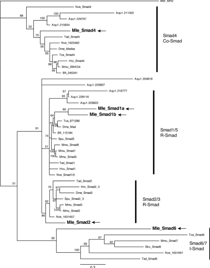
We were also able to detect and isolate five Smad family members. Unlike the TGF-breceptors, these genes all grouped in moderately well-supported Smad families (Figure 5). There are three receptor Smads, two belonging to the Smad1/5 family (MlSmad1a, MlSmad1b) and one Smad2/3 (MlSmad2). There is a single Co-Smad (MlSmad4) and a single inhibitory or I-Smad (MlSmad6).MlSmad4,MlSmad1a,MlSmad1b, andMlSmad2have the predicted MH1 and MH2 domains, characteristic of Smad proteins. MlSmad6 has the MH2 domain, as well as an amino terminal domain that resembles an MH1 domain. We were also able to identify and clone the E3 ubiquitin ligase SMURF, which can bind to receptor Smad proteins and target them for degradation, thereby inhibiting the cascade. Other intracellular components, including Jun, Myc, Max, CBP, and JNK, are present in theMnemiopsisgenome (Table 1). However we were not able to identify an ortholog of SARA, a protein that is involved
with recruiting receptor Smads to the receptor [36]. There is also no apparent TGIF (transforming growth-interacting factor) protein; this homeodomain transcription factor acts with nuclear Smads as a co-repressor [37]. We were also not able to identify Ski/Sno or Fos.
Althoughin silicosearches have discovered von Willebrand-type domains and Follistatin-like domains, we have not been able to find true Chordin, Noggin, Follistatin, or Gremlin orthologs, which are known diffusible antagonists of TGF-b signaling. Interestingly, we were able to identify a Tolloid gene (MlTolloid), which is known for enhancing signaling by cleaving Chordin, as well as other proteins. We also identified a Nodal Modulator (Nomo) ortholog, even though there is no true Nodal gene.
Early TGF-b expression
We examined the expression patterns of TGF-b and Smad genes during development. A set of TGF-b genes (MlBmp5–8,
MlBmp3,MlTgf1a, andMlTgfbA) are expressed relatively early in development, just prior to and during gastrulation (Figure 6). These genes are expressed in staggered domains along all three body axes. MlBmp5–8 is expressed in the most aboral region, surrounding the aboral pole in cells that will form the apical organ
Figure 3. TGF-bprotein structures and motifs.(A) Predicted amino acid sequences of the TGF-bpeptide domain and flanking region. Adjacent to the peptide domain is the cleavage site, showing the conserved RXXR motif. Asterisks below the sequence mark the seven conserved cysteine residues. The arrow indicates the conserved cysteine found in TGF-b-like class of ligands. (B) Conserved protein domains ofMnemiopsisTGF-bligands. The red boxes indicate signal sequences, while the other shaded boxes represent TGF-bpropeptide and TGF-bpeptide domains, as predicted by SMART.
Figure 4. Bayesian analysis of TGF-b receptors. Mnemiopsis members are bolded and marked by arrows. Representative taxa from deuterostomes, protostomes, and non-bilaterians were used (for full list of taxa, see Table S1). Four independent runs of 5 million generations were run using the ‘‘mixed’’ model, with the strict consensus tree shown. Nodes are labeled with posterior probabilities.
doi:10.1371/journal.pone.0024152.g004
Figure 5. Bayesian analysis of Smad proteins. Mnemiopsis members are bolded and marked by arrows. Representative taxa from deuterostomes, protostomes, and non-bilaterians were used (for full list of taxa, see Table S1). Four independent runs of 5 million generations were run using the ‘‘mixed’’ model, with the strict consensus tree shown. Nodes are labeled with posterior probabilities.
(Figure 6A). There is also more extensive staining in cells along the sagittal plane than the tentacular plane (see black arrows).MlTgf1a
begins expression prior to gastrulation at about two hours post
fertilization (hpf) in 12–16 micromeres at the aboral pole (Figure 6B). Unlike all other genes studied here, expression begins confined to the nuclei or the perinuclear region. At gastrulation,
Figure 6. Early TGF-bmRNA expression.Four of the TGF-bgenes are detected early in development, prior to and during gastrulation. The schematic at the top depicts the stages of embryos during cleavage and gastrulation, at 1–2 and 3 hours post fertilization (hpf), respectively. Embryos are lateral views, otherwise oral/aboral as stated. The asterisk marks the position of the blastopore. (A)MlBmp5–8expression in the aboral ectoderm, with more expression detected in the sagittal plane (black arrows). (B)MlTgf1aexpression is detected in late cleavage stages around the nuclei of aboral micromeres. By gastrulation, the aboral expression remains, however there expression is primarily along the tentacular plane (white arrows). (C)MlBmp3is detected in four groups of ectodermal cells from early to mid-gastrulation. (D)MlTGFbAis detected in four groups of ectodermal cells just adjacent to the blastopore at gastrulation.
doi:10.1371/journal.pone.0024152.g006
these cells give rise to portions of the aboral pole, primarily in the tentacular plane (see white arrows). At this time, expression is cytoplasmic, so it is not clear what the significance is of the earlier nuclear expression or how it changes to the cytoplasm. In this stage of development, expression overlaps with that ofMlBmp5–8.
MlBmp3 is expressed in four groups of ectodermal micromeres towards the oral pole at the onset of gastrulation (Figure 6C). This expression is very transient since transcripts cannot be detected in later stages of development. Finally MlTgfbA is expressed in ectodermal micromeres around the blastoporal opening (Figure 6D). These genes are expressed in staggered ectodermal domains along oral-aboral axis, as well as differentially in the tentacular and sagittal planes.
Late TGF-bexpression
In later developmental stages, we were able to examine the expression of four TGF-bgenes (MlBmp5–8,MlTgf1a,MlTgf2, and
MlTgfbB). The primary areas of expression for these genes are within the tentacle bulb and in the pharynx (Figure 7).MlBmp5–8
is expressed in a few cells of the apical organ, which correspond to the cells of the early expression domain and faintly in the pharynx (Figure 7A). It is also expressed in the tentacle bulb, in the most oral region. In cydippid stages, this pharyngeal and tentacular expression is not present, and there is only expression in the apical organ and anal pores. MlTgf1a is primarily expressed in two regions of each tentacle bulb, a larger region in the central part of the bulb and a smaller region (2–4 cells) closer towards the apical organ (Figure 7B). There is also expression in parts of the pharynx. In cydippids, there is an additional expression domain in two small regions of the apical organ in the most sagittal areas. MlTgf2is expressed faintly in the tentacle bulb that overlaps withMlTgf1a
expression (Figure 7C). MlTgfbB has the broadest expression domain, which includes a large portion of the tentacle bulb, the oral and aboral extremes of the pharynx, the muscle cells connecting the tentacle bulbs, and the floor of the apical organ. Although we were able to clone the remaining TGF-b ligands (MlTgf1b,MlTgf3, andMlTgf4) from mixed stage cDNA, we were not able to detect their expression viain situhybridization. We also analyzed the expression of the metalloprotease, MlTolloid, and found that it is expressed after gastrulation around the blastopore and in cells that have entered the blastocoel (Figure 7E). In later stages, it is expressed along the entirety of the pharynx, as well as in a the tentacle bulbs and transtentacular musculature, which overlaps with the expression ofMlTGFbB. However expression is not detected in the apical organ, and in cydippid stages, expression levels appear to be downregulated, in comparison to earlier in development.
TGF-breceptor and Smad expression
The lone Type II receptor is expressed ubiquitously from egg to cydippid stage (Figure 8A). Contrastingly, the three Type I receptors are expressed in relatively non-overlapping regions.
MlTgfRIais expressed initially at gastrulation in aboral ectodermal tissue, then in later stages in the apical organ, in cells around the comb rows, and faintly along the entire pharynx (Figure 8B).
MlTgfRIb is expressed in the muscle that connects the tentacle bulbs, in the most aboral part of the pharynx, in the outer regions of the tentacle bulb, and possibly also in the endoderm (Figure 8C).
MlTgfRIcis expressed initially in ectoderm towards the oral pole (Figure 8D). This expression fades and a later expression domain shows up in the mesoderm, which forms part of the tentacle bulb.
MlSmad6, which is the I-Smad, is expressed in the mesoderm, apical organ, and the aboral part of the pharynx (Figure 9A). In cydippids, only the apical organ expression remains, as well as the
outer portion of the tentacle bulb.MlSmad4, the Co-Smad, is only expressed in the aboral part of the pharynx, which forms the boundary of the ectodermal and endodermal portion of the gut (Figure 9B). In the cydippid stage, there is an additional staining in the apical organ in a few cells in the sagittal plane (Figure 9B).
MlSmad1ais expressed in a somewhat similar region in the tentacle bulb and apical organ as the receptorMlTgfRIa(Figure 9C). We were not able to detect the expression of MlSmad1b via in situ
hybridization. MlSmad2 is expressed ubiquitously from egg to cydippid (Figure 9D).
TGF-binhibitor SB431542
To better understand the function of TGF-bsignaling, we used the drug SB431542 (CAS 301836-41-9) to interfere with the signaling pathway. It has been shown in other animals to inhibit the activity of alk5/TGF-b Type I receptors [38]. Treatment of
Mnemiopsis eggs at concentrations less than 25mM resulted in normal cydippids. Treatment between 25–50mM resulted in consistent morphological defects. Rather than forming eight rows of comb plates, the combs appear to be clustered in two to four groups (Figure 10A–C). In addition to being in clusters, the combs are also not organized in rows, such that they do not beat synchronously. In addition, there is a thickening of the pharyngeal ectoderm, but it does not invaginate inward (Figure 10D). There are also thickenings where the tentacle bulbs are; however, they appear to be smaller than usual, and tentacles never grow out from these bulbs. The apical organ forms normally, and the ectoderm and endoderm also appear to be relatively normal.
In addition to the morphological phenotypes, development is delayed slightly when compared to wild type animals. Raising the animals in SB431542 for longer than 12 hours results in death. When embryos are treated after gastrulation (3–4 hpf), the embryos develop normally, implying that there is a window during which signaling is active. While there is no true alk5/TGF-bRI receptor, the gene that is phylogenetically most closely related to this receptor isMlTgfRIa. It is likely that the effects that we see are authentic, as this gene is expressed in the forming comb rows from gastrulation onward, as well as in the invaginating pharynx.
Discussion
Evolution of the TGF-b signaling pathway
Both the Wnt/b-catenin pathway and the TGF-b pathway likely evolved early in metazoan evolution, with the core components present in all animals studied to date [23–25]. However unlike the Wnt pathway, where some of the proteins (or, at a minimum, specific domains) are found in non-metazoans, including beta-catenin-like and frizzled-like proteins, nearly all of the TGF-b pathway genes are metazoan-specific. There are no known ligands or receptors found outside the metazoa, although serine/threonine kinase domains similar to those in TGF-b receptors are found in other eukaryotes. Additionally, there are no Smad genes in any other eukaryote, although there is a single Smad-like MH2 domain in the choanoflagellate, Monosiga. This domain is coupled with a C2H2 zinc finger, which is unlike all other Smad genes [25]. Searches of the recently sequenced genomes of the eukaryotes Salpingoeca rosetta and Capsaspora owczarzakihave also not revealed any TGF-b ligands, receptors, or Smads. Therefore, the origin of this pathway may have been a key innovation in metazoan evolution.
vertebrates (and teleosts, in particular), which have an expanded set of Smads and receptors, most likely due to lineage-specific genome duplications. In comparison, the number of TGF-bligands is much more variable. This is consistent with the hypothesis that there are more constraints on intracellular relative to the extracellular components of the signaling pathway [5]. The Smads and intracellular regions of the TGF-b receptors can be utilized for multiple purposes and in response to various ligands and signals. On the other hand, the ligands themselves are not so highly constrained, which might explain why there are so many more ligands than receptors and why the sequences of the ligands are much less conserved than those of the receptors and Smads. It is possible that ligands diversified and were co-opted for multiple developmental processes, while the intracellular components were reused.
While the core components of ligand-receptor-downstream mediators appear to have co-evolved, the addition of antagonistic
ligand regulation appear to have arisen later. Similar to the sponge,Amphimedon, theMnemiopsisgenome does not contain any of the known diffusible antagonists (Figure 11). Since both
Amphimedon and Mnemiopsis possess Tolloid orthologues, the ancestral function of this metalloprotease and BMP enhancer must have targeted proteins other than Chordin. Noggin, CAN family members and Follistatin are present in cnidarians,
Trichoplax, and bilaterians, while Chordin is only found in only cnidarians and bilaterians. Chordin-like genes have been found in
AmphimedonandTrichoplax; however, these lack the CHRD domain of true chordin genes [24]. In addition, both SARA and Ski/Sno are present in cnidarians, Trichoplax, and bilaterians, but absent from ctenophores and sponges. Assuming that there was not a secondary loss, these were also later additions to the signaling pathway, giving more support to the early branching position of ctenophores and sponges. Interestingly, there is no obvious
Figure 7. Late TGF-bmRNA expression.MlBmp5–8,MlTgf1a,MlTgf2,MlTGFbBandMlTolloidare detected during later stages of development. The diagram at the top depicts the stages of development in the columns below, identifying some of the major features and structures. Views are lateral, unless otherwise specified as oral or aboral. The asterisks marks the position of the blastopore or mouth. (A)MlBmp5–8expression in the aboral ectoderm and later in the invaginating pharynx. The aboral expression later becomes part of the apical organ and the anal canals. There is also an additional domain of expression in the tentacle bulbs. (B)MlTgf1ais expressed in parts of the tentacle bulbs, pharynx, and apical organ. (C)MlTgf2is expressed faintly in part of the tentacle bulbs, similar to that ofMlTgf1a, however by cydippid stages, expression is barely detectable. (D)MlTGFbBis expressed after gastrulation in a fairly complex pattern. There are expression domains at the oral and aboral ends of the pharynx. There is also expression in parts of the tenacle bulbs and in the apical organ. (E)MlTolloidis expressed around the blastopore and later in the pharynx, as well as in mesodermal derivatives, in transtentacular muscle and parts of the tentacle bulbs.
doi:10.1371/journal.pone.0024152.g007
Figure 8. TGF-breceptor expression patterns. Expression of TGF-breceptors through development, from gastrulation (3 hpf) to cydippid (24 hpf). Views are lateral unless otherwise specified, and asterisks mark the position of the blastopore or mouth. (A) MlTgfRII, the lone Type II receptor, is expressed ubiquitously from egg through cypdippid stages. (B)MlTgfRIais expressed in the aboral ectoderm as well as in the pharynx. The aboral ectoderm expression is confined to the developing comb rows and apical organ. (C)MlTgfRIbis detected in the pharynx, as well as in mesodermal derivatives. Cydippid expression is confined to parts of the tentacle bulb, as well as the endodermal part of the gut. (D)MlTgfRIcis expressed in the ectoderm, more towards the oral pole. Late expression is confined to parts of the tentacle bulbs.
doi:10.1371/journal.pone.0024152.g008
Figure 9. Smad expression patterns.mRNA expression ofMnemiopsisSmad genes during development. All views are lateral, unless otherwise specified. The asterisk marks the position of the blastopore or mouth. (A)MlSmad6, the I-Smad, is expressed in mesodermal derivatives of the tentacle bulb, as well as in the apical organ. (B)MlSmad4, the Co-Smad, is expressed in a discrete domain of the pharynx at the ectoderm-endoderm boundary. There is also late expression in the apical organ in four spots. (C)MlSmad1a, an R-Smad, is expressed in parts of the tentacle bulb and apical organ. (D)MlSmad2, another R-Smad, is expressed ubiquitously from egg to cydippid.
relationship between morphological complexity and signaling pathway complexity, at least in the non-bilaterians. In comparison to sponges andTrichoplax, ctenophores are much more morpho-logically complex, yet they have a similar (if not simpler) TGF-b signaling pathway complement.
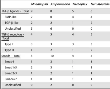
What is also surprising is that the ligand complements of the non-bilaterians are not conserved. Many of the ctenophore and sponge ligands do not group with major bilaterian subclasses (Figure 2) [24,25], but the cnidarian, Nematostella vectensis, and placozoan, Trichoplax adhaerens, sequences do (Figure 2 for
Nematostella; Trichoplax not shown). However, comparing just BMP-like and TGF-b-like classes, we see that all non-bilaterians have at least one TGF-b-like gene, while all non-bilaterians (except sponges) have a BMP-like gene, suggesting that this radiation also occurred early in animal evolution. Whether the unclassified ctenophore and sponge genes represent extremely divergent remnants of other classes or if they are just novel genes that arose in these lineages remains to be seen. Interestingly, if the ligand
orthology does hold to be true, these would be the first orthologs of Bmp3, TGF-b sensu strictu, and Lefty identified outside the deuterostomes. However due to the low support values, we have doubts as to whether this is the case.
Expression patterns
The expression patterns of the TGF-b ligands MlBmp3,
MlBmp5–8,MlTgf1a, andMlTgfbA, are highly suggestive of a role in axial patterning. They are all expressed relatively early in development (at gastrulation) and are expressed differentially along all three body axes. They are expressed in ectodermal micromeres, withMlBmp5–8expressed the most aborally, followed byMlTgf1a,MlBmp3, andMlTgfbAmost orally. Additionally, there is differential expression along the tentacular and sagittal planes, withMlTgf1aexpressed mainly along the tentacular plane, while
MlBmp5–8 is expressed more along the sagittal plane, although there is some overlap with MlTgf1a. However, experimental embryological evidence indicates that even at this early stage of
Figure 10. SB431542 treatment duringMnemiopsisdevelopment.Effects of TGF-binhibitor, SB431542, at 12 hours post fertilization. (A,B, D– F) are treated embryos, while (C, G–I) are controls. (A–C) Confocal projections of embryos stained with anti-tyrosinated tubulin (red) showing the cilia, Alexa-488 phalloidin (green) showing cell borders, and Hoechst 33342 (blue) showing the nuclei. All are aboral views, with the apical organ (ao) in the center. The white arrowheads point to individual comb plates, while the arrows in (C) show the eight comb rowsC. (D–I) are live embryos imaged under DIC. (D) Lateral view of SB431542-treated embryo, showing that pharynx has not invaginated (compare to (G)), and the tentacle bulbs (tb) have formed but are smaller in size. The apical organ appears normal. (E) Aboral view of the same embryo, mid-focal plane, again showing the smaller tentacle bulbs. The ectoderm (ecto) and endoderm (endo) both appear normal. (F) Aboral and surface view, showing the disorganized comb plates (arrowhead), compared to the eight comb rows (arrows) in the control (I).
doi:10.1371/journal.pone.0024152.g010
development, the axes are already specified [39,40], so these ligands may be transducing earlier signals. Possibly low levels of ligand expression during early cleavage stages are undetectable by
in situ hybridization, or perhaps there are maternal proteins that are localized in the egg and early embryo. As early as the four-cell and eight-cell stages, factors that specify different cell types, including comb plates and photocytes, are localized to different
lineages [39]. Whether these determinants are proteins or mRNAs is not known, however we have yet to see differential mRNA expression at these early stages, suggesting that these factors could be maternal proteins. Additionally, at these stages, only the Type II receptor (MlTgfRII) and a TGF-b-like Smad (MlSmad2) are expressed. None of the Type I receptors are expressed at this stage, and neither is the Co-Smad,MlSmad4. The Type I receptors are not detected until 4–5 hours post fertilization, about an hour after the earliest ligand expression. It is also possible that these ligands are initiating signals via Smad-independent pathways, such as the MAP kinase, Rho-like GTPase, or PI3K/AKT pathways [13,41]. One interesting aspect of the expression patterns is that, while the lone Type II receptor is expressed uniformly, the three Type I receptors are expressed in non-overlapping domains. Assuming protein distribution is similar, this would suggest the specificity of the response to ligands is dependent on which Type I receptor is expressed.MlTgfRIais expressed predominantly in the ectoderm, specifically in the forming comb rows and in the apical organ. Meanwhile,MlTgfRIbis expressed broadly in the mesoderm and endoderm, whileMlTgfRIc is expressed in putative mesoderm of the tentacle bulb.
The observation thatMlSmad4, the only Co-Smad, is detected in only a small region of cells, the pharynx at the ectoderm-endoderm boundary and a few cells of the apical organ, implies that it might not be necessary for signaling in other areas. Perhaps the R-Smads can function independently or with other factors to activate transcription of target genes. There is evidence from mammalian systems showing that Smad2/3 can bind to non-Smad proteins, including IKKa[42] and TIF1c[43], to illicit signaling independently of Smad4. Whether this is the case in Mnemiopsis, and what exactly the binding partners are remains to be seen. Functional work is needed to determine whether theMnemiopsis R-Smads are even capable of binding the Co-Smad. The I-Smad,
MlSmad6, is expressed in many areas that are overlapping with other Smads and Type I receptors (i.e., in the mesoderm, apical organ, ectoderm-endoderm boundary, and tentacle bulb). This suggests that there is both active signaling and highly complex regulation in these regions, as both activators and inhibitors of the pathway are co-expressed in the same cells and regions. Given that these are discrete areas of the developing embryo/larva, the observed expression patterns suggest that TGF-bsignaling may be important for germ layer specification or differentiation. For example, the apical organ is highly innervated and the primary sensory structure, and the tentacle bulb, where there is co-expression of activators and an inhibitor, is the site of putative stem cells for tentacle growth. The tentacle bulbs are regions of continual growth, suggesting that TGF-bsignaling is also involved in proliferation and cell cycle regulation. In addition, the ligands
MlBmp5–8, MlTgf1a, MlTgf2, and MlTGFbBare all expressed in regions of the tentacle bulb, suggesting this is an important signaling center of the developing embryo. It is likely thatMlTolloid
is also important forMlTGFbB function because of their highly overlapping expression domains. It is possible that MlTolloid is playing a role in cleavage and activation of this ligand, similar to its role in vertebrates and flies [44–46].
The results of our experiments with the TGF-b inhibitor SB431542 suggest that there is also a role of TGF-bsignaling in comb row organization and morphogenesis.MlTgfRIais expressed in the developing comb rows and is the most similar receptor to alk5/TGF-bRI, the known target of SB431542 [38]. The onset of MlTgfRIa expression (at gastrulation, 2.5–3 hpf) is within the window of sensitivity to SB431542. When exposed to the inhibitor, the comb plates still form at the correct time and display similar morphology, but they are not separated into eight rows and not
Table 2.Non-bilaterian TGF-bpathway components.
Mnemiopsis Amphimedon Trichoplax Nematostella
TGF-bligands - Total 9 8 5 6
BMP-like 2 0 4 4
TGF-b-like 2 2 1 2
Unclassified 5 6 0 0
TGF-breceptors -Total
4 5 4 5
Type I 3 3 3 3
Type II 1 2 1 2
Smads - Total 5 10 4 4
Smad4 1 3 1 1
Smad1/5 2 3 1 1
Smad2/3 1 2 1 1
Smad6/7 1 0 1 1
Unclassified 0 2 0 0
doi:10.1371/journal.pone.0024152.t002
Figure 11. Summary of presence and absence of TGF-b
organized in the same orientation. However, as is the case with any pharmaceutical inhibitor, there is a chance of non-specific effects, so further experiments such as injection of a morpholino antisense oligonucleotide designed againstMlTgfRIais necessary to ensure the drug is acting as we hypothesize. Recently, morpholino to the T-box gene,brachyury, has been shown to specifically inhibit its function during development by blocking pharyngeal invagi-nation [47]. Since we obtained a similar pharyngeal defect using SB431542, it is possible that brachyury is a target of TGF-b signaling, similar to both frog and the chickbrachyurythat are direct targets of Activin-like signaling [48,49]. Thus TGF-b signaling could be playing a role in ctenophore pharyngeal morphogenesis by activating brachyury. Exactly how it is mediating comb row organization has yet to be determined.
In conclusion, the TGF-b signaling pathway was present and most likely active early in metazoan evolution. With few components present in extant non-metazoans, it is highly probable that the emergence of this pathway was a key innovation in the transition to multicellularity in the metazoan ancestor. While a Smad-like gene is present in the choanoflagellates, there is very little similarity of TGF-bsignaling ligands and receptors outside of the metazoa. From expression studies here, it appears that TGF-b signaling is active in the ctenophore embryo. However, it is unlikely that this pathway is involved in early axis specification. The earliest expression of any TGF-b ligand is just prior to gastrulation, after the embryonic axes are already specified. The staggered expression patterns of the ligands at gastrulation is suggestive that TGF-bsignaling is responding to earlier signals. It remains to be seen what these early signals are, but it is possible that proteins for components of this pathway (and other key pathways) could be maternally loaded.
Materials and Methods
Genome search and phylogenetic analyses
We utilized theMnemiopsisdraft genome, which was previously sequenced using 454 and Ilumina sequencing and assembled onto scaffolds [50]. This sequence data was compiled into 10,106 scaffolds (scaffold N-50 of 123 kb), which corresponds to a physical coverage of approximately 506. Searches for TGF-b pathway components are similar to those in searches for Wnt pathway components [51], using a reciprocal Blast approach. Cnidarian and bilaterian gene orthologs were used in tblastn searches of the
Mnemiopsisgenome assembly. Putative positive matches were then aligned to orthologs from other organisms. Alignments were performed using MUSCLE (www.drive5.com/muscle) and then corrected by eye. For TGF-b ligands, only the mature peptide domain was used in phylogenetic analyses. For TGF-breceptors, we used the extracellular receptor, transmembrane, and intracel-lular kinase domains. For the Smad proteins, the MH1 and MH2 domains were used. All alignments are located in the Supporting Information files (Text S1, S2, S3). For all trees, we used Mr.Bayes3.2 [52], using the ‘mixed’ model with four independent runs of five million generations, with trees sampled every 100 generations. Consensus trees and posterior probabilities were calculated once the stationary phase was obtained.
Gene isolation and expression studies
Genes of interest were isolated using RACE PCR (Clontech), with all verified sequences being deposited into GenBank (JN380180–JN380199).In situhybridizations were as previously described [53]. Full-length or partial-length sequences, ranging in size from 800 bp to 2 kb, were used to transcribe digoxigenin-labeled RNA probes. We detected these probes using an alkaline
phosphatase-conjugated digoxigenin antibody, utilizing the substrates NBT and BCIP to then detect the alkaline phosphatase activity (Roche). Specimens were mounted in 70% glycerol, viewed under a Zeiss AxioSkop, and imaged using an AxioCam.
SB431542 treatments
Embryos were obtained from adult animals in Woods Hole, MA during the summers of 2009 and 2010 as previously described [53]. Following collection, they were treated with the pharmaco-logical agent SB431542, a potent inhibitor of TGF-bsignaling that blocks Type I receptor activity [38]. We started soaking one to four-cell stage embryos at concentrations from 25–50mM in 24-well plates (30–50 embryos per 24-well, approximate volume 1.0 ml). Treated embryos were immersed in SB431542 through their entire development and kept in the dark as much as possible. They were monitored periodically and fixed at 9–12 hours post fertilization (hpf).
Antibody staining and confocal microscopy
Embryos were fixed for antibody staining in 4% paraformal-dehyde and 0.02% glutaralparaformal-dehyde, as described previously [53]. Following fixation, embryos were removed from their membranes by gentle pipetting. They were then washed with PBS plus 0.2% Triton (PBT) and then placed in blocking buffer (5% goat serum) for one hour. They were then incubated with anti-tyrosine tubulin (Sigma, T9028) overnight at 4uC. Following six 30-minute washes with PBT, they were then incubated with a secondary antibody, goat anti-mouse conjugated to Alexa-594 (Invitrogen, Molecular Probes). After an overnight incubation, they were again washed with PBT six times for 30 minutes. In the last wash, they were also incubated with Alexa-488 phalloidin (Invitrogen, Molecular Probes) and Hoechst 33342 (Invitrogen, Molecular Probes). Following two 5-minute washes in PBS, they were then mounted on a slide and imaged using a Zeiss 710 confocal microscope. Images were processed using Zen software (Zeiss) and Volocity (Improvision) to create 3D image reconstructions of confocal sections.
Supporting Information
Table S1 Taxa used in phylogenetic analyses. The first column lists the different phyla, the second column lists the species, and the third column lists the abbreviation used in the phylogenetic trees and alignments.
(DOC)
Text S1 Amino acid alignment of TGF-bligands.Shown here are only the mature peptide sequences for taxa shown in Table S1, which was used to generate the tree in Figure 2. They were aligned using Muscle, then corrected by hand.
(NEX)
Text S2 Amino acid alignment of TGF-breceptors used in Figure 4. Shown here are the extracellular receptor, transmembrane, and intracellular kinase domains.
(NEX)
Text S3 Amino acid alignment of Smad proteins used in Figure 5.Shown here are the MH1 and MH2 domains. (NEX)
Acknowledgments
would also like to thank members of the Martindale lab and two anonymous reviewers for comments and suggestions which have greatly improved this manuscript.
Author Contributions
Conceived and designed the experiments: KP JFR ADB MQM. Performed the experiments: KP. Analyzed the data: KP JFR. Contributed reagents/ materials/analysis tools: KP. Wrote the paper: KP MQM.
References
1. Derynck R, Jarrett JA, Chen EY, Eaton DH, Bell JR, et al. (1985) Human transforming growth factor-beta complementary DNA sequence and expression in normal and transformed cells. Nature 316: 701–705.
2. Kingsley DM (1995) The TGF-bsuperfamily: new members, new receptors, and new genetic tests of function in different organisms. Genes Dev 8: 133–146. 3. Massague J (1998) TGF-bsignal transduction. Annu Rev Biochem 67: 753–791. 4. Massague J, Blain SW, Lo RS (2000) TGFB signaling in growth control, cancer,
and heritable disorders. Cell 103: 295–309.
5. Herpin A, Lelong C, Favrel P (2004) Transforming growth factor-b-related proteins: an ancestral and widespread superfamily of cytokines in metazoans. Dev Comp Immunol 28: 461–485.
6. De Robertis EM, Sasai Y (1996) A common plan for dorsoventral patterning in Bilateria. Nature 380: 37–40.
7. De Robertis EM, Kuroda H (2004) Dorsal-ventral patterning and neural induction in Xenopus embryos. Annu Rev Cell Dev Biol 20: 285–308. 8. Massague J, Chen YG (2000) Controlling TGF-bsignaling. Genes Dev 14:
627–644.
9. Dubois CM, Laprise MH, Blanchette F, Gentry LE, Leduc R (1995) Processing of transforming growth factorb1 precursor by human furin convertase. J Biol Chem 270: 10618–10624.
10. Attisano L, Lee-Hoeflich ST (2000) The Smads.Genome Biol2: reviews3010.1– 3010.8.
11. Newfeld SJ, Wisotzkey RG (2006) Molecular evolution of Smad proteins. In: Helden G, ten Dijke P, eds.Smad SignalTransduction. Netherlands: Springer. pp 15–35.
12. Tsukazaki T, Chiang TA, Davidson AF, Attisano L, Wrana JL (1998) SARA, a FYVE domain protein recruits Smad2 to the TGF-breceptor. Cell 95: 779–791. 13. Derynck R, Zhang YE (2003) Smad-dependent and Smad-independent
pathways in TGF-bfamily signaling. Nature 425: 577–584.
14. Balemans W, Van Hul W (2002) Extracellular regulation of BMP signaling in vertebrates: a cocktail of modulators. Dev Biol 250: 231–250.
15. Marques G, Musacchio M, Shimell MJ, Wunnenberg-Stapleton K, Cho KW, et al. (1997) Production of a DPP activity gradient in the early Drosophila embryo through the opposing actions of the SOG and TLD proteins. Cell 91: 417–426.
16. Kessler E, Takahara K, Biniaminov L, Brusel M, Greenspan DS (1996) Bone morphogenetic protein-1: the type I procollagen C-proteinase. Science 5247: 360–362.
17. von Marschall Z, Fisher LW (2010) Decorin is processed by three isoforms of bone morphogenetic protein-1 (BMP1). Biochem Biophys Res Commun 391: 1374–1378.
18. Chen YG, Liu F, Massague J (1997) Mechanism of TGFbreceptor inhibition by FKBP12. EMBO J 16: 3866–3876.
19. Onichtchouk D, Chen YG, Dosch R, Gawantka V, Delius H, et al. (1999) Silencing of TGF-b signaling by the pseudoreceptor BAMBI. Nature 401: 480–485.
20. Lonn P, Moren A, Raja E, Dahl M, Moustakas A (2009) Regulating the stability of TGFbreceptors and Smads. Cell Res 19: 21–35.
21. Zhu H, Kavsak P, Abdollah S, Wrana JL, Thomsen GH (1999) A SMAD ubiquitin ligase targets the BMP pathway and affects embryonic pattern formation. Nature 400: 687–693.
22. Liu X, Sun Y, Weinberg RA, Lodish HF (2001) Ski/Sno and TGF-bsignaling. Cytokine Growth Factor Rev 12: 1–8.
23. Huminiecki L, Goldovsky L, Freilich S, Moustakas A, Ouzounis C, et al. (2009) Emergence, development and diversification of the TGF-bsignaling pathway within the animal kingdom. BMC Evol Biol 9: 28.
24. Richards GS, Degnan BM (2009) The dawn of developmental signaling in the Metazoa. Cold Spring Har Symp Quant Biol 74: 81–90.
25. Srivastava M, Simakov O, Chapman J, Fahey B, Gauthier ME, et al. (2010) The
Amphimedon queenslandicagenome and the evolution of animal complexity. Nature 466: 720–726.
26. Hayward DC, Samuel G, Pontynen PC, Catmull J, Saint R, et al. (2002) Localized expression of a dpp/BMP2/4 ortholog in a coral embryo. Proc Natl Acad Sci USA 99: 8106–8111.
27. Finnerty JR, Pang K, Burton P, Paulson D, Martindale MQ (2004) Origins of bilateral symmetry: Hox and Dpp expression in a sea anemone. Science 304: 1335–1337.
28. Reinhardt B, Broun M, Blitz IL, Bode HR (2004) HyBMP5-8b, a BMP5-8 orthologue, acts during axial patterning and tentacle formation in hydra. Dev Biol 267: 43–59.
29. Matus DQ, Pang K, Marlow H, Dunn CW, Thomsen GH, et al. (2006) Molecular evidence for deep evolutionary roots of bilaterality in animal development. Proc Natl Acad Sci USA 103: 11195–11200.
30. Matus DQ, Thomsen GH, Martindale MQ (2006) Dorso/ventral genes are asymmetrically expressed and involved in germ-layer demarcation during cnidarian gastrulation. Curr Biol 16: 499–505.
31. Rentzsch F, Anton R, Saina M, Hammerschmidt M, Holstein TW, et al. (2006) Asymmetric expression of the BMP antagonists chordin and gremlin in the sea anemone Nematostella vectensis: implications for the evolution of axial patterning. Dev Biol 296: 375–387.
32. Saina M, Genikhovic G, Renfer E, Technau U (2009) BMPs and chordin regulate patterning of the directive axis in a sea anemone. Proc Natl Acad Sci USA 106: 18592–18597.
33. Saina M, Technau U (2009) Characterization of myostatin/gdf8/11 in the starlet sea anemone Nematostella vectensis. J Exp Zool B Mol Dev Evol 312: 780–788.
34. Adamska M, Degnan SM, Green KM, Adamski M, Craigie A, et al. (2007) Wnt and TGF-beta expression in the spongeAmphimedon queenslandicaand the origin of metazoan embryonic patterning. PLoS One 2: e1031.
35. Letunic I, Doerks T, Bork P (2008) SMART 6: recent updates and new developments. Nucleic Acids Res 37: D229–D232.
36. Runyan CE, Schnaper HW, Poncelet A-C (2005) The role of internalization in transforming growth factor-b1 induced Smad2 association with Smad anchor for receptor activation (SARA) and Smad2-dependent signaling in human mesangial cells. J Biol Chem 9: 8300–8308.
37. Wotton D, Knoefpler PS, Laherty CD, Eisenman RN, Massague J (2001) The Smad transcriptional corepressor TGIF recruits mSin3. Cell Growth Diff 12: 457–463.
38. Inman GJ, Nicolas FJ, Callahan JF, Harling JD, Gaster LM, et al. (2002) SB-431542 is a potent and specific inhibitor of transforming growth factor-b
superfamily type I activin receptor-like kinase (ALK) receptors ALK4, ALK5, and ALK7. Molecular Pharmacology 62: 65–74.
39. Freeman G (1976) The role of cleavage in the localization of developmental potential in the ctenophore Mnemiopsis leidyi. Dev Biol 49: 143–177. 40. Freeman G (1977) The establishment of the oral-aboral axis in the ctenophore
embryo. J Embryol Exp Morph 42: 237–260.
41. Zhang YE (2009) Non-Smad pathways in TGF-b signaling. Cell Res 19: 128–139.
42. Descargues P, Sil AK, Sano Y, Korchynskyi O, Han G, et al. (2008) IKKalpha is a critical coregulator of a Smad4-independent TGFb-Smad2/3 signaling pathway that controls keratinocyte differentiation. Proc Natl Acad Sci USA 105: 2487–2492.
43. He W, Dorn DC, Erdjument-Bromage H, Tempst P, Moore MAS, et al. (2006) Hematopoiesis controlled by distinct TIF1gamma and Smad4 branches of the TGFbpathway. Cell 125: 929–941.
44. Wolfman NM, McPherron AC, Pappano WN, Davies MV, Song K, et al. (2003) Activation of latent myostatin by the BMP-1/tolloid family of metalloprotei-nases. Proc Natl Acad Sci USA 100: 15842–15846.
45. Ge G, Hopkins DR, Ho WB, Greenspan DS (2005) GDF11 forms a bone morphogenetic protein 1-activated latent complex that can modulate nerve growth factor-induced differentiation of PC12 cells. Mol Cell Biol 25: 5846–5858.
46. Serpe M, O’Connor MB (2006) The metalloprotease Tolloid-related and its TGF-b-like substrate Dawdle regulate Drosophila motoneuron axon guidance. Development 133: 4969–4979.
47. Yamada A, Martindale MQ, Fukui A, Tochina S (2010) Highly conserved functions of the Brachyury gene on morphogenetic movements: insights from the early-diverging phylum Ctenophora. Dev Biol 339: 212–222.
48. Latinkic BV, Umbhauer M, Neal KA, Lerchner W, Smith JC, et al. (1997) The Xenopus brachyury promoter is activated by FGF and low concentrations of activin and suppressed by high concentrations of activin and by paired-type homeodomain proteins. Genes Dev 11: 3265–3276.
49. Kispert A, Ortner H, Cooke J, Herrmann BG (1995) The chick brachyury gene: developmental expression pattern and response to axial induction by localized activin. Dev Biol 168: 406–415.
50. Ryan JF, Pang K, NISC Comparative Sequencing Program, Mullikin JC, Martindale MQ, et al. (2010) The homeodomain complement of the ctenophore Mnemiopsis leidyi suggests that Ctenophora and Porifera diverged prior to ParaHoxozoa. Evo Devo 1: 9.
51. Pang K, Ryan JF, NISC Comparative Sequencing Program, Mullikin JC, Baxevanis AD, et al. (2010) Genomic insights into Wnt signaling in an early diverging metazoan, the ctenophore Mnemiopsis leidyi. Evo Devo 1: 10. 52. Rohnquist F, Huelsenbeck JP (2003) MrBayes 3: Bayesian phylogenetic
inference under mixed models. Bioinformatics 19: 1572–1574.