UPDATE ON BARRETT’S OESOPHAGUS
Claudia Tarlarini,
1Enzo Grossi,
2*Silvana Penco
11. Department of Laboratory Medicine, Medical Genetics, Niguarda Ca’ Granda Hospital, Milan, Italy 2. Centro Diagnostico Italiano, Milan, Italy
*Correspondence to [email protected]
Disclosure: No potential conlict of interest.
Received: 27.06.14 Accepted: 15.09.14
Citation: EMJ Gastroenterol. 2014;3:64-72.
ABSTRACT
Barrett’s oesophagus (BO) is a precancerous lesion associated with the development of oesophageal adenocarcinoma (OAC). Although diferent types of metaplasia have been described in BO, only the presence of intestinal metaplasia with goblet cells seems to be indispensable for an accurate diagnosis. Surveillance in BO is still controversial and, to date, the endoscopic screening is recommended only for patients who have at least one risk factor for OAC in addition to chronic gastroesophageal relux disease (GERD), including being 50 years of age, male gender, Caucasian ethnicity, hiatal hernia, increased body mass index, intra-abdominal distribution of fat, nocturnal relux symptoms, and tobacco use. Moreover, genetic factors play an important and critical role in the development of BO. In particular, genes related to inlammation, DNA repair, and xenobiotic metabolism have been investigated. To date, relatively little is known about the mechanisms that confer susceptibility to BO carcinogenesis even though several risk factors, genetic and acquired, have been identiied. Since BO is a complex disease we support the use of advanced intelligent systems to integrate all the variables involved in this complex pathology and in its progression to cancer. In this review we summarise some of the most interesting controversial topics about the diagnosis, pathogenesis, management, and treatment of BO.
Keywords: Barrett’s oesophagus, pathogenesis, management, clinical features.
BARRETT’S OESOPHAGUS (BO)
OVERVIEW
BO is deined as a change in the tissue lining the oesophagus. In this condition the normal squamous epithelium (SE) of the oesophagus is replaced with specialised columnar-lined epithelium, a type of tissue that is very similar to the intestinal lining. This process, called metaplasia, usually depends on the gastroesophageal relux disease (GERD), and it is thought to be an adaptation to chronic acid exposure from relux since columnar cells are more resistant to acid than squamous cells. After BO identiication, patients should undergo a periodic surveillance endoscopy in order to identify early dysplasia: the best histological markers for cancer risk. Diferent studies have established an association between the presence of BO and the risk of progression to the oesophageal
signiicance of BO is its strong association (about 0.5% per patient-year) with OAC, very often a deadly cancer.1,2 The prevalence of the disease varies from 0.45-2.2% in patients who undergo upper endoscopy and is >12% when the indication is for relux symptoms. The prevalence has progressively increased in recent years, mainly in the Western world, where it is actually higher at 5.5%.3-5 The male/female ratio for BO patients is about 5:1; the diference in distribution of fat among men (more central) and women (more peripheral) may explain the increased risk observed in males.6
Symptoms, Diagnosis, and Deinition
particular salmon-pink colour and a coarse texture in the distal oesophagus extending up from the gastroesophageal junction (GEJ), compared with the pale, glossy features of the normal tissue of an oesophagus (Figure 1).7 Endoscopy detects most, but not all, cases of BO because of the individual variations in the anatomy of the oesophagus and the diferences in the squamocolumnar junction location in patients with BO. During the OGD, a biopsy is performed; guideline recommendations provide four quadrant biopsies every 2 cm for nondysplastic BO, as well as four quadrant biopsies every 1 cm for dysplastic BO.8 However, this protocol investigated only a small portion of metaplastic epithelium (5%) and skipped areas with ambiguous and unapparent BO.9
The histological spectrum of BO includes one or a combination of three types of columnar epithelium: gastric fundic-type, junctional-type, and specialised intestinal metaplasia (SIM).10 SIM means intestinal metaplasia with goblet cells, this is the oesophageal epithelial type usually associated with OAC, and has been considered the precondition for BO diagnosis in past years.8 In the USA, the presence of intestinal goblet cells is widely accepted as a BO diagnostic criterion, even if this deinition could recently include the presence of columnar-lined oesophagus without goblet cells. Once the diagnosis is conirmed, it is the diicult task of the pathologist to distinguish whether or not dysplasia is present and even the diferent grade
of dysplasia.11 The American Gastroenterological Association8 has deined BO as: “The condition in which any extent of metaplastic columnar epithelium that predisposes to cancer development replaces the stratiied SE that normally lines the distal oesophagus.”12
Screening Strategies
Screening modalities to detect epithelial changes could be divided into endoscopic and non-endoscopic. Speciically, BO can be diagnosed by endoscopic biopsy, endoscopic white-light visual inspection, or high-deinition endoscopy (chromoendoscopy) - a newer endoscope with trimodal imaging capacity. However, white-light endoscopy, as well as chromoendoscopy, is expensive and unsuitable for a population-based screening. Therefore, transnasal endoscopy is a cheaper alternative strategy that is well tolerated and speciic in BO detection.9 Recently, new molecular imaging technologies have been developed. Sturm et al.13 have produced a peptide that binds speciically to BO presenting with high-grade dysplasia (HGD) and BO associated OAC. This peptide proved to be quite safe and useful for addressing both tissue biopsies and the early detection of BO.13 New advances have been studied in order to detect precancerous lesions, reducing invasive diagnostic examinations such as targeted imaging with novel luorescent dye, next generation molecular imaging with proteomics, and novel biomarkers.14-16
Figure 1: Endoscopic images of Barrett’s oesophagus.
A) Great evidence of salmon-pink colour in metaplastic columnar epithelium; B) slight diference of staining between squamous and columnar epithelium.
Among non-endoscopic strategies, we focus on a capsule sponge device (Cytosponge) that has been recently approved by the Medical Health Regulatory Agency in the UK;17 it consists of a polyurethane sponge, contained within a gelatin capsule, which is attached to a string. To clearly distinguish Barrett’s cells from normal cell population, the device is coupled with trefoil factor 3, an immunohistochemical diagnostic biomarker of BO.17,18 Kadri et al.19 demonstrated that the Cytosponge test is simple, safe, and well tolerated by patients; the sensitivity and speciicity for BO segments of 1 cm or longer are 73.3% and 93.8%, respectively.19
Risk Factors
According to the latest guidelines the endoscopic screening for BO may be appropriate only for patients who have at least one risk factor for OAC, in addition to chronic GERD, including being 50 years old, male gender, Caucasian ethnicity, hiatal hernia, increased body mass index (BMI), intra-abdominal distribution of fat, nocturnal relux symptoms, and tobacco use (Figure 2).8,12,20,21
GERD
GERD is the most important risk factor for BO,
is a chronic form of gastroesophageal relux, characterised by regurgitation of stomach contents back into the oesophagus. Acid relux can cause heartburn, a burning sensation in the midchest, behind the breastbone, or in the upper part of the abdomen, and damage the cells in the oesophagus, causing diiculty swallowing (though this is rare). The features of GERD are diferent according to short-segment BO (SSBO <3 cm) or long-segment BO (LSBO >3 cm). Approximately 50% of patients with SSBO do not show any GERD symptoms5 or have symptoms for only a short duration. Conversely, patients with GERD in LSBO tend to have a longer duration of relux symptoms; in addition 40% of OAC patients have no history of GERD.23
Obesity
A strong positive association between BMI and the risk of OAC has been reported;24 a stronger association of OAC with central abdominal obesity than BMI alone, and a strong association between central obesity and BO has been reported too.25,26 Central obesity may predispose to GERD by increasing intra-abdominal pressure, and obesity may alter circulating levels of pro-proliferative factors so as to promote oesophageal carcinogenesis.27 Inlammatory
Figure 2: Some of the major risk factors for Barrett’s oesophagus.
GERD: gastroesophageal relux disease.
Alcohol and smoking Genetic background
GERD
Obesity
which will produce other cytokines, inducing chronic inlammation systematically.28
Alcohol and smoking
Diferent studies on smoking and BO/OAC have shown contradictory results: a greater number of smokers were identiied in BO patients compared to the population-based controls; in addition, a dose-response efect linked to cigarette consumption was present.29 Conversely, Smith et al.30 found that smoking was associated with an increased risk of BO and BO with dysplasia, but no dose-response efect was found. Other small studies found no clear association.31 BO studies have generally reported null indings for alcohol consumption; however, results among studies reporting beverage-speciic efects have been conlicting. While some have reported an inverse association with wine consumption, others have found lower risk associated with beer, and some evidence for higher risk associated with liquor.32 These contrasting indings may be due to measurement error; one study captured lifetime alcohol exposure, whilst others used recent alcohol exposure which may be afected by disease status in case–control studies. In one study only wine seemed to be protective33,34 and perhaps constituents of wine may prevent metaplastic progression to cancer.35
Human papillomavirus (HPV)
HPV has been previously investigated in aetiology and progression of BO and OAC with either negative data or positive results of doubtful clinical/ aetiological signiicance.36,37 Recently, a discovery of a strong association of transcriptionally active high-risk HPV with Barrett’s dysplastic tissue has been demonstrated; in addition, viral cancer protein activity was detected more frequently with disease progression. The results strongly indicate that HPV is a common denominator in a signiicant proportion of pre-malignant oesophageal tissue (Barrett’s dysplasia [BD]) and oesophageal cancer.38
Genetic risk factors
Several genetic studies have been performed to identify diferent genomic regions or candidate genes associated with BO.39-42 Genes related to inlammation, DNA repair, and xenobiotic metabolism have been associated with risk of BO.43 In 2012, the irst genome-wide association (GWA) study on BO was performed in the UK, comprising 1,852 cases and 5,172 controls in the discovery
stage and 5,986 cases and 12,825 controls in the replication stage. This study identiied two single nucleotide polymorphisms (SNPs) associated with BO: on chromosome 6p21 (rs9257809), within the major histocompatibility complex locus, and on chromosome 16q24 (rs9936833), a locus near the FOXF1 gene that is involved in oesophageal development and structure.44 In 2013, another group carried out, for the irst time, a GWA of OAC together with the precancerous lesion BO: three new associated loci have been identiied. The irst, on chromosome 19p13 (rs10419226), is associated with oncogenic activity. The second, in BARX1 gene, on chromosome 9q22 (rs11789015), encodes a homeobox transcription factor involved in oesophageal diferentiation. Finally, the third, in FOXP1 gene, on chromosome 3p14 (rs2687201), regulates the oesophageal development. The authors conclude that much of the genetic basis for OAC lies in the development of BO, rather than in its progression from a precancerous lesion to cancer.45 Very recently, Ren and colleagues46 identiied three SNPs and one haplotype in the CDK1 gene, as well as two SNPs in the CDK2 gene associated with BO.
Protection Factors and Prevention
Helicobacter pylori
H. pylori infection as well as a ‘healthy’ diet may decrease the risk of developing BO.47 Likely H.
pylori infection decreases gastric acid secretion and thus prevents the development of GERD.48 While the bacteria damages the stomach and the tissue in the duodenum, some researchers believe the bacteria can actually make the stomach contents less damaging to the oesophagus when GERD is present.
Chemoprevention
patients to undergo treatment with aspirin and esomeprazole, a proton pump inhibitor (PPI). Up to now, the treatment appears to be well tolerated and without many side-efects.51
Endoscopic surveillance
Dysplasia remains the only validated marker for identifying BO patients at risk, and forms the basis of OAC surveillance. Gaddam et al.52 recruited a large cohort of 1,401 patients with non-dysplastic BO who were followed-up for ~5 years; the risk of cancer decreased over time, with every subsequent endoscopy, from 0.32% in patients with only one surveillance to 0.11% for patients who had ive endoscopies. The largest BO study in the world, the BOSS study,53 is randomising 3,600 individuals with BO in the UK to evaluate the efectiveness
of the surveillance endoscopy; the results are still ongoing.
Artiicial Neural Networks and Genetic
Predisposition to BO
Relatively little is known about the mechanisms that confer susceptibility to BO carcinogenesis, and the data available are rather controversial due to diferent methodological issues (e.g. inappropriate control group, lack of population-based DNA collections, small study size, etc.). These indings prompted us to carry out a genetic study.54 74 BO patients and 67 controls coming from 6 gastrointestinal (GI) Italian units were evaluated for 6 polymorphisms in 4 genes: XPC, XPDnucleotide excision repair (NER) genes, XRCC1 (BER gene), and glutathione S-transferase P1.
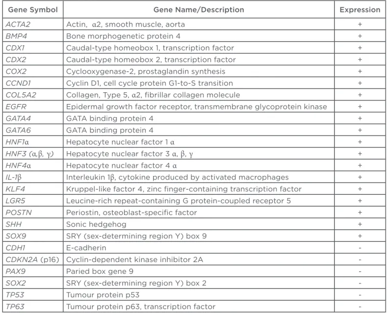
Table 1: Some of the genes implicated in the development of Barrett’s oesophagus.
Gene Symbol Gene Name/Description Expression
ACTA2 Actin, α2, smooth muscle, aorta +
BMP4 Bone morphogenetic protein 4 +
CDX1 Caudal-type homeobox 1, transcription factor +
CDX2 Caudal-type homeobox 2, transcription factor +
COX2 Cyclooxygenase-2, prostaglandin synthesis +
CCND1 Cyclin D1, cell cycle protein G1-to-S transition +
COL5A2 Collagen, Type 5, α2, ibrillar collagen molecule +
EGFR Epidermal growth factor receptor, transmembrane glycoprotein kinase +
GATA4 GATA binding protein 4 +
GATA6 GATA binding protein 4 +
HNF1α Hepatocyte nuclear factor 1 α +
HNF3 (α,β, γ) Hepatocyte nuclear factor 3 α, β, γ +
HNF4α Hepatocyte nuclear factor 4 α +
IL-1β Interleukin 1β, cytokine produced by activated macrophages +
KLF4 Kruppel-like factor 4, zinc inger-containing transcription factor +
LGR5 Leucine-rich repeat-containing G protein-coupled receptor 5 +
POSTN Periostin, osteoblast-speciic factor +
SHH Sonic hedgehog +
SOX9 SRY (sex-determining region Y) box 9 +
CDH1 E-cadherin
-CDKN2A (p16) Cyclin-dependent kinase inhibitor 2A
-PAX9 Paried box gene 9
-SOX2 SRY (sex-determining region Y) box 2
-TP53 Tumour protein p53
-Smoking status was analysed together with the genetic data. Since the linear correlation among genetic variants distribution and BO diagnosis was extremely low, with no R-squared values higher than 0.02, we decided to employ for data analysis artiicial neural networks, particularly suitable to handle non-linear relations among variables, rather than classical statistical tests. Using artiicial neural networks, it was possible to explain two-thirds of the variance related to cases, and control diference through the adaptive selection on nine polymorphisms, with a sensitivity near to 80%.
Molecular Pathogenesis
The metaplastic conversion of SE to specialised columnar epithelium in the distal oesophagus may originate from two diferent mechanisms.55 Transdiferentiation seems to be wrong, since new SE can develop after ablation treatment in which the BO epithelium has been completely removed.56 The best pathogenic hypothesis regarding BO is likely the altered diferentiation of stem cells.57 Diferent experimental data support four potential origins of these altered metaplastic stem cells: SE, GEJ, the neck, and bone marrow.58,59 In addition, acid and bile salts, alone or together, might also be involved in the pathogenesis through an increase in reactive oxygen species, causing oxidative stress that results in DNA damage and cell death.60,61 Chen and colleagues62 suggest that when gastroesophageal stem cells are stimulated by GERD, the squamous diferentiation programme may be inactivated through a loss or downregulation of squamous transcription factors; at the same time the overexpression of the transcription factors related to intestinal development may be activated (Table 1).
Experimental Models
In recent years, diferent approaches have been used to ind a model for BO, but as of yet, no one model ofers an ideal system for the study of environmental exposure, genetic risk, and prevention strategies. Cell culture based methods lack the complexity of a multicell system and this aspect can be overcome through the use of organotypic culture that mimics the in vivo interplay between the epithelium and underlying stoma. However, animal models provide a better solution to study such a complex disease since they ofer the opportunity to evaluate clinical and environmental risk factors in a controlled setting. Furthermore, since several genes and pathways
have been implicated in the development of BO, genetic manipulation can also be applied. Mouse, rats, dogs, opossum, guinea pigs, baboons, and pigs have all been used to study BO; however, the lack of spontaneous development of BO in animals presents a strong limitation.63
Treatment
The target of treatment is the control of relux symptoms in order to stop the impairment of the oesophageal lining. This goal could be achieved through a dietary change, removing foods that increase the risk of relux (e.g. chocolate, cofee and tea, peppermint, orange juice). Alternatively, the use of acid-suppressing medications (PPIs, e.g. omeprazole, lansoprazole, pantoprazole) can be applied. Although the acid suppression is important, the dose to use is still controversial.8,64 Recently, while continuous PPI therapy may be a symptomatic treatment at best, it could potentially promote dysplastic progression and adenocarcinoma, rather than prevent it.65 A recent study observed an increased risk for developing HGD and adenocarcinoma in the oesophagus with long-term PPI usage. Therefore, PPI may not protect against malignant progression in BO patients and in selected high-risk patients, and clinicians may consider adding or replacing long-term medical treatment with other modalities.66 Anti-relux surgery (ARS) may be considered for people with GERD symptoms. This therapy seems to promote the resolution of BO metaplasia; a meta-analysis demonstrated that 15.4% of patients who had undergone ARS had a regression of BO, compared with 1.9% of patients who were medication treated.67 In some papers the ARS is even associated to a lower cancer risk progression.68,69 Dysplasia is the typical precursor of OAC in BO patients and some studies have demonstrated that surgical or endoscopic removal of the dysplastic tissue can prevent its progression to cancer.8 In the recent years diferent endoscopic therapies have been established.
Endoscopic ablative therapies
REFERENCES
1. Shaheen NJ, Richter JE. Barrett’s oesophagus. Lancet. 2009;373(9666):850-61.
2. Koppert LB et al. The molecular biology of esophageal adenocarcinoma. J Surg
3. Zagari RM et al. Gastro-oesophageal relux symptoms, oesophagitis and Barrett’s oesophagus in the general population: the Loiano-Monghidoro study. Gut. 2008;57(10):1354-9.
esophagus in the general population: an endoscopic study. Gastroenterology. 2005;129(6):1825-31.
5. Rex DK et al. Screening for Barrett’s esophagus in colonoscopy patients with destroys the mucosa of BO patients. RFA appears
as the better treatment in eradicating dysplasia and cancer prevention, with greater simplicity management and fewer serious adverse efects compared with PDT.7,72 The problems of RFA regarded the recurrence rate of intestinal metaplasia ranging from 0-9%73,74 to 30%.75,76 Both PDT and RFA have been proven to be superior to eradicate dysplastic BO compared to routine anti-relux measures and pharmacological anti-relux measures in randomised trials. Nevertheless, the relative eicacy and safety of the promising endoscopic ablation treatment modalities remain unclear, since no previous head-to-head comparison of PDT versus RFA exists. In a recent study, the two modalities were compared with regards to complete eradication of BO and BD, adverse events, and costs. Both resulted in successfully eradicating dysplasia in BO. However, the overall success rate of RFA was higher than PDT, and RFA was very well tolerated without any major complications and fewer side-efects.77,78
Endoscopic mucosal resection (EMR)
EMR is increasingly being utilised as an alternative to surgery in the management of high-grade intraepithelial neoplasia, intramucosal cancer of the GI tract, dysplasia, and some small, very early-stage cancers of the oesophagus. It is less invasive than surgery and, unlike ablative therapies, it provides tissue for histological assessment. EMR is a technique where a piece of the inner lining of the oesophagus is removed with instruments passed down the endoscope. The most common side-efect of EMR is bleeding in the oesophagus, which is usually not serious. Less common, but more serious, side-efects can include oesophageal strictures (areas of narrowing) that might need to be treated with dilation, and puncture (perforation) of the oesophagus wall.79-81 Both ablative and mucosal resection are often combined in order to reach a better outcome.
Oesophagectomy
This procedure is associated with high morbidity and mortality and causes detrimental efects on the quality of life. Thus, it should be reserved only for patients in which ablation or resection eradication is not complete or durable, and only when endoscopic screening or surveillance revealed HGD. The risk of progression to cancer in BO patients with HGD is considered high enough to determine an intervention through endoscopic eradication therapy. This method includes the use of one or any combination of endoscopic strategies to remove all of the Barrett metaplasia - dysplastic or not.8,12 Conversely, the low-grade dysplasia (LGD) data of management are contradictory. One study on 147 patients revealed a risk of neoplastic progression of 85%, whereas another one carried out on 210 patients, described a rate of progression to HGD or cancer of only 1.83% per year.82,83 After this controversial evidence, the guidelines suggested either a more intensive programme of endoscopic surveillance or endoscopic ablation. In addition the diagnosis of LGD should be conirmed by at least two expert GI pathologists.8,12 Despite the wide variability for cancer risk in the LGD patients, novel speciic biomarkers (e.g. abnormal presence of p53 or a number of dysplastic glands) are able to recognise the patients at risk.84
CONCLUSIONS
2003;125(6):1670-7.
6. Rodríguez-Díjesús A et al. [Prevalence and epidemiology of Barrett’s esophagus in the province of Barcelona]. Gastroenterol Hepatol. 2014;37(7): 397-401.
7. Dunbar KB, Spechler SJ. Controversies in Barrett esophagus. Mayo Clin Proc. 2014;89(7):973-84.
8. Spechler SJ et al. American Gastroenterological Association technical review on the management of
Barrett’s esophagus. Gastroenterology. 2011;140(3):e18-52; quiz e13.
9. Chandra S et al. Barrett’s esophagus in 2012: updates in pathogenesis, treatment, and surveillance. Curr Gastroenterol Rep. 2013;15(5):322.
10. Paull A et al. The histologic spectrum of Barrett’s esophagus. N Engl J Med. 1976;295(9):476-80.
11. Goldblum JR. Controversies in the diagnosis of Barrett esophagus and Barrett-related dysplasia: one pathologist’s perspective. Arch Pathol Lab Med. 2010;134(10):1479-84.
12. Evans JA et al; ASGE Standards of Practice Committee. The role of endoscopy in Barrett’s esophagus and other premalignant conditions of the esophagus. Gastrointest Endosc. 2012;76(6):1087-94.
13. Sturm MB et al. Targeted imaging of esophageal neoplasia with a luorescently labeled peptide: irst-in-human results. Sci Transl Med. 2013;5(184):184ra61. 14. von Holzen U, Enders GH. A surprise cell of origin for Barrett’s esophagus. Cancer Biol Ther. 2012;13(8):588-91. 15. Franks I. Barrett esophagus: new insights into the stem cell organization of Barrett esophagus. Nat Rev Gastroenterol Hepatol. 2012;9(3):125.
16. Ko KH et al. Recent advances in molecular imaging of premalignant gastrointestinal lesions and future application for early detection of barrett esophagus. Clin Endosc. 2014;47(1):7-14. 17. Lao-Sirieix P et al. Non-endoscopic screening biomarkers for Barrett’s oesophagus: from microarray analysis to the clinic. Gut. 2009;58(11):1451-9. 18. Benaglia T et al. Health beneits and cost efectiveness of endoscopic and nonendoscopic cytosponge screening for Barrett’s esophagus. Gastroenterology. 2013;144(1):62-73.e6.
19. Kadri SR et al. Acceptability and accuracy of a non-endoscopic screening test for Barrett’s oesophagus in primary care: cohort study. BMJ. 2010;341:c4372. 20. Wang KK, Sampliner RE; Practice Parameters Committee of the American College of Gastroenterology. Updated guidelines 2008 for the diagnosis, surveillance and therapy of Barrett’s
esophagus. Am J Gastroenterol. 2008;103(3):788-97.
21. Katz PO et al. Guidelines for the diagnosis and management of gastroesophageal relux disease. Am J Gastroenterol. 2013;108(3):308-28. 22. Phillips WA et al. Barrett’s esophagus. J Gastroenterol Hepatol. 2011;26(4): 639-48.
23. Chak A et al. Gastroesophageal relux symptoms in patients with adenocarcinoma of the esophagus or cardia. Cancer. 2006;107(9):2160-6. 24. Kubo A, Corley DA. Body mass index and adenocarcinomas of the esophagus or gastric cardia: a systematic review and meta-analysis. Cancer Epidemiol Biomarkers Prev. 2006;15(5):872-8. 25. El-Serag HB et al. Abdominal obesity and the risk of Barrett’s esophagus. Am J Gastroenterol. 2005;100(10):2151-6. 26. Murray L, Romero Y. Role of obesity in Barrett’s esophagus and cancer. Surg Oncol Clin N Am. 2009;18(3):439-52. 27. Trayhurn P, Wood IS. Adipokines: inlammation and the pleiotropic role of white adipose tissue. Br J Nutr. 2004;92(3):347-55.
28. Wajchenberg BL. Subcutaneous and visceral adipose tissue: their relation to the metabolic syndrome. Endocr Rev. 2000;21(6):697-738.
29. Cook MB et al. Cigarette smoking increases risk of Barrett’s esophagus: an analysis of the Barrett’s and Esophageal
Adenocarcinoma Consortium. Gastroenterology. 2012;142(4):744-53.
30. Smith KJ et al. Interactions among smoking, obesity, and symptoms of acid relux in Barrett’s esophagus. Cancer Epidemiol Biomarkers Prev. 2005;14(11 Pt 1):2481-6.
31. Gray MR et al. The role of smoking and alcohol in metaplasia and cancer risk in Barrett’s columnar lined oesophagus. Gut. 1993;34(6):727-31.
32. Thrift AP et al. Lifetime alcohol consumption and risk of Barrett’s Esophagus. Am J Gastroenterol. 2011;106(7):1220-30.
33. Lagergren J et al. The role of tobacco, snuf and alcohol use in the aetiology of cancer of the oesophagus and gastric cardia. Int J Cancer. 2000;85(3):340-6. 34. Wiseman EF, Ang YS. Risk factors for neoplastic progression in Barrett’s esophagus. World J Gastroenterol. 2011;17(32):3672-83.
35. Yates M et al. Body mass index, smoking, and alcohol and risks of Barrett’s esophagus and esophageal adenocarcinoma: a UK prospective cohort study. Dig Dis Sci. 2014;59(7):1552-9. 36. Rajendra S, Robertson IK. Similar immunogenetics of Barrett’s oesophagus and cervical neoplasia: is HPV the
common denominator? J Clin Pathol. 2010;63(1):1-3.
37. El-Serag HB et al. Human papillomavirus and the risk of Barrett’s esophagus. Dis Esophagus. 2013;26(5):517-21.
38. Rajendra S et al. Transcriptionally active human papillomavirus is strongly associated with Barrett’s dysplasia and esophageal adenocarcinoma. Am J Gastroenterol. 2013;108(7):1082-93. 39. Orlof M et al. Germline mutations in MSR1, ASCC1, and CTHRC1 in patients with Barrett esophagus and esophageal adenocarcinoma. JAMA. 2011;306(4): 410-9.
40. Moons LM et al. A pro-inlammatory genotype predisposes to Barrett’s
esophagus. Carcinogenesis. 2008;29(5):926-31.
41. Kala Z et al. Polymorphisms of glutathione S-transferase M1, T1 and P1 in patients with relux esophagitis and Barrett’s esophagus. J Hum Genet. 2007;52(6):527-34.
42. McElholm AR et al. A population-based study of IGF axis polymorphisms and the esophageal inlammation, metaplasia, adenocarcinoma sequence. Gastroenterology. 2010;139(1):204-12.e3. 43. Robertson EV, Jankowski JA. Genetics of gastroesophageal cancer: paradigms, paradoxes, and prognostic utility. Am J Gastroenterol. 2008;103(2):443-9.
44. Su Z et al; Esophageal Adenocarcinoma Genetics Consortium; Wellcome Trust Case Control Consortium 2. Common variants at the MHC locus and at chromosome 16q24.1 predispose to Barrett’s esophagus. Nat Genet. 2012;44(10):1131-6.
45. Levine DM et al. A genome-wide association study identiies new susceptibility loci for esophageal adenocarcinoma and Barrett’s esophagus. Nat Genet. 2013;45(12):1487-93.
46. Ren D et al. Single nucleotide polymorphisms of caudal type homeobox 1 and 2 are associated with Barrett’s esophagus. Dig Dis Sci. 2014;59(1):57-63. 47. Winberg H et al. Risk factors and chemoprevention in Barrett’s esophagus--an update. Scand J Gastroenterol. 2012;47(4):397-406.
48. Rokkas T et al. Relationship between Helicobacter pylori infection and esophageal neoplasia: a meta-analysis. Clin Gastroenterol Hepatol. 2007;5(12):1413-7.
Gastroenterol Hepatol. 2012;24(8):917-23. 51. Das D et al. Chemoprevention of oesophageal cancer and the AspECT trial. Recent Results Cancer Res. 2009;181: 161-9.
52. Gaddam S et al. Persistence of nondysplastic Barrett’s esophagus identiies patients at lower risk for esophageal adenocarcinoma: results from a large multicenter cohort. Gastroenterology. 2013;145(3):548-53.e1. 53. Jankowski J, Barr H. Improving surveillance for Barrett’s oesophagus: AspECT and BOSS trials provide an evidence base. BMJ. 2006;332(7556):1512. 54. Tarlarini C et al. Role of XPC, XPD, XRCC1, GSTP genetic polymorphisms and Barrett’s esophagus in a cohort of Italian subjects. A neural network analysis. Clin Exp Gastroenterol. 2012;5:159-66.
55. Fitzgerald RC. Molecular basis of Barrett’s oesophagus and oesophageal adenocarcinoma. Gut. 2006;55(12): 1810-20.
56. Titi M et al. Development of subsquamous high-grade dysplasia and adenocarcinoma after successful radiofrequency ablation of Barrett’s
esophagus. Gastroenterology. 2012;143(3):564-6.e1.
57. Wang DH, Souza RF. Biology of Barrett’s esophagus and esophageal adenocarcinoma. Gastrointest Endosc Clin N Am. 2011;21(1):25-38.
58. Shaheen NJ. Advances in Barrett’s esophagus and esophageal adenocarcinoma. Gastroenterology. 2005;128(6):1554-66.
59. Jankowski JA et al. Barrett’s metaplasia. Lancet. 2000;356(9247):2079-85.
60. Fang Y et al. Cellular origins and molecular mechanisms of Barrett’s esophagus and esophageal adenocarcinoma. Ann N Y Acad Sci. 2013;1300:187-99.
61. Aichler M, Walch A. In brief: the (molecular) pathogenesis of Barrett’s oesophagus. J Pathol. 2014;232(4):383-5. 62. Chen H et al. Molecular mechanisms of Barrett’s esophagus. Dig Dis Sci. 2011;56(12):3405-20.
63. Garman KS et al. Review: Experimental models for Barrett’s esophagus and esophageal adenocarcinoma. Am J Physiol Gastrointest Liver Physiol. 2012;302(11):G1231-43.
64. Estores D, Velanovich V. Barrett esophagus: epidemiology, pathogenesis, diagnosis, and management. Curr Probl Surg. 2013;50(5):192-226.
65. Rosch PJ. Letter: proton pump inhibitors, GERD and oesophageal adenocarcinoma. Aliment Pharmacol Ther. 2014;40(3):319.
66. Hvid-Jensen F et al. Proton pump inhibitor use may not prevent high-grade dysplasia and oesophageal adenocarcinoma in Barrett’s oesophagus: a nationwide study of 9883 patients. Aliment Pharmacol Ther. 2014;39(9): 984-91.
67. Chang EY et al. The efect of antirelux surgery on esophageal carcinogenesis in patients with barrett esophagus: a systematic review. Ann Surg. 2007;246(1):11-21.
68. Gurski RR et al. Barrett’s esophagus can and does regress after antirelux surgery: a study of prevalence and predictive features. J Am Coll Surg. 2003;196(5):706-12.
69. Bowers SP et al. Clinical and histologic follow-up after antirelux surgery for Barrett’s esophagus. J Gastrointest Surg. 2002;6(4):532-8; discussion 539.
70. Prasad GA et al. Predictors of stricture formation after photodynamic therapy for high-grade dysplasia in Barrett’s esophagus. Gastrointest Endosc. 2007;65(1):60-6.
71. Gray NA et al. Buried metaplasia after endoscopic ablation of Barrett’s esophagus: a systematic review. Am J Gastroenterol. 2011;106(11):1899-908; quiz 1909.
72. Shaheen NJ et al. Radiofrequency ablation in Barrett’s esophagus with dysplasia. N Engl J Med. 2009;360(22):2277-88.
73. Shaheen NJ et al. Durability of radiofrequency ablation in Barrett’s esophagus with dysplasia. Gastroenterology. 2011;141(2):460-8.
74. Fleischer DE et al. Endoscopic radiofrequency ablation for Barrett’s esophagus: 5-year outcomes from a prospective multicenter trial. Endoscopy. 2010;42(10):781-9.
75. Vaccaro BJ et al. Detection of intestinal metaplasia after successful eradication of Barrett’s esophagus with radiofrequency ablation. Dig Dis Sci. 2011;56(7):1996-2000.
76. Gupta M et al. Recurrence of esophageal intestinal metaplasia after endoscopic mucosal resection and radiofrequency ablation of Barrett’s esophagus: results from a US Multicenter
Consortium. Gastroenterology. 2013;145(1):79-86.e1.
77. Ertan A et al. Photodynamic therapy vs radiofrequency ablation for Barrett’s dysplasia: eicacy, safety and cost-comparison. World J Gastroenterol. 2013;19(41):7106-13.
78. Akiyama J et al. Managing Barrett’s esophagus with radiofrequency ablation. Gastroenterol Rep (Oxf). 2013;1(2): 95-104.
79. Pech O et al. Long-term eicacy and safety of endoscopic resection for patients with mucosal adenocarcinoma of the esophagus. Gastroenterology. 2014;146(3):652-660.e1.
80. van Vilsteren FG et al. Learning to perform endoscopic resection of esophageal neoplasia is associated with signiicant complications even within a structured training program. Endoscopy. 2012;44(1):4-12.
81. Alvarez Herrero L et al. Safety and eicacy of multiband mucosectomy in 1060 resections in Barrett’s esophagus. Endoscopy. 2011;43(3):177-83.
82. Curvers WL et al. Low-grade dysplasia in Barrett’s esophagus: overdiagnosed and underestimated. Am J Gastroenterol. 2010;105(7):1523-30.
83. Wani S et al. Risk factors for progression of low-grade dysplasia in patients with Barrett’s esophagus. Gastroenterology. 2011;141(4):1179-86, 1186.e1.