1 23
European Radiology
ISSN 0938-7994
Eur Radiol
DOI 10.1007/s00330-016-4673-z
European society of urogenital radiology
(ESUR) guidelines: MR imaging of pelvic
endometriosis
M. Bazot, N. Bharwani, C. Huchon,
K. Kinkel, T. M. Cunha, A. Guerra,
L. Manganaro, L. Buñesch, A. Kido,
1 23
UROGENITAL
European society of urogenital radiology (ESUR) guidelines:
MR imaging of pelvic endometriosis
M. Bazot1&N. Bharwani2&C. Huchon3&K. Kinkel4&T. M. Cunha5&A. Guerra6&
L. Manganaro7&L. Buñesch8&A. Kido9&K. Togashi9&I. Thomassin-Naggara1&
A. G. Rockall10
Received: 3 June 2016 / Revised: 5 October 2016 / Accepted: 23 November 2016 #The Author(s) 2016. This article is published with open access at Springerlink.com
Abstract
Endometriosis is a common gynaecological condition of un-known aetiology that primarily affects women of reproductive age. The accepted first-line imaging modality is pelvic ultra-sound. However, magnetic resonance imaging (MRI) is in-creasingly performed as an additional investigation in
complex cases and for surgical planning. There is currently no international consensus regarding patient preparation, MRI protocols or reporting criteria. Our aim was to develop clinical guidelines for MRI evaluation of pelvic endometriosis based on literature evidence and consensus expert opinion. This work was performed by a group of radiologists from the
Electronic supplementary materialThe online version of this article (doi:10.1007/s00330-016-4673-z) contains supplementary material, which is available to authorized users.
* M. Bazot
marc.bazot@tnn.aphp.fr
N. Bharwani
nishat.bharwani@imperial.nhs.uk
C. Huchon
cyrillehuchon@yahoo.fr
K. Kinkel
karen.kinkel-trugli@wanadoo.fr
T. M. Cunha
tmargarida@gmail.com
A. Guerra
aguerra@hospitaldaluz.pt
L. Manganaro
lucia.manganaro@uniroma1.it
L. Buñesch
lbunesch@clinic.ub.es
A. Kido
akikido@kuhp.kyoto-u.ac.jp
K. Togashi
ktogashi@kuhp.kyoto-u.ac.jp
I. Thomassin-Naggara isabelle.thomassin@tnn.aphp.fr
A. G. Rockall
a.rockall@imperial.ac.uk
1 Department of Radiology, Tenon Hospital, 58 Avenue Gambetta,
Paris 75020, France
2
Department of Radiology, St Mary’s Hospital, Imperial College Healthcare NHS Trust, 3rd Floor Queen Elizabeth the Queen Mother Building, Praed Street, London W2 1NY, UK
3
Department of Obtetrics and Gynaecology, CHI Poissy Saint-Germain en Laye,Versailles University France, 10 rue du champ Gaillard, 78300 Poissy, France
4 Institut de radiologie, Clinique des Grangettes, 7, chemin des
Grangettes, CH 1224 Chêne-Bougeries, Switzerland
5
Serviço de Radiologia, Instituto Português de Oncologia de Lisboa Francisco Gentil, Rua Prof. Lima Basto, 1099-023 Lisboa, Portugal
6
Department of Radiology, Hospital da Luz, Lisbon, Portugal
7 Department of Radiological Sciences, Sapienza University of Rome,
Vle. Regina Elena 324, 00162 Rome, Italy
8
Department of Radiology (Urogenital Section), Hospital Clínic Barcelona, Villarroel, 170, Barcelona 08036, Spain
9
Department of Diagnostic Radiology, Kyoto University Hospital, 54 Shogoin-Kawahara-cho, Sakyo-ku, Kyoto 606-8507, Japan
10 Department of Radiology, The Royal Marsden Hospital, Fulham
Road, London SW3 6JJ, UK Eur Radiol
European Society of Urogenital Radiology (ESUR), experts in gynaecological imaging and a gynaecologist expert in meth-odology. The group discussed indications for MRI, technical requirements, patient preparation, MRI protocols and criteria for the diagnosis of pelvic endometriosis on MRI. The expert panel proposed a final recommendation for each criterion using Oxford Centre for Evidence Based Medicine (OCEBM) 2011 levels of evidence.
Key Points
•This report provides guidelines for MRI in endometriosis.
• Minimal and optimal MRI acquisition protocols are provided.
•Recommendations are proposed for patient preparation,
best MRI sequences and reporting criteria.
Keywords Endometriosis . Guidelines . Magnetic resonance imaging (MRI) . Protocols . Evidence-based Medicine/ standards
Abbreviations
2D Two-dimensional 3D Three-dimensional DPE Deep pelvic endometriosis DWI Diffusion-weighted imaging
ESUR European Society of Urogenital Radiology
FPI-ESUR
Female Pelvic Imaging working group of the European Society of Urogenital Radiology GPP Good practice point
HASTE Half-Fourier acquisition single shot turbo-spin-echo
IM Imtramuscular IV Intravenous LE Level of evidence
MRI Magnetic resonance imaging
OCEBM Oxford Centre for Evidence Based Medicine SC Subcutaneous
SNR Signal-to-noise ratio
SWI Susceptibility-weighted imaging
T Tesla
T1W T1-weighted T2W T2-weighted
TVS Transvaginal sonography US Ultrasound
Introduction
Endometriosis is a common gynaecological condition that is defined as functional ectopic endometrial glands and stroma outside the uterus. This disease affects women of reproductive age, with a prevalence of
approximately 10% [1]. Patients can be asymptomatic or present with chronic pelvic pain and/or infertility. The combination of laparoscopy and histological verifi-cation of endometrial glands and/or stroma is considered to be the gold standard for diagnosis of the disease [2]. Ultrasonography (US) is the first-line imaging modal-ity for the assessment of pelvic endometriosis but has limitations with respect to field-of-view and operator dependence [3–8]. Magnetic resonance imaging (MRI) is usually performed as an additional examination in complex cases or prior to surgery and is highly accurate in the evaluation of endometriosis [9–18]. However, there is no international consensus on patient prepara-tion, best MRI sequences or reporting criteria.
The aim of this work was to develop guidelines for optimal MRI protocols and image interpretation in en-dometriosis based on a detailed literature review and consensus expert opinion from the Female Pelvic Imaging working group of the European Society of Urogenital Radiology (FPI-ESUR).
Methods
MRI protocols for the diagnosis of pelvic endometriosis were collected from one non-European and seven European institutions. Inclusion criteria to participate in the guideline development process were: to be a mem-ber of the FPI-ESUR working group, and to perform regular MRI examinations and/or publications related to endometriosis. All but one investigators included in this guidelines process were experts in gynaecological imaging, the last being a gynaecologist expert in methodology.
A questionnaire was established containing the following information: patient preparation, magnet field strength, type of coil, type of MRI sequences, use of intravenous contrast in-jection, and vaginal and/or rectal opacification.
The summary of valid scientific data for each question analysed by the experts included a level of evidence (LE) based on the quality of available data and defined according to the rating scheme developed by the French National Authority for Health:
LE1: high-power randomized comparative trials or meta-analyses of randomized comparative trials;
LE2: low-power randomized trials, well-conducted non-randomized comparative studies and cohort studies; LE3: non-consecutive studies or studies without consis-tently applied reference standards;
LE4: non-randomized comparative studies with substan-tial bias, retrospective studies, cross-sectional studies and case series.
The practice guidelines were summarised from the re-sponses provided by the experts, and grades were attributed as follows: Grade A: established scientific evidence (LE1); Grade B: scientific presumption (LE2); Grade C: based on a low level of evidence (LE3 or LE4). Recommendations based on professional consensus were reduced to a minimum (Good Practice Point: GPP) and were used in the queries when liter-ature was lacking in evidence.
Results
Indications for MRI in endometriosis
No data exist in the literature about the indications for MR imaging for pelvic endometriosis: evaluation of pelvic pain, infertility or indeterminate adnexal mass. In accordance with
the analysis performed in our different ESUR centres, more than 90% of MRI examinations are performed for staging deep pelvic endometriosis that is the main indication. Hence, an indeterminate adnexal mass represents an ancillary indica-tion of MR imaging.
No consensus exists in the literature regarding the use of MRI in comparison to US. In practice, radiological papers tend to favour MRI whereas gynaecological publi-cations underline the value of US [19, 20]. This assess-ment is in line with recent published meta-analyses [21–24]. The first confirmed that transvaginal sonography (TVS) should remain the first-line method in the evalua-tion of patients with suspicion of deep pelvic endometri-osis (DPE) [21]. Two further meta-analyses demonstrated that the overall diagnostic performance of TVS for detect-ing DPE is fair but a high specificity is present for all locations [22, 23]. Based on the results of these meta-analyses, further investigations, especially MRI, are rec-ommended in a symptomatic patient in the presence of negative US findings (LE1) [22,23]. Finally, an addition-al meta-anaddition-alysis suggested that MRI is a useful preopera-tive test for predicting the diagnosis of multiple sites of DPE (LE1) [24].
In patients with equivocal US, MRI is recommended as a second-line technique in the preoperative workup of DPE (grade A).
Technical requirements
Technical requirements for each centre are presented in Table1. A summary of literature review is provided for each specific criterion.
Table 1 Technical requirements
Paris London Geneva Lisbon Lisbon Roma Barcelona Kyoto
Device (Tesla) 1.5 1.5/3.0 3.0 1.5 1.5/3.0 3.0 1.5/3.0 1.5/3.0
Phased-array Yes Yes Yes Yes Yes Yes Yes Yes
Endocavitary probe No No No No No No No No
Timing of MRI No No > Day 8 No No No No No
Fasting 3h No 6h 6h 4h 6h 4h 4h
Special diet No No No No No No No No
Bowel enema Yes No Yes No Yes Yes No No
Bladder emptying 2h No No 2h 1h 1h No No
IV catheter No (Option) Yes Yes Yes Yes Yes Yes No
Anti-peristaltic agent SC IV IM IV IV IV IV SC
Belt strapping Yes No Yes Yes No No No Yes
Vaginal opacification No No Yes* Yes* Yes* No No No
Rectal opacification No Yes Yes* Yes* Yes* Yes* No No
Supine position Yes Yes Yes Yes Yes Yes Yes Yes
Prone position Yes** No No No No No No No
IMintramuscular,IVintravenous,SCsubcutaneous
*
If doubt or symptoms present (i.e. dyspareunia, dyschezia)
**
1.5 Tesla versus 3.0 Tesla systems
The majority of published studies use a 1.5T magnet. Only four publications used 3.0T but suggested promising results [11,16,25,26]. At 3.0T, improved signal-to-noise ratio results in the acquisition of high-spatial resolution images and accu-rate depiction of all locations of DPE [11,16,25]. However, there is increased image heterogeneity at 3.0T when compared to 1.5T, which can have a negative effect on the fat-saturation techniques routinely utilised in the evaluation of endometri-osis [16,27]. The application of the Dixon technique may overcome this and achieve stronger fat-suppression [27].
Both 1.5T and 3.0T seem valuable for the evaluation of DPE; however, studies comparing the systems are lacking. Therefore, no recommendation can be made for the use of a specific device and further work is necessary to perform this comparison (Fig.1).
Array type
In line with different publications, pelvic phased-array coils provide a higher SNR than is possible with a body coil (LE3) [28,29]. In addition, three studies have reported additional value of endocavitary coils in conjunction with pelvic phased-array [9,30,31]. Drawbacks in terms of cost and ac-ceptability limit its potential use in the evaluation of DPE.
Pelvic phased array coils are recommended in the evalua-tion of DPE at both 1.5T and 3.0T (grade C).
Timing of MRI examination
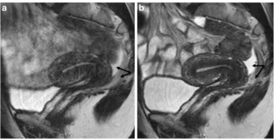
Several studies reported discrepant results regarding timing of MRI evaluation. Fiaschetti et al. examined patients between days 8 and 12 of the menstrual cycle, due to the possibility of spontaneous T1W -signal intensity of blood prior to day 8 of the menstrual cycle [32]. Bazot et al. suggested that the pres-ence of pelvic free fluid (e.g. menstruation, post-ovulatory phase) is a useful aid to image interpretation (Fig.2) [33,
34]. Tamai et al. reported that during menstruation the uterus can demonstrate marked pseudo-thickening of the junctional zone, suggesting an inappropriate diagnosis of adenomyosis [35]. They went on to suggest that MRI should be avoided in the menstrual phase [35]. In addition, these authors reported that the evaluation of uterine peristalsis is optimal during the peri-ovulatory phase [36]. Solak et al. reported no significant difference in size of lesions in the early days of menstruation compared to the mid-menstrual period for abdominal wall endometriosis [37]. Finally, Botterill et al. did not observe a significant difference in disease extent between menstruating and non-menstruating scans [38].
No recommendation can be proposed for timing of MRI in relation to the menstrual cycle in the evaluation of DPE.
Patient preparation
There was no consensus regarding patient preparation before MRI. The committee felt that the protocol should be tailored to the main indication for pelvic MRI (diagnosis/staging of DPE, indeterminate adnexal mass).
Fasting
When fasting prior to the MRI study was mentioned, the length of fast was variable at 3, 4 or 6 h (LE2) [16,17,19,
20,32,33,39]. However, the majority of studies did not men-tion this pre-imaging preparamen-tion.
Fasting is recommended in the evaluation of DPE (grade B).
Bowel preparation
Most studies did not mention the use of bowel preparation prior to pelvic MRI. Where authors advocated the use of bow-el preparation, the type of preparation varied. The most com-monly utilised method was bowel enema with either rectal suppository pills (e.g. bisacodyl) or water [39,40]. In addition, there was variable use of dietary preparation, ranging from
nothing to low-residue diet 3 days prior to MRI accompanied by enema, magnesium sulphate and fluid re-hydration the day before the study [41].
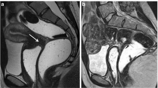
Bowel preparation is advocated as‘best practice’for the detection of DPE (GPP) (Fig.3).
Bladder emptying
No studies have been published in the medical literature addressing the importance of bladder distension for de-tection of anterior DPE. When bladder distension is discussed, authors describe a moderately filled or full bladder in order to correct the angle of uterine anteversion and thereby improve visualisation of the re-gion, allowing detection of small nodules located ante-rior to or in the vesicouterine pouch and to displace the bowel superiorly reducing artefact caused by bowel mo-tion (LE4) [12, 16, 39, 42–46]. Excessive bladder
distension is not recommended as associated detrusor contractions may cause artefact and can complicate the identification of small parietal nodules (LE4) (Fig. 3) [42, 44, 46]. To achieve the appropriate distension, au-thors mainly ask their patients not to empty their blad-der for 1 h prior to the examination [16, 39].
A moderately full bladder is recommended in the evalua-tion of DPE (grade C).
Patient position
All centres performed MRI with a patient in the supine position.
A recent systematic review specifically looked at possible interventions aimed to reduce anxiety, distress and the need for sedation in adults undergoing MRI exams, and confirmed evidence for the benefit of prone scanning in reducing claus-trophobia (LE2) [47].
Fig. 2 Sagittal 2D T2-weighted MR images performed at 1.5 Tesla showing the benefits of anti-peristaltic agents on image quality. Imaging performed in the same patient before (a) and after (b) administration of glucagon demonstrating a dramatic improvement in image quality. Note the presence of pelvic fluid in the pouch of Douglas underlining a clear demarcation between peritoneal and posterior subperitoneal compartments (double arrow) (reprinted with permission - Bazot M. Ed. Lavoisier-Paris 2016)
Fig. 3 Sagittal 2D T2-weighted MR images performed at 1.5 Tesla showing the benefits of patient preparation on image quality. (a) Imaging performed with a full urinary bladder and without bowel preparation is sub-optimal for interpretation and disease may be
The supine position is recommended in the evaluation of pelvic endometriosis (GPP). The prone position is an‘option’
in claustrophobia (grade B).
Abdominal strapping
A few papers recommend the use of a broad abdominal belt in MRI examinations for the evaluation of endometriosis (Fig.1) [34,48,49]. The purpose is to reduce artefact caused by re-spiratory movement and it has been recommended to apply the belt at the end of expiration (LE3) [50,51].
Abdominal strapping is recommended in the evaluation of pelvic endometriosis (grade C).
Anti-peristaltic agent
The use of an anti-peristaltic agent (e.g. glucagon, butyl-sco-polamine), unless contraindicated (e.g. diabetes or phaeochromocytoma), is the most efficient way to limit bowel motion artefact (Fig.2) (LE4) [52]. Recently, Gutzeit et al. suggested that intravenous spasmolysis is more reliable than intramuscular administration, and glucagon is better than butyl-scopolamine [52].
An anti-peristaltic agent is recommended in the evaluation of DPE (grade C).
Vaginal opacification
Four studies provided discrepant results on the value of vaginal opacification with gel in the diagnosis of posterior DPE (Fig.4) (LE4) [14,32,45,53]. The first reported an improvement in
sensitivity between pre- and post-contrast MRI in the diagnosis of DPE; however, this improvement was only significant for junior radiologists [45]. The second did not find any significant difference in the diagnosis of vaginal or rectal endometriosis with or without vaginal opacification, whatever the level of expertise of readers (LE4) [14]. The third reported better eval-uation for the detection of vaginal and uterosacral endometri-osis but not for pouch of Douglas or rectovaginal septum dis-ease (LE4) [32]. Finally, the most recent study reported a sig-nificant improvement in the diagnosis of pouch of Douglas obliteration in the presence of vaginal opacification (LE4) [53]. Vaginal opacification with sonographic gel is considered as an‘option’in the evaluation of DPE (GPP).
Rectal opacification
No consensus exists in the literature on the value of rectal opacification in the diagnosis of DPE (Fig.4). In practice, two different types of contrast medium are used (sonographic gel or water) [11,14,45,53]. Discrepant results are available with some authors claiming that rectal opacification provides a better evaluation of pouch of Douglas and rectosigmoid colon endometriosis [32,41,43,53], while several other stud-ies argued that this technique was useless in the evaluation of posterior DPE locations [10,11,14]. In this setting, different arguments against systematic rectal opacification are sug-gested including time, patient discomfort, movement artefact and rectosigmoid colon spasm [10].
Rectal opacification is suggested as an‘option’in the eval-uation of pelvic endometriosis (GPP).
Fig. 4 Sagittal 2D T2-weighted MR images performed in two different patients at 1.5 Tesla following vaginal and rectal opacification with sonographic gel and with (a) or without (b) bowel preparation. Vaginal distension demonstrates thickening of the posterior vaginal fornix (white arrow) without involvement of the pouch of Douglas or rectum
MRI protocol
MRI sequences (Table2)
There is significant variability in the literature regarding the MRI protocols used [11,16–18,31–33,44,45,54–56].
T2-weighted MRI
T2W MR sequences without fat-suppression technique are the best sequences for detecting pelvic endometriosis (LE2) [33]. Most MRI studies are performed using at least two orthogonal T2W planes [11, 16–18, 31–33, 44, 45,
54–56]. Further studies are required to clarify the field-of-view used for the axial acquisition and which additional T2W MR plane should be used. Axial 2D-T2W MRI from renal hila to pubic bone, allowing a systematic visualisa-tion of kidneys and potential analysis of the right iliac fossa (i.e. caecum, appendix, small bowel) should be rec-ommended [33]. The use of thin section-oblique 2D-T2W imaging improves the success of conventional MRI (sag-ittal and axial) for assessment of uterosacral and parametrial endometriosis (LE3) [13, 15]. In addition, several authors have recently reported the potential value of 3D-T2W imaging in the evaluation of DPE [16,54]. In contrast, no studies have demonstrated the value of coro-nal 2D-T2W MRI sequence in the evaluation of pelvic endometriosis.
Three 2D-T2W MRI sequences (sagittal, axial, oblique) are recommended in the evaluation of DPE (grade B).
The addition of 3D-T2W MRI sequence is proposed as an
‘option’(grade C).
T1-weighted MRI
Several studies have underlined that T1W MRI is the gold standard for the diagnosis of endometriotic cysts (LE2) [57,
58]. The 2D or 3D Dixon technique providing four simulta-neous different T1W contrasts during the same acquisition and a stronger fat suppression in the female pelvis when com-pared to a 3D-FSPGR sequence should progressively become the reference technique (LE4) [58]. There has been no compar-ative study between conventional fat saturated T1W sequences and the Dixon technique in the identification of endometrial implant. The reduced spatial resolution of currently available Dixon techniques might prevent the identification of small peri-toneal implants compared to conventional fat-saturated T1W sequences. This hypothesis requires further research.
Data are lacking for the evaluation of DPE using T1W MRI. Preliminary papers have suggested fat-suppressed T1W MRI to be of value in the diagnosis of peritoneal endometri-osis, but this finding must be confirmed [59,60].
T1W MRI sequences without and with fat suppression are recommended in the evaluation of adnexal endometriosis (grade B)
The ‘Dixon technique’ may be used as an alternative to standard T1W sequence (grade C).
Intravenous contrast-enhanced MRI
Few data are available regarding the value of gadolinium in the evaluation of endometriosis. A clear distinction must be made regarding the indication for MRI (diagno-sis/staging of endometriosis/characterisation of US-indeterminate adnexal mass).
Three studies reported data for the evaluation of different DPE locations [14,61,62]. Firstly, Onbas et al. suggested that
Table 2 MRI sequences
MRI sequence Paris London Geneva Lisbon Lisbon Roma Barcelona Kyoto
2DT2W sagittal Yes Yes Yes Yes Yes Yes Yes Yes 2DT2W axial LP* P* LP* LP* LP* P* P* P*
2DT2W coronal No No Yes No Yes Yes Yes No 2DT2W oblique Yes Yes Yes Yes Yes Yes No No
3DT2W Yes No No No No Yes No No
T2* No No No No No No No No
SSFSE/Haste Yes No Yes No No No No Yes
2D/3D T1W 3D 2D 2D 2D 3D 3D 2D 2D
T1W no FS§ Yes Yes Yes Yes Yes Yes Yes Yes
T1W with FS§ Yes Yes Yes Yes Yes Yes Yes Yes
Gadolinium ± ± ± ± ± ± ± ±
Peristalsis ± No No No No No No Yes
DWI No No No No No Yes No Yes
T1WT1-weighted,T2WT2-weighted,2Dtwo-dimensional,3Dthree-dimensional LP*: from renal hila to pubic bone
P*: from iliac crests to pubic bone T2*: susceptibility-weighted MR sequence FS§: fat-saturation technique
dynamic MRI could be useful to depict abdominal wall endo-metriosis [61]. Secondly, Scardapane et al. underlined that the combination of MR colonography and 3D-T1W MRI allows easier recognition of colorectal endometriosis and higher inter-observer agreement (LE3) [62]. Finally, Bazot et al. sug-gested the absence of benefit of intravenous gadolinium for the diagnosis of rectosigmoid colon, vaginal and bladder en-dometriosis, whatever the level of expertise of readers (LE3) (Fig.5) [14].
These variable results contrast with the usefulness of gad-olinium when dealing with endometriotic adnexal masses. Using conventional T2W and T1W sequences, MRI has only moderate accuracy in distinction of endometrial cysts from other haemorrhagic adnexal lesions [63,64]. The use of gad-olinium may help to distinguish an endometrioma from a lu-teal ovarian cyst or tubo-ovarian abscess displaying intense wall enhancement [65]. Moreover, gadolinium enhancement is crucial for depicting strongly enhancing mural nodules if Fig. 5 Sagittal 2D MR images performed at 1.5 Tesla demonstrating the
use of sonographic gel to opacify and distend the vagina. (a) Sagittal 2D T2-weighted image demonstrating an endometriotic plaque involving the posterior vaginal fornix (white arrow). Following distension of the vagina
with sonographic gel, the plaque is better delineated on both T2-weighted (b) and fat-suppressed T1-weighted (c) sequences (white arrows) (reprinted with permission - Bazot M. Ed. Lavoisier-Paris)
atypical features suggest potential malignancy on either ultra-sound (US) or T2W (LE4) (Fig.6) [66,67]. Finally, endome-triosis and pelvic inflammatory disease are two conditions that can be easily confused, especially in situations where they co-exist. Hence, the presence of a strong wall enhancement with-in adnexal masses is useful to suggest pelvic with-inflammatory disease (LE3) [65,68].
No recommendation can be achieved regarding the use of gadolinium in the evaluation of DPE (Fig.5).
The use of gadolinium is recommended as an‘option’in the evaluation of indeterminate adnexal endometriosis (grade C).
Diffusion-weighted MRI (DWI)
No data are available on DWI for the evaluation of DPE. Two recent studies have suggested its potential value in the evalu-ation of abdominal wall and sacral nerve root abnormalities endometriosis (LE4) [37,69,70].
A role for DWI has also been suggested in differentiation of endometriomas from haemorrhagic cysts with significantly lower ADC values in endometriomas when compared with haemorrhagic ovarian cysts at all b values [71].
No recommendation can be achieved for the use of DWI in the evaluation of DPE.
Susceptibility-weighted MRI (SWI)
Two recent studies using SWI suggest that the presence of signal voids reflecting acute to chronic haemorrhage are very sensitive in the diagnosis of extra-ovarian endometriosis, es-pecially abdominal wall endometriosis (LE4) [37,72].
Endometriotic cysts contain blood degradation products secondary to recurrent cyclic bleeding from ectopic endome-trial tissue giving rise to punctate or curvilinear signal voids along the cyst wall on SWI [57, 73]. However, a pitfall of imaging with SWI is susceptibility artefact caused by intesti-nal gas, particularly at 3.0T.
No recommendation can be proposed for the use of susceptibility-weighted MR imaging in the evaluation of deep endometriosis.
Half-Fourier acquisition single shot turbo spin echo
Half-Fourier acquisition single shot turbo-spin-echo (SSFSE, HASTE) enables multiphase and multislice image acquisition producing kinematic images for the evaluation of pelvic ad-hesions [74].
HASTE imaging is used to evaluate uterine function by assessing uterine peristalsis, identifiable as rhythmic and sub-tle wave-like endometrial and subendometrial myometrium movements [75–77]. During the peri-ovulatory phase, uterine peristalsis is significantly reduced in subjects with endometri-osis when compared to normal controls that may be due to
increased, sustained contractions in endometriosis patients (LE4) [36,78]. This abnormal uterine peristalsis in endome-triosis patients could interfere with fertility [79].
Half-Fourier acquisition single shot turbo spin echo is rec-ommended for the evaluation of uterine peristalsis (grade C).
Reporting criteria
A consensus exists in the ESUR group and in the literature about the criteria used in the diagnosis of endometrial cysts [57] and different locations of DPE [15,33] (Appendix 1).
Conclusion
These recommendations argue that:
1. MRI should be considered as a second-line technique
ex-amination after US in the evaluation of pelvic endometriosis
2. MRI is recommended before surgery for optimal
preoper-ative staging
3. Some requirements for the acquisition of MR images have
to be observed to provide optimal studies.
Future research for the evaluation of endometriosis using MRI:
1. Patients with clinical suspicion and negative US 2. Intra-individual comparison between 1.5T and 3.0T 3. Management of medical treatment
4. Diagnostic performance without and with bowel
prepara-tion to evaluate DPE
5. Evaluation of the clinical impact of MRI as a
pre-operative test.
Acknowledgements The committee would like to thank Mr Arnold Stipsits, ESUR secretary, for his help with co-ordination of the committee members. The scientific guarantor of this publication is Pr. Marc Bazot. The authors of this manuscript declare no relationships with any compa-nies whose products or services may be related to the subject matter of the article. The authors state that this work has not received any funding. No complex statistical methods were necessary for this paper. Institutional Review Board approval was not required because this manuscript pre-sents guidelines only and has not required the use of patient-sensitive data. Some study subjects or cohorts have not been previously reported. Methodology: Guidelines based on expert opinion and literature review
References
1. Clement MD (2002) Endometriosis, lesions of the secondary müllerian system, and pelvic mesothelial proliferations. In: Kurman RJ (ed) Blaustein's pathology of the female genital tract, 5th edn. Springer-Verlag, New York, pp 516–559
2. Dunselman GA, Vermeulen N, Becker C et al (2014) ESHRE guideline: management of women with endometriosis. Hum Reprod 29(3):400–412
3. Fedele L, Bianchi S, Raffaelli R, Portuese A (1997) Pre-operative assessment of bladder endometriosis. Hum Reprod 12(11):2519–2522 4. Chapron C, Dumontier I, Dousset B et al (1998) Results and role of rectal endoscopic ultrasonography for patients with deep pelvic endometriosis. Hum Reprod 13(8):2266–2270
5. Bazot M, Detchev R, Cortez A, Amouyal P, Uzan S, Darai E (2003) Transvaginal sonography and rectal endoscopic sonography for the assessment of pelvic endometriosis: a preliminary comparison. Hum Reprod 18(8):1686–1692
6. Bazot M, Thomassin I, Hourani R, Cortez A, Darai E (2004) Diagnostic accuracy of transvaginal sonography for deep pelvic endometriosis. Ultrasound Obstet Gynecol 24(2):180–185 7. Piketty M, Chopin N, Dousset B et al (2009) Preoperative work-up
for patients with deeply infiltrating endometriosis: transvaginal ul-trasonography must definitely be the first-line imaging examina-tion. Hum Reprod 24(3):602–607
8. Hudelist G, Ballard K, English J et al (2011) Transvaginal sonogra-phy vs. clinical examination in the preoperative diagnosis of deep infiltrating endometriosis. Ultrasound Obstet Gynecol 37(4):480–487 9. Kinkel K, Chapron C, Balleyguier C, Fritel X, Dubuisson JB, Moreau JF (1999) Magnetic resonance imaging characteristics of deep endometriosis. Hum Reprod 14(4):1080–1086
10. Chamie LP, Blasbalg R, Goncalves MO, Carvalho FM, Abrao MS, de Oliveira IS (2009) Accuracy of magnetic resonance imaging for diagnosis and preoperative assessment of deeply infiltrating endo-metriosis. Int J Gynaecol Obstet 106(3):198–201
11. Hottat N, Larrousse C, Anaf V et al (2009) Endometriosis: contri-bution of 3.0-T pelvic MR imaging in preoperative assessment–
initial results. Radiology 253(1):126–134
12. Grasso RF, Di Giacomo V, Sedati P et al (2010) Diagnosis of deep infiltrating endometriosis: accuracy of magnetic resonance imaging and transvaginal 3D ultrasonography. Abdom Imaging 35(6):716–725 13. Bazot M, Gasner A, Ballester M, Darai E (2011) Value of thin-section oblique axial T2-weighted magnetic resonance images to assess uterosacral ligament endometriosis. Hum Reprod 26(2):346–353 14. Bazot M, Gasner A, Lafont C, Ballester M, Darai E (2011) Deep
pelvic endometriosis: limited additional diagnostic value of postcontrast in comparison with conventional MR images. Eur J Radiol. doi:10.1016/j.ejrad.2010.12.006
15. Bazot M, Jarboui L, Ballester M, Touboul C, Thomassin-Naggara I, Darai E (2012) The value of MRI in assessing parametrial involve-ment in endometriosis. Hum Reprod 27(8):2352–2358
16. Manganaro L, Fierro F, Tomei A et al (2012) Feasibility of 3.0T pelvic MR imaging in the evaluation of endometriosis. Eur J Radiol 81(6): 1381–1387
17. Saba L, Guerriero S, Sulcis R et al (2012) MRI and "tenderness guided" transvaginal ultrasonography in the diagnosis of recto-sigmoid endometriosis. J Magn Reson Imaging 35(2):352–360 18. Di Paola V, Manfredi R, Castelli F, Negrelli R, Mehrabi S, Pozzi
Mucelli R (2015) Detection and localization of deep endometriosis by means of MRI and correlation with the ENZIAN score. Eur J Radiol 84(4):568–574
19. Abrao MS, Goncalves MO, Dias JA Jr, Podgaec S, Chamie LP, Blasbalg R (2007) Comparison between clinical examination, transvaginal sonography and magnetic resonance imaging for the diagnosis of deep endometriosis. Hum Reprod 22(12):3092–3097
20. Bazot M, Lafont C, Rouzier R, Roseau G, Thomassin-Naggara I, Darai E (2009) Diagnostic accuracy of physical examination, transvaginal sonography, rectal endoscopic sonography, and mag-netic resonance imaging to diagnose deep infiltrating endometri-osis. Fertil Steril 92(6):1825–1833
21. Noventa M, Saccardi C, Litta P et al (2015) Ultrasound techniques in the diagnosis of deep pelvic endometriosis: algorithm based on a sys-tematic review and meta-analysis. Fertil Steril 104(2):366–383,e362
22. Guerriero S, Ajossa S, Orozco R et al (2015) Diagnostic accuracy of transvaginal ultrasound for diagnosis of deep endometriosis in the recto-sigmoid: a meta-analysis. Ultrasound Obstet Gynecol. doi:10.1002/uog.15662
23. Guerriero S, Ajossa S, Minguez JA et al (2015) Diagnostic accura-cy of transvaginal ultrasound for diagnosis of deep endometriosis regarding locations other than recto-sigmoid: systematic review and meta-analysis. Ultrasound Obstet Gynecol. doi:10.1002/uog.15667 24. Medeiros LR, Rosa MI, Silva BR et al (2015) Accuracy of magnetic resonance in deeply infiltrating endometriosis: a systematic review and meta-analysis. Arch Gynecol Obstet 291(3):611–621 25. Rousset P, Peyron N, Charlot M et al (2014) Bowel endometriosis:
preoperative diagnostic accuracy of 3.0-T MR enterography–initial results. Radiology 273(1):117–124
26. Thomeer MG, Steensma AB, van Santbrink EJ et al (2014) Can mag-netic resonance imaging at 3.0-Tesla reliably detect patients with endo-metriosis? Initial results. J Obstet Gynaecol Res 40(4):1051–1058 27. Cornfeld D, Weinreb J (2008) Simple changes to 1.5-T MRI
abdo-men and pelvis protocols to optimize results at 3 T. AJR Am J Roentgenol 190(2):W140–W150
28. McCauley TR, McCarthy S, Lange R (1992) Pelvic phased array coil: image quality assessment for spin-echo MR imaging. Magn Reson Imaging 10(4):513–522
29. Kier R, Wain S, Troiano R (1993) Fast spin-echo MR images of the pelvis obtained with a phased-array coil: value in localizing and stag-ing prostatic carcinoma. AJR Am J Roentgenol 161(3):601–606 30. Balleyguier C, Chapron C, Dubuisson JB et al (2002) Comparison of
magnetic resonance imaging and transvaginal ultrasonography in diag-nosing bladder endometriosis. J Am Assoc Gynecol Laparosc 9(1):15–
23
31. Roy C, Balzan C, Thoma V, Sauer B, Wattiez A, Leroy J (2009) Efficiency of MR imaging to orientate surgical treatment of posterior deep pelvic endometriosis. Abdom Imaging 34(2):251–259 32. Fiaschetti V, Crusco S, Meschini A et al (2012) Deeply infiltrating
endometriosis: evaluation of retro-cervical space on MRI after vag-inal opacification. Eur J Radiol 81(11):3638–3645
33. Bazot M, Darai E, Hourani R et al (2004) Deep pelvic endometri-osis: MR imaging for diagnosis and prediction of extension of dis-ease. Radiology 232(2):379–389
34. Macario S, Chassang M, Novellas S et al (2012) The value of pelvic MRI in the diagnosis of posterior cul-de-sac obliteration in cases of deep pelvic endometriosis. AJR Am J Roentgenol 199(6):1410–1415 35. Tamai K, Togashi K, Ito T, Morisawa N, Fujiwara T, Koyama T (2005) MR imaging findings of adenomyosis: correlation with histo-pathologic features and diagnostic pitfalls. Radiographics 25(1):21–40 36. Kido A, Togashi K, Nishino M et al (2007) Cine MR imaging of uterine peristalsis in patients with endometriosis. Eur Radiol 17(7): 1813–1819
37. Solak A, Sahin N, Genc B, Sever AR, Genc M, Sivrikoz ON (2013) Diagnostic value of susceptibility-weighted imaging of abdominal wall endometriomas during the cyclic menstrual changes: a prelim-inary study. Eur J Radiol 82(9):e411–e416
38. Botterill EM, Esler SJ, McIlwaine KT et al (2015) Endometriosis: does the menstrual cycle affect magnetic resonance (MR) imaging evaluation? Eur J Radiol. doi:10.1016/j.ejrad.2015.08.003 39. Chamie LP, Pereira RM, Zanatta A, Serafini PC (2011)
endometriosis: protocol, imaging appearances, and laparoscopic correlation. Radiographics 30(5):1235–1249
40. Yoon JH, Choi D, Jang KT et al (2010) Deep rectosigmoid endo-metriosis: "mushroom cap" sign on T2-weighted MR imaging. Abdom Imaging 35(6):726–731
41. Faccioli N, Foti G, Manfredi R et al (2010) Evaluation of colonic involvement in endometriosis: double-contrast barium enema vs. magnetic resonance imaging. Abdom Imaging 35(4):414–421 42. Zanardi R, Del Frate C, Zuiani C, Bazzocchi M (2003) Staging of
pelvic endometriosis based on MRI findings versus laparoscopic classification according to the American Fertility Society. Abdom Imaging 28(5):733–742
43. Takeuchi H, Kuwatsuru R, Kitade M et al (2005) A novel technique using magnetic resonance imaging jelly for evaluation of rectovaginal endometriosis. Fertil Steril 83(2):442–447
44. Del Frate C, Girometti R, Pittino M, Del Frate G, Bazzocchi M, Zuiani C (2006) Deep retroperitoneal pelvic endometriosis: MR imaging appearance with laparoscopic correlation. Radiographics 26(6):1705–1718
45. Chassang M, Novellas S, Bloch-Marcotte C et al (2010) Utility of vaginal and rectal contrast medium in MRI for the detection of deep pelvic endometriosis. Eur Radiol 20(4):1003–1010
46. Coutinho A Jr, Bittencourt LK, Pires CE et al (2011) MR imaging in deep pelvic endometriosis: a pictorial essay. Radiographics 31(2):549–567
47. Munn Z, Jordan Z (2013) Interventions to reduce anxiety, distress and the need for sedation in adult patients undergoing magnetic resonance imaging: a systematic review. Int J Evid Based Healthc 11(4):265–274
48. Kunz G, Beil D, Huppert P, Leyendecker G (2000) Structural ab-normalities of the uterine wall in women with endometriosis and infertility visualized by vaginal sonography and magnetic reso-nance imaging. Hum Reprod 15(1):76–82
49. Bazot M, Darai E (2008) Evaluation of pelvic endometriosis: the role of MRI. J Radiol 89(11 Pt 1):1695–1696
50. Yang RK, Roth CG, Ward RJ, deJesus JO, Mitchell DG (2010) Optimizing abdominal MR imaging: approaches to common prob-lems. Radiographics 30(1):185–199
51. Ishida M, Schuster A, Takase S et al (2011) Impact of an abdominal belt on breathing patterns and scan efficiency in whole-heart coro-nary magnetic resonance angiography: comparison between the UK and Japan. J Cardiovasc Magn Reson 13:71
52. Gutzeit A, Binkert CA, Koh DM et al (2012) Evaluation of the anti-peristaltic effect of glucagon and hyoscine on the small bowel: comparison of intravenous and intramuscular drug administration. Eur Radiol 22(6):1186–1194
53. Kikuchi I, Kuwatsuru R, Yamazaki K, Kumakiri J, Aoki Y, Takeda S (2014) Evaluation of the usefulness of the MRI jelly method for diag-nosing complete cul-de-sac obliteration. BioMed Res Int 2014:437962 54. Bazot M, Stivalet A, Darai E, Coudray C, Thomassin-Naggara I, Poncelet E (2013) Comparison of 3D and 2D FSE T2-weighted MRI in the diagnosis of deep pelvic endometriosis: preliminary results. Clin Radiol 68(1):47–54
55. Kruger K, Behrendt K, Niedobitek-Kreuter G, Koltermann K, Ebert AD (2013) Location-dependent value of pelvic MRI in the preop-erative diagnosis of endometriosis. Eur J Obstet Gynecol Reprod Biol 169(1):93–98
56. Scardapane A, Lorusso F, Scioscia M, Ferrante A, Stabile Ianora AA, Angelelli G (2014) Standard high-resolution pelvic MRI vs. low-resolution pelvic MRI in the evaluation of deep infiltrating endometri-osis. Eur Radiol 24(10):2590–2596
57. Togashi K, Nishimura K, Kimura I et al (1991) Endometrial cysts: diagnosis with MR imaging. Radiology 180(1):73–78
58. Cornfeld DM, Israel G, McCarthy SM, Weinreb JC (2008) Pelvic imaging using a T1W fat-suppressed three-dimensional dual echo Dixon technique at 3T. J Magn Reson Imaging 28(1):121–127
59. Ha HK, Lim YT, Kim HS, Suh TS, Song HH, Kim SJ (1994) Diagnosis of pelvic endometriosis: fat-suppressed T1-weighted vs conventional MR images. AJR Am J Roentgenol 163(1):127–131 60. Tanaka YO, Itai Y, Anno I, Matsumoto K, Ebihara R, Nishida M (1996) MR staging of pelvic endometriosis: role of fat-suppression T1-weighted images. Radiat Med 14(3):111–116
61. Onbas O, Kantarci M, Alper F et al (2007) Nodular endometriosis: dynamic MR imaging. Abdom Imaging 32(4):451–456
62. Scardapane A, Bettocchi S, Lorusso F et al (2011) Diagnosis of colorectal endometriosis: contribution of contrast enhanced MR-colonography. Eur Radiol. doi:10.1007/s00330-011-2079-5 63. Outwater E, Schiebler ML, Owen RS, Schnall MD (1993)
Characterization of hemorrhagic adnexal lesions with MR imaging: blinded reader study. Radiology 186(2):489–494
64. Lopes Dias J, Veloso Gomes F, Lucas R, Cunha TM (2015) The shading sign: is it exclusive of endometriomas? Abdom Imaging. doi:10.1007/s00261-015-0465-1
65. Suzuki S, Yasumoto M, Matsumoto R, Andoh A (2012) MR find-ings of ruptured endometrial cyst: comparison with tubo-ovarian abscess. Eur J Radiol 81(11):3631–3637
66. Tanaka YO, Yoshizako T, Nishida M, Yamaguchi M, Sugimura K, Itai Y (2000) Ovarian carcinoma in patients with endometriosis: MR imaging findings. AJR Am J Roentgenol 175(5):1423–1430 67. Tanaka YO, Okada S, Yagi T et al (2010) MRI of endometriotic
cysts in association with ovarian carcinoma. AJR Am J Roentgenol 194(2):355–361
68. Grammatikakis I, Evangelinakis N, Salamalekis G et al (2009) Prevalence of severe pelvic inflammatory disease and endometriotic ovarian cysts: a 7-year retrospective study. Clin Exp Obstet Gynecol 36(4):235–236
69. Busard MP, Mijatovic V, van Kuijk C, Pieters-van den Bos IC, Hompes PG, van Waesberghe JH (2010) Magnetic resonance imaging in the evaluation of (deep infiltrating) endometriosis: the value of diffusion-weighted imaging. J Magn Reson Imaging 32(4):1003–1009 70. Manganaro L, Porpora MG, Vinci V et al (2014) Diffusion tensor
imaging and tractography to evaluate sacral nerve root abnormalities in endometriosis-related pain: a pilot study. Eur Radiol 24(1):95–101 71. Balaban M, Idilman IS, Toprak H, Unal O, Ipek A, Kocakoc E (2015) The utility of diffusion-weighted magnetic resonance imag-ing in differentiation of endometriomas from hemorrhagic ovarian cysts. Clin Imaging 39(5):830–833
72. Takeuchi M, Matsuzaki K, Harada M (2015) Susceptibility-weighted MRI of extra-ovarian endometriosis: preliminary results. Abdom Imaging. doi:10.1007/s00261-015-0378-z
73. Takeuchi M, Matsuzaki K, Nishitani H (2008) Susceptibility-weighted MRI of endometrioma: preliminary results. AJR Am J Roentgenol 191(5):1366–1370
74. Katayama M, Masui T, Kobayashi S et al (2001) Evaluation of pelvic adhesions using multiphase and multislice MR imaging with kinematic display. AJR Am J Roentgenol 177(1):107–110 75. de Vries K, Lyons EA, Ballard G, Levi CS, Lindsay DJ (1990)
Contractions of the inner third of the myometrium. Am J Obstet Gynecol 162(3):679–682
76. Lyons EA, Taylor PJ, Zheng XH, Ballard G, Levi CS, Kredentser JV (1991) Characterization of subendometrial myometrial contrac-tions throughout the menstrual cycle in normal fertile women. Fertil Steril 55(4):771–774
77. Chalubinski K, Deutinger J, Bernaschek G (1993) Vaginosonography for recording of cycle-related myometrial contractions. Fertil Steril 59(1):225–228
78. Nakai A, Togashi K, Kosaka K et al (2004) Uterine peristalsis: comparison of transvaginal ultrasound and two different sequences of cine MR imaging. J Magn Reson Imaging 20(3):463–469 79. Leyendecker G, Kunz G, Wildt L, Beil D, Deininger H (1996)
Uterine hyperperistalsis and dysperistalsis as dysfunctions of the mechanism of rapid sperm transport in patients with endometriosis and infertility. Hum Reprod 11(7):1542–1551