299 Fetal cervical lymphangioma: correlation between MRI and US
Radiol Bras. 2009 Set/Out;42(5):299–302 Original Article • Artigo Original
Evaluation of fetal cervical lymphangioma by magnetic
resonance imaging and correlation with sonographic
findings*
Avaliação de linfangiomas cervicais fetais por ressonância magnética e correlação com achados ultrassonográficos
Erika da Gloria Antunes1, Heron Werner Junior2, Pedro Augusto Daltro3, Leise Rodrigues4, Bruno Amim5, Fernando Guerra6, Romeu Côrtes Domingues7, Emerson Leandro Gasparetto8
OBJECTIVE: To evaluate three cases of cervical lymphangioma with magnetic resonance imaging and correlating with sonographic findings. MATERIALS AND METHODS: Three pregnant women between the 24th and 35th gestational weeks, with sonographic findings suggestive of fetal cystic hygroma, were submitted to magnetic resonance and subsequently to a new ultrasonography for correlation of imaging findings. Tumors size, location, content and extent were evaluated both at magnetic resonance imaging and ultrasonography. RESULTS: Findings regarding tumor location, size and content were similar for both methods. All the lesions were found in the posterior and lateral cervical space. As regards the tumors content, two of the lesions were predominantly cystic, with thin septations, and the other was heterogeneous. Lesions extent and adjacent structures invasion were better characterized by magnetic resonance imaging, with appropriate demonstration of invasion of the pinna in one case and invasion of the superior mediastinum in another. CONCLUSION: Fetal magnetic resonance imaging can be a useful adjuvant to obstetric ultrasonography in cases of lymphangioma because of its higher accuracy in the determination of these tumors extent and adjacent structures invasion, allowing a better postnatal surgical planning.
Keywords: Fetus; Lymphangioma; Cystic hygroma; Ultrasonography; Fetal magnetic resonance imaging.
OBJETIVO: Avaliar três casos de linfangioma cervical por ressonância magnética e correlacionar com os achados da ultrassonografia. MATERIAIS E MÉTODOS: Três pacientes com idade gestacional entre 24 e 35 semanas, com suspeita de higromas císticos cervicais fetais na ultrassonografia obstétrica de rotina, foram submetidas a ressonância magnética e, posteriormente, a nova ultrassonografia para correlação dos acha-dos. Em ambos os métodos de imagem foram avaliadas as dimensões, a localização, o conteúdo e a exten-são das lesões. RESULTADOS: Tanto a ultrassonografia quanto a ressonância magnética avaliaram de modo semelhante a localização, o tamanho e o conteúdo dos tumores. As três lesões localizavam-se na região cervical posterior e lateral. Quanto ao conteúdo, duas eram predominantemente císticas com finos septos em seu interior e uma era heterogênea. A extensão e invasão das estruturas adjacentes foram mais bem caracterizadas na ressonância magnética do que na ultrassonografia, demonstrando de forma adequada o acometimento do pavilhão auditivo do feto em um caso e do mediastino superior em outro. CONCLUSÃO: A ressonância magnética fetal pode ser um complemento útil da ultrassonografia em fetos portadores de linfangiomas, avaliando de forma mais precisa a extensão e invasão de estruturas vizinhas, permitindo me-lhor planejamento cirúrgico pós-natal.
Unitermos: Feto; Linfangioma; Higroma cístico; Ultrassonografia; Imagem por ressonância magnética fetal.
Abstract
Resumo
* Study developed at clinics Multi-Imagem and Clínica de Diagnóstico por Imagem (CDPI), Rio de Janeiro, RJ, Brazil.
1. MD, Radiologist at Clínica de Diagnóstico Por Imagem (CDPI), Trainee at Multi-Imagem, Rio de Janeiro, RJ, Brazil.
2. MD, Responsible for the Unit of Gynecologic and Obstetric Radiology at Clínica de Diagnóstico Por Imagem (CDPI), Rio de Janeiro, RJ, Brazil.
3. MD, Radiologist, Responsible for the Division of Pediatric Radiology at Clínica de Diagnóstico por Imagem (CDPI), Rio de Janeiro, RJ, Brazil.
4. MD, Radiologist at Clínica de Diagnóstico Por Imagem (CDPI), Rio de Janeiro, RJ, Brazil.
5. Fellow Master degree, Course of Post-Graduation in
Radi-INTRODUCTION
Lymphangiomas are congenital malfor-mations of lymphatic vessels and constitute approximately 5% to 6% of all benign le-sions in the childhood and adolescence(1,2),,
occurring most frequently in the head, neck Antunes EG, Werner Junior H, Daltro PA, Rodrigues L, Amim B, Guerra F, Domingues RC, Gasparetto EL. Evaluation of fetal cervical lymphangioma by magnetic resonance imaging and correlation with sonographic findings. Radiol Bras. 2009; 42(5):299–302.
0100-3984 © Colégio Brasileiro de Radiologia e Diagnóstico por Imagem
ology, Universidade Federal do Rio de Janeiro (UFRJ), MD, Trainee at Multi-Imagem, Rio de Janeiro, RJ, Brazil.
6. MD, Surgeon, Instituto Fernandes Figueiras, Rio de Janei-ro, RJ, Brazil.
7. MD, Radiologist, Director of clinics Multi-Imagem and Clí-nica de Diagnóstico Por Imagem (CDPI), Rio de Janeiro, RJ, Brazil.
8. Associate Professor of Radiology, Universidade Federal do Rio de Janeiro (UFRJ), MD, Radiologist at Clínica de Diagnóstico por Imagem (CDPI), Rio de Janeiro, RJ, Brazil.
Mailing address: Dra. Erika da Gloria Antunes. Avenida das Américas, 4666, sala 325, Barra da Tijuca. Rio de Janeiro, RJ, Brazil, 22649-900. E-mail: [email protected]
300
Antunes EG et al.
Radiol Bras. 2009 Set/Out;42(5):299–302
or axilla, although they may occur any-where in the developing lymphatic system. No racial or sex predominance is reported, and the entity is subdivided into four his-tological types, cystic hygroma being the most frequently found(1,2). Although these
lesions tend to involve and, sometimes, in-vade adjacent structures, they do not present malignant potential(1). In cases of isolated
presentation, lymphangiomas have a good prognosis and most of times should be sur-gically resected. However, in cases of pre-natal detection, it is important to perform a fetal karyotyping considering that lym-phatic dysplasias are associated with chro-mosomal abnormalities(3). In such cases the
prognosis is more reserved, so the prena-tal diagnosis of lymphangiomas becomes extremely important, allowing a more ap-propriate counseling and treatment .
Ultrasonography (US) is the method of choice for fetal evaluation because of its low cost, real-time capability and non-in-vasiveness, besides being easy to per-form(4–7). Most of times, lymphangiomas
are diagnosed at the second and third tri-mesters of gestation, and identified as multiseptated, thin-walled cystic masses in the fetal head and neck region(8). Magnetic
resonance imaging (MRI) has been utilized as a valuable complementary method to US in the diagnosis of fetal malformations. The developments in the techniques of this imaging modality have revolutionized the assessment of pregnant women and fetuses, with increasingly rapid sequences and re-duction of fetal motion artifacts(9). The
examination can be performed without se-dation and provides an excellent resolution of the fetal anatomy(4). In cases of
lymphan-giomas, the method determines more accu-rately the lesion extent and its relationship with the adjacent structures, allowing a more appropriate surgical planning. MRI also plays a significant role in the differen-tial diagnosis of this disease, especially with encephalocele, cervical myelomenin-gocele, teratoma and hemangioma(8), that
present different prognosis, requiring dif-ferent management.
The objective of the present study is to report three cases of cervical lymphangio-mas in fetuses evaluated by routine US, and correlating the findings with alterations ob-served at fetal MRI.
MATERIALS AND METHODS
The present study evaluated three preg-nant women referred by Instituto Fernandes Figueira, Rio de Janeiro, after the finding of fetal cervical cystic mass at obstetric US. The patients were 20 years old (patient 1), 29 years old (patient 2) and 34 years old (patient 3), with mean age of 27 years, and underwent MRI one week after US with, respectively 24, 35 and 33 gestational weeks (mean 30.6 weeks). Subsequently, a new US study was performed for compara-tive purposes. The three patients were pre-viously informed about the procedure and signed a term of free and informed consent. A 1.5 T Magnetom Avanto MRI unit (Siemens Medical Systems; Erlangen, Ger-many) was utilized, with the patients in dorsal or left lateral decubitus, feet first entry, and with a surface coil placed on their abdomen. The total examination time was, on average, 20 minutes, with acqui-sition of T1-weighted sequences (repetition time [TR], 201 ms; echo time [TE], 4.72 ms; field of view (FOV), 250–400 mm; matrix, 256 × 90–256) and T2-weighted HASTE (half-Fourier acquired single shot turbo spin echo) sequences (TR, 1000 ms; TE, 85–87 ms; FOV, 250–380 mm; matrix, 256 × 112–256) in the axial, coronal and sagittal planes of the fetuses with 3.0–7.0 mm-thick slices. The subsequent sono-graphic studies were performed immedi-ately after the MRI studies, with Logic 500 and Voluson 730 US units (General Elec-tric; Wisconsin, USA) with 3.5 MHz, 5.0 MHz and volumetric (3D and 4D) trans-ducers.
The MRI images were independently analyzed by the radiologist who performed the comparative US study and by a second radiologist, both with experience in fetal medicine, to evaluate the lesions site, size and extent, seeking a consensus on the es-tablishment of a correlation between the findings of the two imaging methods uti-lized.
The three patients had a Cesarean sec-tion between their 37th and 38th gesta-tional weeks, and underwent postnatal fol-low-up. The newborns (two girls and one boy) were submitted to surgery right after the birth, except for one of them who was evaluated by MRI after one week of life to
re-study the lesion extent, confirming the pre-natal findings. The three newborns were successfully submitted to surgery and had the diagnosis confirmed by means of histopathological evaluation.
RESULTS
All the lesions under suspicion of being cervical lymphangiomas at obstetric US were confirmed by MRI that, similarly to the previous and comparative US studies, evaluated the lesions site, size and content. All the lesions were located in the poste-rior cervical region, two of them at left (fe-tuses 1 and 3), and one at right (fetus 2). The lesions dimensions (lateral × trans-verse × anteroposterior) were 6.0 × 2.5 × 4.0 cm, 4.5 × 2.5 × 6.5 cm, and 11.0 × 2.0 × 5.0 cm, respectively in the fetuses 1, 2 and 3. As regards the lesions content, US demonstrated predominantly cystic lesions with thin septations inside (fetuses 2 and 3), and one was heterogeneous (fetus 1) (Figure 1). At MRI, two lesions presented hypointense signal on T1-weighted se-quence and hyperintense on T2-weighted sequence, with hypointense thin septations inside on both sequences (fetuses 2 and 3), and one was heterogeneous, with a pre-dominantly hyperintense signal on the T2-weighted sequence (fetus 1) (Figure 2).
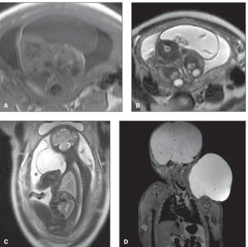
Additionally, MRI demonstrated in-volvement of the auricula in the case of fetus 2, and intrathoracic extension of the lesion towards the upper mediastinum in the case of fetus 1, which had not been demonstrated at US. By request of the assisting physician, the fetus 3 was submit-ted to a second MRI study after one week of life (Figure 2D). No other structure was involved by the lesion as compared with the prenatal MRI and obstetric US.
In the three cases, airways involvement and associated chromosomal abnormalities were not observed, which has allowed a safe surgical planning and a good progno-sis. All the three lesions were successfully resected without any impairment to the future life of the neonates (Figure 3).
DISCUSSION
man-301 Fetal cervical lymphangioma: correlation between MRI and US
Radiol Bras. 2009 Set/Out;42(5):299–302
agement of pregnant patients, representing significant medical and ethical dilemmas(1).
The knowledge on the specific types of tumors and respective biological character-istics is necessary for an appropriate
coun-seling and management of these patients(1).
Lymphangiomas are congenital malforma-tions of the lymphatic system caused by a failure in the drainage from the primordial lymphatic sac(1,3). This failure leads to an
enlargement of lymphatic channels, this theory being the one most commonly ac-cepted, that of cystic hygroma. There are other theories regarding lymphangiomas, but the authors have opted for considering that of cystic hygromas, since this is the entity approached by the present study. There are four histological types of lym-phangiomas: cystic hygroma, cavernous lymphangioma, capillary lymphangioma or lymphangioma simplex and vasculolym-phatic malformation. All these types are formed by lymphatic channels with endot-helial lining separated by a conjunctive tis-sue stroma(1). Frequently a combination of
these four histological types may be found in a single lesion, and the difference among them is based merely on the size of lym-phatic spaces and on their composition(1).
Cystic hygroma is the most common type o lymphangioma and consists of huge cystic dilatations of the lymphatic space, occurring in the posterior cervical space in 70% of cases, 63% at left(1–3). Only 5% of
these lesions are located below the dia-phragm(3). No sex predominance is
re-ported, usually occurring in children less than two years of age(2). These lesions
present an infiltrative nature, usually invad-ing adjacent structures (1,2), extending from
Figure 1. Sonographic findings of cystic hygromas. On A, axial US demonstrating heterogeneous septate lesion (arrows), affecting the cervical region (fetus 2), predominantly at left. On B, 3D US image of the same fetus demonstrating cervical mass.
A B
Figure 2. MRI findings of cystic hygromas. On A (fetus 2), axial, T1-weighted sequence demonstrating heterogeneous lesion with predominantly hypointense signal. On B, axial, T2-weighted sequence show-ing lesion with cystic content (hyperintense signal) with septations inside. On C (fetus 3), parasagittal T2-weighted sequence demonstrating hyperintense lesion with septation inside. On D, postnatal, coro-nal MRI T2-weighted sequence (fetus 3) confirming the previously described findings.
A B
C D
Figure 3. On A, image of the newborn 3 imme-diately before the surgery, demonstrating volumi-nous cystic hygroma at left. On B, image of the same newborn immediately after the surgery.
A
302
Antunes EG et al.
Radiol Bras. 2009 Set/Out;42(5):299–302
the posterior cervical triangle to the mouth floor and tongue in the majority of cases, presenting extension towards the upper mediastinum in 10% of cases(1). In most of
cases, the condition is asymptomatic and the size of the lesions is variable(2). Their
growth is slow, but these lesions may present a rapid increase in size in cases of hemorrhage or trauma(2). Large masses may
cause airways compression resulting in death(1). Small to middle-sized lesions may
regress along the gestation(10). A correct
prenatal diagnosis is extremely important, considering that, although cystic hygromas may be isolated malformations(1), these
le-sions are frequently associated with other chromosomal abnormalities, such as Turner syndrome and Down syndrome(1,2,10), that
should be immediately investigated in case of suspicious diagnosis(8). In cases of
iso-lated presentation, lymphangiomas have a good prognosis and most of times should be surgically resected.
US is the method of choice for screen-ing fetal malformations(11). Most of times,
cystic hygroma presents like a predomi-nantly cystic, multilocular mass with septa of variable thickness(2), and rarely with
some solid component(1). The most
echo-genic portion of the lesion correlates with groups of small and abnormal lymphatic channels(2). In the three cases evaluated in
the present series, the lesions were cystic hygromas, two of them predominantly cys-tic with thin septations inside, and one het-erogeneous. All of the lesions were located in the posterior cervical region, two of them at left and one at right, with mean size of 7.0 × 2.3 × 5.2 cm (lateral × transverse × anteroposterior).
MRI has been recognized as a useful complementary method, especially in cases where US results are dubious or undeter-mined(5,7,11,12). The advantages inherent to
this imaging method include higher accu-racy in the evaluation of soft tissues be-cause of the better tissue contrast resolution besides the multiplanar capability(11),
play-ing a significant role in the evaluation of lesions extent, most of times providing a more comprehensive and better fetal
evalu-ation when performed in the sagittal plane(9).
MRI is a safe method in fetal evaluation after the first gestational trimester(6,13) and
provides additional information on lesions extent, demonstrating the presence or not of invasion of adjacent structures(13–15), and
allowing a more appropriate, safe and in-dividualized surgical planning. The most frequently observed pattern of cystic hygro-mas is that of a hygro-mass with iso- or hypoin-tense signal on T1-weighted sequences, depending on the protein content of the lymphatic fluid, and hyperintense on T2-weighted sequences(1,2). Fluids with high
protein content present hyperintense signal on T1-weighted sequences(1). Fluid level
may be observed in cases of hemorrhage within the lesion(1–3). In two cases of the
present study, the lesions were hypointense on T1-weighted sequences, and hyperin-tense on T2-weighted sequences, with thin septations inside. In the third case, the le-sion was heterogeneous with predominant hyperintense signal on T2-weighted se-quences. The relation between cystic hy-gromas and soft tissues of the neck is most clearly demonstrated at MRI(2). This fact
was confirmed by the present cases where MRI was more accurate than US in the evaluation of the lesions extent, demon-strating the invasion of adjacent structures, with extension towards the pinna of the fetus in one case, and towards the upper mediastinum in another one.
CONCLUSION
In the present three-case series evaluat-ing cervical lymphangiomas, the authors considered that MRI was complementary to US, with a similar role in the evaluation of the lesions site, size and content. However, MRI demonstrated to be superior in the correct and more accurate demonstration of lymphangiomas extent, allowing an appro-priate and individualized surgical planning. After the analysis of the three cases of cys-tic hygromas by US and MRI, the authors have concluded that MRI plays a signifi-cant role as an ally of obstetric US in the evaluation of fetal lesions because of its
higher accuracy in the determination of these tumors extent and adjacent structures invasion, allowing a better surgical plan-ning and, therefore improving the quality of life of these patients.
REFERENCES
1. Woodward PJ, Sohaey R, Kennedy A, et al. A comprehensive review of fetal tumors with patho-logic correlation. Radiographics. 2005;25:215– 42.
2. Koeller KK, Alamo L, Adair CF, et al. Congeni-tal cystic masses of the neck: radiologic-patho-logic correlation. Radiographics. 1999;19:121– 46.
3. Kaminopetros P, Jauniaux E, Kane P, et al. Pre-natal diagnosis of an extensive fetal lymphan-gioma using ultrasonography, magnetic reso-nance imaging and cytology. Br J Radiol. 1997; 70:750–3.
4. Levine D, Barnes PD, Edelman RR. Obstetric MR imaging. Radiology. 1999;211:609–17.
5. Coakley FV, Hricak H, Filly RA, et al. Complex fetal disorders: effect of MR imaging on manage-ment – preliminary clinical experience. Radiol-ogy. 1999;213:691–6.
6. Shinmoto H, Kashima K, Yuasa Y, et al. MR im-aging of non-CNS fetal abnormalities: a pictorial essay. Radiographics. 2000;20:1227–43.
7. Weinreb JC, Lowe TW, Santos-Ramos R, et al. Magnetic resonance imaging in obstetric diagno-sis. Radiology. 1985;154:157–61.
8. Hertzberg BS, Bowie JD, Carroll BS, et al. Normal sonographic appearance of the fetal neck late in the first trimester: the pseudomembrane. Radiology. 1989;171:427–9.
9. McCarthy SM, Filly RA, Stark DD, et al. Obstet-rical magnetic resonance imaging: fetal anatomy. Radiology. 1985;154:427–32.
10. Sanders RC, Blakemore K. Lethal fetal anoma-lies: sonographic demonstration. Radiology. 1989;172:1–6.
11. Kubik-Huch RA, Huisman TAGM, Wisser J, et al. Ultrafast MR imaging of the fetus. AJR Am J Roentgenol. 2000;174:1599–606.
12. TekÕam M, Ozyer U, McKinney A, et al. MR im-aging and ultrasound of fetal cervical cystic lym-phangioma: utility in antepartum treatment plan-ning. Diagn Interv Radiol. 2005;11:87–9.
13. Frates MC, Kumar AJ, Benson CB, et al. Fetal anomalies: comparison of MR imaging and US for diagnosis. Radiology. 2004;232:398–404. 14. Kathary N, Bulas DI, Newman KD, et al. MRI
imaging of fetal neck masses with airway com-promise: utility in delivery planning. Pediatr Radiol. 2001;31:727–31.