Angle Class III malocclusion with anteroposterior
and vertical discrepancy in the final stage of growth
Marcelo B. P de Arruda1
Angle Class III malocclusion is characterized by an anteroposterior dental discrepancy with or without anteroposterior and vertical skeletal changes. Patients usually seek orthodontic treatment because facial appearance is compromised in most cases. The present study describes the clinical case of a 12-year and 6-month-old girl in her final stage of pubertal growth presenting Class III malocclusion with anteropos-terior and vertical discrepancies. Initial treatment consisted of maxillary expansion using a Hass expander followed by the use of a Petit facemask for a minimum of 16 hours a day. During corrective treatment, Class III elastics were used to complement protraction. At the end of the treatment, skeletal discrepancy had improved, and the ANB angle increased from 0 to 2o. Angle Class III malocclusion, anterior
crossbite and open bite were corrected. This case was presented to the Committee of the Brazilian Board of Orthodontics and Facial Orthopedics (BBO) as part of the requisites to become a BBO Diplomate.
Keywords: Angle Class III malocclusion. Open bite. Maxilla.
How to cite this article: Arruda MBP. Angle Class III malocclusion with anteroposterior and vertical discrepancy in the final stage of growth. Dental Press J Orthod. 2017 May-June;22(3):109-18.
DOI: https://doi.org/10.1590/2177-6709.22.3.109-118.bbo
Submitted: March 15, 2017 - Revised and accepted: April 20, 2017
Contact address: Marcelo B. P de Arruda Rua Rio Grande do Sul, 805
Campo Grande/MS – CEP: 79.020-011 E-mail: [email protected] » Patients displayed in this article previously approved the use of their facial and
in-traoral photographs.
» The author reports no commercial, proprietary or financial interest in the products or companies described in this article.
1 Universidade Federal de Mato Grosso do Sul, Orthodontics Department,
(Campo Grande/MS, Brazil). Diplomate, Brazilian Board of Orthodontics and Facial Orthopedics.
DOI: https://doi.org/10.1590/2177-6709.22.3.109-118.bbo
INTRODUCTION
A healthy 12-year and 6-month-old girl presented for an initial clinical examination. Her main complaint was that her mandibular teeth were in an anterior po-sition in relation to her maxillary teeth, which com-promised her facial esthetics (Fig 1). According to her mother, the girl had undergone previous dental exami-nations, and all the other dentists had indicated surgery.
All the patient’s teeth were intact, but she had anterior crossbite and open bite and discrete tongue thrust. Ac-cording to the mother, malocclusion had a hereditary component of paternal origin.
DIAGNOSIS
The patient had both vertical and anteroposterior skeletal discrepancies, with an ANB angle of zero
de-A má oclusão de Classe III de de-Angle tem como característica uma discrepância dentária anteroposterior, que pode ou não estar acompa-nhada por alterações esqueléticas tanto no sentido anteroposterior quanto no vertical. O aspecto facial fica comprometido na maioria dos casos, levando o paciente a procurar o tratamento ortodôntico. O presente artigo descreve o caso clínico de uma paciente com doze anos e seis meses de idade, portadora de má oclusão de Classe III, com discrepância anteroposterior e vertical, em fase final de crescimento. O tratamento inicial consistiu de expansão maxilar com o disjuntor de Haas e utilização da máscara facial de Petit por no mínimo 16 horas/ dia. Na fase de tratamento corretivo, foram usados elásticos Classe III como complemento à tração reversa. Ao término do tratamento, obteve-se melhora na desarmonia esquelética, com aumento do ângulo ANB de 0o para 2o. Quanto ao padrão dentário, corrigiu-se a
relação de Classe III de Angle, o leve cruzamento entre os incisivos e a mordida aberta anterior. Esse caso foi apresentado à Diretoria do Board Brasileiro de Ortodontia e Ortopedia Facial (BBO) como parte dos requisitos para a obtenção do título de Diplomado pelo BBO.
Figure 1 - Baseline facial and intraoral photographs.
grees, overgrowth of the mandible and a vertical growth
pattern (SN.GoGn = 43o, FMA = 28o). Both the
max-illa, more markedly, and the mandible were retruded in relation to the cranial base, which resulted in a con-cave proile (SNA = 77o and SNB = 77o, convexity
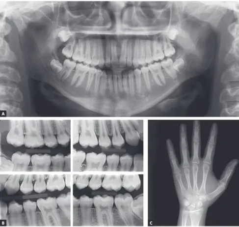
an-gle = -2o) (Figs 1 and 4). A wrist and hand radiograph
revealed that the patient was at the end of her pubertal growth spurt, and her bone age was close to 14 years, which may have led other dentists to indicate orthogna-thic surgery (Fig 3).
Dental examination (Fig 2) revealed that the pa-tient had Angle Class III malocclusion, maxillary and
mandibular incisor protrusion (1-NA = 7 mm and
1-NB = 8 mm), mandibular midline deviation of 2.5 mm to the let, negative overjet and overbite, both of -2 mm, mandibular anterior crowding of -2 mm and anterior crossbite. The patient had no CO-CR deviation.
The analysis of panoramic and interproximal radio-graphs (Fig 3) conirmed that she had all teeth, includ-ing third molars, and no abnormalities that might com-promise orthodontic treatment.
Figure 2 - Baseline casts.
Figure 3 - Baseline panoramic (A), interproxi-mal (B) and hand and wrist (C) radiographs.
B C
The following results were deined as the objectives of orthodontic treatment: to harmonize proile by improv-ing skeletal positionimprov-ing, to correct negative mandibular discrepancy and to retract mandibular incisors; to expand the maxilla to ensure a more efective protraction and to maintain facial height and prevent its increase. The treat-ment also included the restoration of ANB balance and the correction of the Class III relation. As the patient’s mandibular growth was limited, the plan included the preservation of cranial base positioning to avoid increas-ing the mandibular plane angle. For that purpose, the fundamental role of cooperation — particularly in the use of Class III elastics and the Petit facemask for protrac-tion — was emphasized for the patient and her family. Midline correction was also planned. Finally, treatment was expected to signiicantly improve esthetics, as well as dental and skeletal patterns.
TREATMENT PLAN
A two-phase treatment plan was prepared. The irst phase consisted of maxillary expansion using a Hass ex-pander, followed by the use of the Petit facemask for a minimum of 16 hours a day. In the second phase, Class III
elastics would be used as a complement to protraction; in case the response was not positive, orthognathic surgery might still be used as an alternative treatment.
TREATMENT PROGRESSION
Initially, a Haas expander was placed in the maxillary arch for rapid palate expansion for 21 days. Ater that, the protraction facemask was connected to the same Haas ex-pander, with the center of resistance placed at the canines, and application of a force of 350 g. The patient should wear the facemask for at least 16 hours a day. A ixed standard Edgewise appliance was placed in the maxilla (slot = 0.022 x 0.028-in), and 0.014-in, 0.016-in, 0.018-in and 0.020-in wires were used for alignment and leveling.
Six months later, the Haas expander was removed, and ixed appliance placement was completed: double tubes were bonded to the maxillary irst molars and single tubes, to the maxillary second molars. Alignment and leveling were achieved using 0.018-in and 0.020-in wires with e-loops between the maxillary lateral 0.020- in-cisors and the canines, to which the facemask elastics were connected. The patient wore the facemask only at night, for an average of 10 to 12 hours a day.
Eleven months later, the use of the facemask was discontinued, and control with Class III elastics start-ed. A space-closing archwire was produced using a bull loop. For completion, a rectangular 0.019 x 0.026-in archwire was placed, and Class III (1/4-in light) elastics were used for 12 hours to improve intercuspation, in addition to vertical elastics (1/8-in heavy) placed in the premolar region, which should be used during sleep. Ater the ixed appliance was removed, a wraparound retainer was adapted to the mouth.
Six months ater the beginning of the treatment, a ixed standard Edgewise (0.022 x 0.028-in) appliance was placed in the mandibular arch. Simple tubes were bonded to the irst molars and lower tubes, to the sec-ond molars, and alignment and leveling were achieved with stainless steel 0.014-in, 0.016-in, 0.018-in and 0.020-in wires and a space-closing archwire with a 0.019 x 0.025-in bull loop. For completion, a rect-angular 0.019 x 0.026-in archwire was used. Class III (¼-in light) elastics were used for 12 hours to improve intercuspation, and vertical elastics (1/8-in heavy) in the premolar region were used during sleep. Ater the ixed appliance was removed, a 3 x 3 retainer made with 0.7-mm wire was bonded.
TREATMENT RESULTS
The examination of inal records revealed that the initially planned objectives were achieved. In the max-illa, anterior protraction was achieved with the use of the Petit facemask, which improved SNA angle from
77 to 80o, although the patient had already reached
the inal stage of her pubertal growth spurt. The pat-tern remained vertical because the ‘y’ axis remained at
60o from the beginning to the end of the treatment.
Maxillary intermolar distance, 56 mm from the begin-ning to the end of the treatment, was preserved despite initial palate expansion. The analysis of dental pattern revealed that the 1.NA angle improved, as there was a
reduction from 29 to 22o and changes in the linear
po-sitioning of incisors (1-NA), which went from 7 mm to 8 mm (Table 1). The changes were beneicial and improved the anteroposterior maxilla-mandible rela-tion, as well as the inter-relation between maxillary and mandibular incisors.
In the mandible, there was a discrete change in the anteroposterior position of the cranial base, with a slight anterior movement followed by backward movement of incisors. A decrease of 1-NB from 8 mm to 6 mm was probably a result of the use of Class III elastics during treatment. This also led to a discrete improvement of skeletal discrepancy, with an increase of the ANB angle
from 0 to 2o (Table 1). Angle Class III malocclusion,
crossbite between incisors and anterior open bite were corrected (Figs 5 and 6). Figure 7 shows the successfully achieved parallel position of the roots. A recommenda-tion was made for extracrecommenda-tion of third molars.
Figure 6 - Final casts.
Figure 7 - Final panoramic (A) and interproximal (B) radiographs.
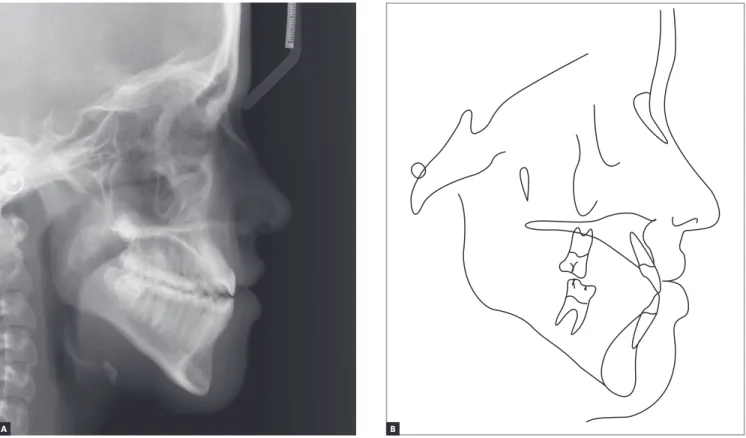
Figure 8 - Final cephalometric profile radiograph (A) and cephalometric tracing (B).
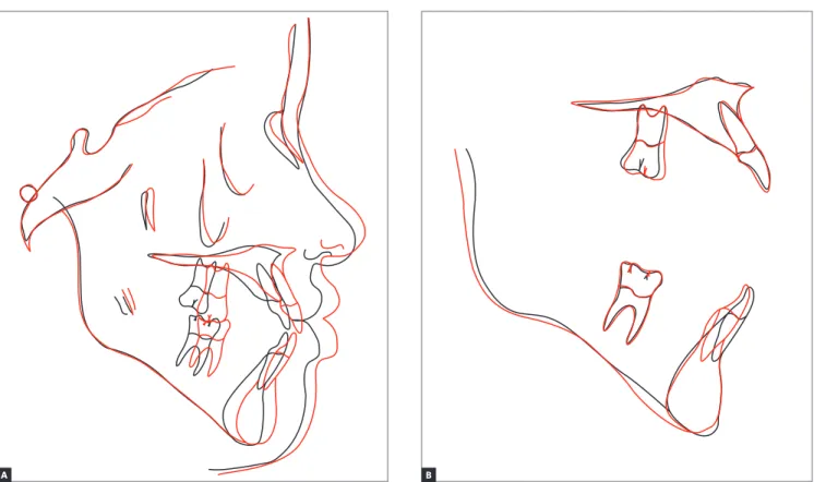
Figure 9 - Total (A) and partial (B) superimpositions of baseline (black) and final (red) cephalometric tracings. B
B A
Table 1 - Baseline (A) and final (B) cephalometric values.
Measurements Normal A B A/B Dif.
Skeletal pattern
SNA (Steiner) 82o 77o 80o 3
SNB (Steiner) 80o 77o 78o 1
ANB (Steiner) 2o 0o 2o 2
Wits (Jacobson) ♀ 0 ±2 mm
♂ 1 ±2 mm 0mm 2mm 2
Angle of convexity (Downs) 0o -2o 4o 6
Y-axis (Downs) 59o 60o 60o 0
Facial angle (Downs) 87o 89o 88o 1
SN-GoGn (Steiner) 32o 43o 42o 1
FMA (Tweed) 25o 28o 30o 2
Dental pattern
IMPA (Tweed) 90o 87o 88o 1
1.NA (degrees) (Steiner) 22o 29° 22o 7
1-NA (mm) (Steiner) 4 mm 7 8 1
1.NB (degrees) (Steiner) 25o 25o 28o 3
1-NB (mm) (Steiner) 4 mm 8mm 6mm 2
1
1- Interincisal angle (Downs) 130o 126o 128° 2
1-APo (Ricketts) 1 mm 6mm 6mm 0
Profile
Upper lip — S-line (Steiner) 0 mm -2mm 2mm 4
Lower lip — S-line (Steiner) 0 mm 1mm 4mm 3
FINAL CONSIDERATIONS
Angle Class III malocclusion, based on an anteropos-terior dental relation, is more serious when associated with skeletal discrepancies resulting from maxillary de-iciency, mandibular excess, or a combination of both. These changes may compromise facial proile. Treat-ment planning requires the use of lateral radiographs and other routine radiographic studies, as well as the evaluation of dental characteristics using clinical exami-nation and analysis of diagnostic casts. The analysis of genetic factors should take into consideration not only facial characteristics — such as cephalometric character-istics of parents, siblings and other relatives —, but also information about possible previous interventions that
other members of the family might have undergone2.
Treatment options to correct this anomaly involve several factors. When the patient has not reached pu-bertal growth spurt, an early intervention is indicated, with the use of a facemask for maxillary protraction,
usually together with palatal expansion.3-7
Palatal expansion8 is essential when a facemask is
used, as it favors the achievement of a more anterior placement of the maxilla and improves the relation with the mandible, resulting in satisfactory occlusion.
However, patient collaboration is fundamental9,10,11.
patient cooperation. To stimulate cooperation, the patient was told that the facemask would substan-tially improve her esthetic and facial profile.
The analysis of dental characteristics revealed that the patient had Class III molar and canine rela-tions, and that these relations were more marked in the left side. At the end of the treatment, the patient was advised to get her third molars extracted in the future. The records obtained at treatment comple-tion (Figs 7 and 8) and the superimposicomple-tions of ini-tial, final and control cephalometric tracings (Fig. 9) revealed that the result achieved after the removal of the orthodontic appliance was satisfactory, which illustrates the efficiency of the treatment that was planned and executed.
REFERENCES
1. Angle EH. Treatment of malocclusion of the teeth. 7th ed. Philadelphia: SSW; 1907.
2. Araújo EA, Araújo CR. Abordagem clínica não-cirúrgica no tratamento da má oclusão de Classe III. Rev Dental Press Ortod Ortop Facial. 2008;13(6):128-57. 3. Baccetti T, McGill JS, Franchi L, McNamara JA Jr, Tollaro I. Skeletal efects of
early treatment of Class III malocclusion with maxillary expansion and face-mask therapy. Am J Orthod Dentofacial Orthop. 1998 Mar;113(3):333-43. 4. Dale HC. Morphologic skeletal asymmetry, with a Class III skeletal
discrepancy, treated without surgical intervention. World J Orthod. 2005 Winter;6(4):391-7.
5. Jiang J, Lin J, Ji C. Two-stage treatment of skeletal Class III malocclusion during the early permanent dentition. Am J Orthod Dentofacial Orthop. 2005 Oct;128(4):520-7.
6. Kokich VG, Kokich VO. Congenitally missing mandibular second premolars:
clinical options. Am J Orthod Dentofacial Orthop. 2006 Oct;130(4):437-44. 7. Macey-Dare LV. The early management of Class III malocclusions using
protraction headgear. Dent Update. 2000 Dec;27(10):508-13. 8. Haas AJ. Rapid expansion of maxillary dental arch and nasal cavity by
opening the midpalatal suture. Angle Orthod. 1961;31(2):73-90. 9. Pangrazio-Kulbersh V, Berger JL, Janisse FN, Bayirli B. Long-term stability
of Class III treatment: rapid palatal expansion and protraction facemask vs LeFort I maxillary advancement osteotomy. Am J Orthod Dentofacial Orthop. 2007 Jan;131(1):7.e9-19.
10. Waring D, Henley E. Growth modiication treatment in Class III malocclusions: an orthodontic case report. Dent Update. 2006 Nov;33(9):546-8, 551-2, 554.