Neuropsychological differences
between frontotemporal lobar
degeneration and Alzheimer’s disease
Claudia Sellitto Porto
1, Valeria Santoro Bahia
2,
Sonia Maria Dozzi Brucki
2, Paulo Caramelli
3, Ricardo Nitrini
2Abstract – Memory impairment is the main clinical feature in Alzheimer disease (AD), whereas in frontotemporal lobar degeneration (FTLD) behavioral and language disorders predominate.Objectives: To investigate possible differences between the neuropsychological performance in FTLD and AD. Methods: Fifty-six AD patients (mean age=72.98±7.43; mean schooling=9.62±4.68; 35 women and 21 men), 17 FTLD patients (mean age=67.64±7.93; mean schooling=12.12±4.77; 9 women and 8 men), and 60 controls (mean age=68.90±7.48; mean school-ing=10.72±4.74; 42 women and 18 men) were submitted to a Dementia Rating Scale (DRS) and a comprehensive neuropsychological evaluation composed of tasks assessing attention, visuoperceptual abilities, constructive abilities, executive functions, memory and language. Results: DRS total score and subscales were not able to differentiate FTLD from AD patients. However, FTLD and AD patients showed statistically significant differences in performance in tests of verbal (Logical Memory, Rey Auditory Verbal Learning Test) and visual (Visual Reproduction, recall of the Rey Complex Figure) episodic memory, verbal immediate memory (Logical Memory), attention with interference (Trail Making Test – Part B), verbal fluency (semantic and phonemic) and concept formation (WCST). Conclusion: Con-trary to expectations, only a few tasks executive function tasks (Trail Making Test – Part B, F.A.S. and WCST) and two memory tests (verbal and visual episodic memory tests) were able to differentiate between FTLD and AD patients.
Key words: neuropsychological assessment, memory, executive functions, Alzheimer disease, frontotemporal lobar degeneration.
Diferenças neuropsicológicas entre degeneração lobar frontotemporal e doença de Alzheimer
Resumo – Comprometimento de memória é a principal característica clínica na doença de Alzheimer (DA), enquanto na degeneração lobar fronto-temporal (DLFT) alterações de linguagem e comportamentais são pre-dominantes. Objetivos: Investigar possíveis diferenças entre o desempenho neuropsicológico de pacientes com DLFT e pacientes com DA. Métodos: 56 pacientes com DA (média de idade=72,98±7,43; média de escolarida-de=9,62±4,68; 35 mulheres e 21 homens), 17 pacientes com DLFT (média de idade=67,64±7,93; média de es-colaridade=12,12±4,77; 9 mulheres e 8 homens) e 60 controles (média de idade=68,90±7,48; média de escola-ridade=10,72±4,74; 42 mulheres e 18 homens) foram submetidos à Escala de Avaliação de Demência (Dementia Rating Scale – DRS) e à extensa avaliação neuropsicológica composta de tarefas que examinam atenção, habili-dades visuais-perceptuais, habilihabili-dades construtivas, funções executivas, memória e linguagem. Resultados: O es-core total e as subescalas da DRS não foram capazes de diferenciar pacientes com DA de DLFT. Pacientes com DLFT e DA demonstraram diferenças estatisticamente significativa em testes de memória episódica verbal (Me-mória Lógica, Teste de Aprendizagem Verbal Auditiva de Rey) e visual (Reprodução Visual e evocação da Figu-ra Complexa de Rey), memória imediata verbal (Memória Lógica), atenção com interferência (Trail Making Test – Parte B) e fluência verbal (semântica e fonêmica). Conclusão: Ao contrário do esperado, somente algu-mas tarefas de função executiva (Trail Making Test – Parte B, F.A.S. e WCST) e dois testes de memória (testes de memória episódica verbal e visual) foram capazes de diferenciar pacientes com DLFT de pacientes com DA.
Palavras-chave: avaliação neuropsicológica, memória, funções executivas, doença de Alzheimer, degeneração lobar frontotemporal.
1PhD. Behavioral and Cognitive Neurology Unit, Department of Neurology of the University of São Paulo School of Medicine and Cognitive Disorders
Reference Center (CEREDIC), Hospital das Clínicas of the University of São Paulo School of Medicine, São Paulo (SP), Brazil. 2MD, PhD. Behavioral and
Cognitive Neurology Unit, Department of Neurology of the University of São Paulo School of Medicine and Cognitive Disorders Reference Center (CE-REDIC), Hospital das Clínicas of the University of São Paulo School of Medicine, São Paulo (SP), Brazil. 3MD, PhD. Behavioral and Cognitive Neurology
Unit, Department of Internal Medicine, Faculty of Medicine, Federal University of Minas Gerais, Belo Horizonte (MG), Brazil.
Claudia Sellitto Porto – Rua Itapeva, 378 / cj. 92 - 01332-000 São Paulo SP - Brazil. E-mail: [email protected]
Memory impairment is the most prominent deficit in
Alzheimer disease (AD). A more heterogeneous pattern of
cognitive impairment, however, is seen in frontotemporal
lobar degeneration (FTLD), a neurodegenerative disorder
characterized by progressive behavioral and/or language
disorders or semantic memory changes.
1Neary et al.
1distin-guished three variants of FTLD: the frontal variant of
fron-totemporal dementia (FTD), semantic dementia (SD) and
progressive non-fluent aphasia (PNFA). In FTD, behavioral
symptoms are predominant, while oral production and
se-mantic deficits are observed in PNFA and SD, respectively.
Clinical differentiation between FTLD and AD remains
a great challenge, especially in the clinical setting. Mendez
et al.
2demonstrated that neuropsychological evaluation
did not distinguish frontotemporal dementia (FTD) from
other causes of dementia while some studies advocate the
use of behavioral scales over neuropsychological tests to
differentiate\ FTD from AD patients.
3In a recent study, Liscic et al.
4investigated clinical and
psychometric differences between neuropathogically
con-firmed FTLD (without or with concomitant AD
pathologi-cal features) and AD, finding that behavioral and language
features were good discriminators between the two
condi-tions. However, FTLD patients or their relatives can also
report memory loss complaints, although – in most cases
– this is related to attention and working memory deficits.
The main objective of this study was to investigate
pos-sible differences between the performance of patients with
FTLD and AD on neuropsychological tests.
Methods
The study involved 73 patients (44 women and 29 men),
aged 50 to 84 years (mean=71.73±7.83), with schooling
ranging from 3 to 17 years (mean=10.21±4.79), attended
by members of the Behavioral and Cognitive Neurology
Unit of the Department of Neurology of the University of
São Paulo School of Medicine, in Brazil. All patients were
submitted to appropriate laboratory tests and to structural
neuroimaging (computed tomography (CT) or magnetic
resonance (MR) of the skull), the Mini-Mental State
Ex-amination (MMSE)
5,6and the Brief Cognitive Screening
Battery (BCSB).
7Information on performance in daily life
activities was obtained through the Pfeffer Functional
Ac-tivities Questionnaire,
8which was applied to an informant.
The probable AD group was composed of 56
individu-als, aged 54 to 84 years (mean=72.98±7.43), with schooling
ranging from 3 to 17 years (mean=9.62±4.68), comprising
35 women and 21 men. The clinical diagnosis of mild
de-mentia was based on the Diagnostic and Statistical Manual
of Mental Disorders, Third Edition, revised (DSM-III-R)
criteria;
9whereas the diagnosis of probable AD was based on
the National Institute of Neurological Disorders and
Com-municative Disorders and Stroke-Alzheimer’s Disease and
Related Disorders Association (NINCDS-ADRDA) criteria.
10The FTLD group was composed of 17 patients (SD=
3; PNFA=4; FTD=10), aged 50 to 80 years (mean
67.64±7.93), with schooling ranging from 4 to 16 years
(mean=12.12±4.77), 9 women and 8 men. The diagnosis
of FTLD was based on the criteria of Neary et al.
1The control group (60 subjects; mean age=68.90±7.48;
mean schooling=10.72±4.74; 42 women and 18 men) was
composed of spouses or consorts of the patients, or
volun-teers from the community, with no memory disorders and
groups
FTLD AD
controls
Trail Making Test - Part
B
500
400
300
200
100
0
-100
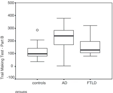
Figure 1. Performance among AD, FTLD patients and controls on
the Trail Making Test – Part B.
groups
FTLD AD
controls
Logical memory - immediate
50
40
30
20
10
0
-10
Figure 2. Performance among AD, FTLD patients and controls in
who were fully independent in terms of daily living
activi-ties. Subjects with neurological disease, history of
alcohol-ism, depression, or any other psychiatric disorder,
non-corrected visual or auditory disorders, motor disorders,
or users of psychotropic drugs that could affect cognitive
functions were excluded. Chronic diseases such as arterial
hypertension, diabetes mellitus and cardiac disorders, if
adequately controlled, were not criteria for exclusion. All
controls were submitted to the MMSE, BCSB and Memory
Complaint Questionnaire (MAC-Q)
11or to the Informant
Questionnaire on Cognitive Decline in the Elderly
(IQ-CODE),
12,13administered to an informant.
Patients and controls were submitted to the Dementia
Rating Scale
14-16and to a comprehensive
neuropsychologi-cal evaluation, which included the following tests: visual
and verbal memory tests (Visual Reproduction subtest of
the Wechsler Memory Scale – Revised (WMS-R),
17Rey
Complex Figure – delayed recall,
18Logical Memory subtest
(WMS-R),
17Rey Auditory Verbal Learning Test (RAVLT)
19),
constructive abilities (Block Design subtest – Wechsler
Adult Intelligence Scale (WAIS),
20Rey Complex Figure
copy
18), visual perception (Hooper Visual Organization
Test
21and Raven’s Progressive Matrices
22), language
(Bos-ton Naming Test)
23), and executive functions (Trail Making
Test versions A and B,
24Stroop Test,
24Wisconsin Card
Sort-ing Test (WCST)
24and phonemic verbal fluency (F.A.S.)
24).
The study was approved by the Research and Ethics
Committee of Hospital das Clínicas from the University of
São Paulo School of Medicine. All subjects who agreed to
participate signed a written informed consent.
Statistical analysis
In order to evaluate associations between the categorical
variables and the results, the Pearson Chi-Squared test was
performed. When the variables were continuous, the
com-parisons were made for two samples by the Mann-Whitney
test, and for more than two, by the Kruskall-Wallis test.
Alpha risk was considered to be less than or equal to
5% for type 1 error, and beta risk greater than or equal to
20% for type II error.
All statistical analysis was carried out using the Statistical
Package for the Social Sciences (SPSS) program, version 10.0.
Results
No differences related to schooling (p=0.105) or gender
(p=0.394) were found between control and patient groups,
but a statistically significant difference related to age was
observed (p=0.004).
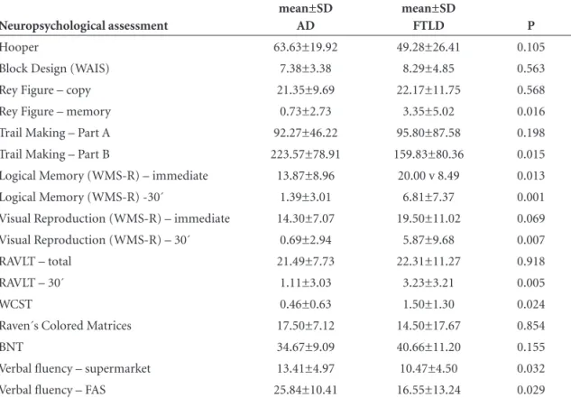
Table 1. Performance on neuropsychological tests in AD and FTLD patients.
Neuropsychological assessment
mean±SD AD
mean±SD
FTLD P
Hooper 63.63±19.92 49.28±26.41 0.105
Block Design (WAIS) 7.38±3.38 8.29±4.85 0.563
Rey Figure – copy 21.35±9.69 22.17±11.75 0.568
Rey Figure – memory 0.73±2.73 3.35±5.02 0.016
Trail Making – Part A 92.27±46.22 95.80±87.58 0.198 Trail Making – Part B 223.57±78.91 159.83±80.36 0.015 Logical Memory (WMS-R) – immediate 13.87±8.96 20.00 v 8.49 0.013 Logical Memory (WMS-R) -30´ 1.39±3.01 6.81±7.37 0.001 Visual Reproduction (WMS-R) – immediate 14.30±7.07 19.50±11.02 0.069 Visual Reproduction (WMS-R) – 30´ 0.69±2.94 5.87±9.68 0.007
RAVLT – total 21.49±7.73 22.31±11.27 0.918
RAVLT – 30´ 1.11±3.03 3.23±3.21 0.005
WCST 0.46±0.63 1.50±1.30 0.024
Raven´s Colored Matrices 17.50±7.12 14.50±17.67 0.854
BNT 34.67±9.09 40.66±11.20 0.155
Verbal fluency – supermarket 13.41±4.97 10.47±4.50 0.032 Verbal fluency – FAS 25.84±10.41 16.55±13.24 0.029
There was also a significant difference between the
per-formance of both patient groups and controls on the DRS,
in total score (p<0.001) and all subscales. No differences
between AD and FTLD patients were seen in total score
(p=0.881) or on the DRS subscales.
Regarding the comparison between AD and FTLD
patients, there were statistically significant differences in
visual and verbal episodic memory tasks, in verbal fluency,
as well as in tests evaluating attention and executive
func-tions (Table 1).
AD patients showed worse performance than FTLD
patients in immediate recall on the Logical Memory test
(p=0.013) and on the Trail Making Test – Part B (p=0.015).
Discussion
Memory impairment is the hallmark feature of AD
while in FTLD episodic memory remains relatively
pre-served,
25,26that could explain the results of our study
dem-onstrating that verbal and visual episodic memory tests
were able to differentiate between the two patient groups.
Wicklund et al.
26compared AD patients, frontal variant of
FTLD patients, and controls on two memory tests: story
memory and word list recall. The results demonstrated
pa-tients with frontal variant of FTLD recalled more
informa-tion from the story and more words after a delay than AD
patients. Heidler-Gary et al.
27also demonstrated that AD
was characterized by severe impairment in verbal learning,
delayed recall and that two variants of FTLD (FTD and
PNFA) were characterized by relatively normal scores on
verbal learning and recall.
AD and FTLD patients performed differently in
imme-diate story recall (the Logical Memory subtest of WMS-R),
with greater impairment shown by AD patients. Wicklund
et al.
26found that individuals with the behavioral variant
of FTLD were able to immediately recall more information
from the story than AD patients. Studies
28,29have shown
low performance in verbal short- term memory tasks in
AD. This deficit is attributed to problems in attention,
co-ordination and integration processes stemming from
im-paired executive control processes. Lezak
30reported that
immediate story recall remained stable in middle age and
declined progressively thereafter. In our study age
differ-ences between groups may have influenced the results.
Alescio-Lautier et al.
31affirmed that certain attentional
mechanisms are impaired early in AD. Patients with AD
showed greater impairment on the divided attention test,
evaluated through the Trail Making Test – Part B, than
FTLD and controls. Belleville et al.
32demonstrated that
mild AD patients presented severe impairment in divided
attention, manipulation capacities and inhibition.
Only two executive function tasks, both of which
as-sess executive function, were able to differentiate AD from
FTDL patients: phonemic verbal fluency and WCST. In the
study by Liscic et al.,
4the FTLD group performed
signifi-cantly worse on word fluency than the AD group.
In our study, the DRS, using either the total or
sub-scale scores, was not effective in discriminating between
AD and FTLD patients, suggesting that this scale is not
useful to differentiate these two groups. This finding was
somewhat unexpected because the subscales of the DRS
evaluate specific items of cognition such as
Initiation/Per-severation (I/P) that are usually more disturbed in FTLD
or memory, which is more involved in AD. On the I/P
sub-scale, the semantic verbal fluency test accounted for 75% of
the total score of this subscale. Verbal fluency impairment
is associated to initial stages of AD and also to FTLD.
25,33The tasks of the Memory subscale proved to easy or poor
to differentiate between AD and FTLD groups.
In this study, verbal and visual episodic memory tests
were better discriminators of the two groups whereas
com-prehensive neuropsychological evaluation was unable to
clearly distinguish AD from FTLD individuals.
References
1. Neary D, Snowden JS, Gustafson L, et al. Frontotemporal lo-bar degeneration. A consensus on clinical diagnostic criteria. Neurology 1998;51:1546-1554.
2. Mendez MF, Shapira JS, McMurtray A, Licht E, Miller B. Ac-curacy of the clinical evaluation for frontotemporal dementia. Arch Neurol 2007;64:830-835.
3. Bahia VS. Underdiagnosis of frontotemporal lobar degenera-tion in Brazil. Dement Neuropsychol 2007;1:361-365. 4. Liscic RM, Storandt M, Cairns NJ, Morris JC. Clinical and
psychometric distinction of frontotemporal and Alzheimer dementias. Arch Neurol 2007;64:535-540.
5. Folstein MF, Folstein SE, McHugh PR. “Mini-mental state”. A practical method for grading the cognitive state of patients for the clinician. J Psychiatr Research 1975;12:189-198. 6. Brucki SMD; Nitrini R; Bertolucci PHP; Caramelli P;
Oka-moto IH. Normas sugeridas para o uso do Mini-Exame do Estado Mental (MEEM) em nosso meio. Arq Neuropsiquiatr 2003;60:46-47.
7. Nitrini R, Caramelli P, Porto CS, et al. Avaliação Cognitiva Breve no diagnóstico de doença de Alzheimer leve. Arq Neu-ropsiquiatr 2005;63:27.
8. Pfeffer RI, Kusosaki TT, Harrah Jr CH, Chance JM, Filos S. Measurement of functional activities in older adults in the community. J Gerontol 1982;37:323-329.
9. American Psychiatric Association. Diagnostic and Statistical Manual of Mental Disorders. 3rd ed. Ver. Washington, DC:
American Psychiatric Association, 1987.
Stadlan EM. Clinical Diagnosis of Alzheimer’s disease: report of the NINCDS-ADRDA work group under the auspices of department of health and human services task force on Al-zheimer’s disease. Neurology 1984;34:939-944.
11. Mattos P, Lino V, Rizo L, et al. Memory complaints and test performance in health elderly persons. Arq Neuropsiquiatr 2003;61:920-924.
12. Jorm AF. A short-form of the Informant Questionnaire on Cognitive Decline in the Elderly (IQCODE): development and cross-validation. Psychological Medicine 1994; 24:145-153. 13. Bustamante SEZ, Bottino CMC, Lopes MA, et al. Instrumen-tos combinados na avaliação de demência de idosos. Arq Neu-ropsiquiatr 2003;61:601-606.
14. Mattis S. Mental Status Examination for Organic Mental Syn-drome in the Elderly Patient. In: Bellak L, Karasu TB, editors. Geriatric Psychiatry. A Handbook for Psychiatrists and Prima-ry Care Physicians. New York: Grune & Stratton;1976:77-121. 15. Mattis S. Dementia Rating Scale. Professional Manual.
Flor-ida: Psychological Assessment Resources, Inc; 1988. 16. Porto CS, Charchat-Fichman H, Caramelli P, Bahia VS,
Ni-trini R. Brazilian version of the Mattis Dementia Rating Scale. Diagnosis of mild dementia in Alzheimer’s disease. Arq Neu-ropsiquiatr 2003;61:339-345.
17. Wechsler D. Wechsler Memory Scale. Manual The Psychologi-cal Corporation Harcourt Brace Jovanovich; 1987.
18. Rey A. Figuras Complexas de Rey. São Paulo: Casa do Psicólo-go; 1998.
19. Diniz LFM, Cruz MF, Torres VM, Consenza RM. O teste de aprendizagem auditivo-verbal de Rey: normas para uma população brasileira. Rev Bras Neurol 2000;36:79-83. 20. Wechsler D. Test de Inteligencia para adultos (WAIS). Manual.
2a Ed. Buenos Aires, Argentina: Editorial Paidos;1993.
21. Hooper Visual Organization Test (VOT) Manual. Western Psychological Services. 1983.
22. Raven JC, Raven J, Court JH. Manual Matrizes Progressivas Col-oridas. São Paulo: Casa do Psicólogo. Divisão Editorial; 1988. 23. Radanovic M, Mansur LL, Scaff M. Normative data for
the Brazilian population in the Boston Diagnostic Aphasia
Examination: influence of schooling. Braz J Med Biol Res 2004;37:1731-1738.
24. Spreen O, Strauss E. A Compendium of Neuropsychological Tests. Administration, Norms, and Commentary. Second Edi-tion. Oxford University Press; 1998.
25. Hodges JR, Patterson K, Ward R, et al. The differentiation of semantic dementia and frontal lobe dementia (temporal and frontal variants of frontotemporal dementia) from early Alzheimer’s disease: a comparative neuropsychological study. Neuropsychology 1999;13:31-40.
26. Wicklund AH, Johnson N, Rademaker A, Weitner BB, Wein-traub S. Word list versus story memory in Alzheimer disease and frontotemporal dementia. Alzheimer Dis Assoc Disord 2006;20:86-92.
27. Heidler-Gary J, Gottesman R, Newhart M, Chang S, Ken L, Hillis AE. Utility of behavioral versus cognitive measures in differentiating between subtypes of frontotemporal lobar degeneration and Alzheimer’s disease. Dement Geriatr Cogn Disord. 2007;23:184-93.
28. Caza N, Belleville S. Reduced short-term memory capacity in Alzheimer’s disease: The role of phonological, lexical, and semantic processing. Memory 2008;16:341-50.
29. Peters F, Majerus S, Olivier L, van der Linden M, Salmon E, Collette F. A multicomponent exploration of verbal short-term storage deficits in normal aging and Alzheimer’s disease. J Clin Exp Neuropsychol. 2007;29:405-417.
30. Lezak MD. Neuropsychological Assessment. 3a Edição. New
York: Oxford University Press; 1995.
31. Alescio-Lautier B, Michel BF, Herrera C, et al. Visual and vi-suospatial short-term memory in mild cognitive impairment and Alzheimer disease: role of attention. Neuropsychologia. 2007;45:1948-1960.