ARTICLE DOI: 10.1590/0004-282X20130038
Cognitive performance of neuromyelitis optica
patients: comparison with multiple sclerosis
Desempenho cognitivo de pacientes com neuromielite óptica: comparação com
esclerose múltipla
Sandra Vanotti1,2, Evangelina Valeria Cores1,2, Barbara Eizaguirre1,2, Luciana Melamud1,2, Raúl Rey2,Andrés Villa1,2
Neuromyelitis optica (NMO) is an uncommon disease that afects the optic nerves and spinal cord1. he recent
iden-tiication of a speciic antibody for aquaporin 4 (AQP4) wa-ter channel at the blood-brain barrier in NMO patients (IgG-NMO) makes it the irst central nervous system autoimmune channelopathy2.
Little is known about cognitive dysfunction in NMO, its frequency, and its relationship with clinical variables. Blanc et al.3 reported impairment of attention, speed of information
processing, and word generation in NMO patients. Nilsson et al.4 also found cognitive impairment (CI) in patients
diag-nosed with isolated optic neuritis (ON) from 24 to 31 years
earlier. Finally, He et al.5 evidenced cognitive alterations in
NMO patients after an acute relapse, and encountered an as-sociation between neuropsychological performance and rou-tine activities6.
During the past 20 years, numerous researches have indi-cated frequent CI in multiple sclerosis (MS) patients. Working memory, attention, verbal luency, and speed of information processing are often afected7-9.
he diferences between MS and NMO neuropathology suggest distinct cognitive patterns, but only one previous study compared these patients and found no signiicant vari-ations in cognitive measures3.
1Neuroimmunology and Electrophysiology Section, J. M. Ramos Mejia Hospital, Buenos Aires, Argentina;
2University Centre of Neurology “Dr. Jose Maria Ramos Mejia” School of Medicine, Buenos Aires, Argentina.
Correspondence: Sandra Vanotti; Urquiza, 609; C1221ADC, Buenos Aires, Argentina; E-mail: [email protected]
Conflict of interest: There is no conflict of interest to declare.
Received 09 November 2012; Received in final form 01 November 2012; Accepted 09 November 2012.
ABSTRACT
The aim of the present research was to investigate cognitive pattern of patients with neuromyelitis optica (NMO) and to compare it with mul-tiple sclerosis (MS) patients’ performance. Methods: Fourteen NMO, 14 relapsing remitting mulmul-tiple sclerosis (RRMS), and 14 healthy control patients participated in the investigation. Neuropsychological functions were evaluated with the Brief Repeatable Neuropsychological Battery for MS; Symbol Digit Modalities Test; Digit Span; and Semantic Fluency. Results: Fifty-seven percent of NMO patients and 42.85% of the MS ones had abnormal performance in at least two cognitive tests. The NMO Group showed abnormal performance in verbal fluency, verbal and visual memories, with greater attention deficits. NMO patients outperformed healthy control in the paced auditory serial addition test (PASAT). However, no difference was found between NMO and RRMS patients. Conclusions: The NMO Group showed more dysfunction in attention and verbal fluencies than in verbal and visual memories. When compared with the MS patients, a similar dysfunction pattern was found.
Key words: neuropsychology, neuromyelitis optica, depression, multiple sclerosis, autoimmune diseases.
RESUMO
O objetivo da presente pesquisa foi investigar o padrão cognitivo de pacientes com neuromielite óptica (NMO) e compará-lo com o desem-penho de pacientes com esclerose múltipla (EM). Métodos: Quatorze pacientes com NMO, 14 com esclerose múltipla recorrente remitente (EMRR) e 14 participantes do Controle saudáveis participaram da presente investigação. As funções neuropsicológicas foram avaliadas com a Bateria Breve de Testes Neuropsicológicos de Rao, Teste Símbolo Digit e a Fluência Semântica. Resultados: Cinquenta e sete por cento dos pacientes com NMO e 42,85% daqueles com EM apresentaram desempenho anormal em pelo menos dois testes cognitivos. O Grupo NMO apresentarou desempenho anormal na fluência verbal e nas memórias visual e verbal, com maiores déficits de atenção. Pacientes com NMO superaram os controles saudáveis em PASAT. No entanto, não foi encontrada diferença entre os pacientes com NMO e aqueles com EMRR. Conclusões: O Grupo NMO mostrou mais disfunção nas fluências de atenção e verbais do que nas memórias verbal e visual. Quando compa-rados com os pacientes com EM, um padrão de disfunção semelhante foi encontrado.
he aim of the present study was to describe the cogni-tive pattern of NMO patients and compare it with that of MS ones, considering pathophysiological mechanisms, diagnos-tic criteria, evolution, and treatments of these diseases.
METHODS
Participants
Fourteen patients diagnosed with NMO, 14 with relapsing remitting MS (RRMS), and 14 healthy controls were evaluat-ed. Twelve females and 12 males were recruited in all groups. NMO patients were diagnosed in the Neurology Service at the J. M. Ramos Mejia Hospital, Buenos Aires, Argentina. he RRMS patients were selected, considering gender, age, and ed-ucation to match them with NMO participants: (±) 3 in age and (±) 2 in education. he inclusion or exclusion criteria were: conirmed diagnosis as deined by Wingerchuk’s10 in the NMO
Group, and as by McDonald’s of RRMS; 18 years or older; being in remission period; absence of psychiatric illness; history of alcohol or drug abuse; no physical disability that could impair performance of tests; no uncontrolled systemic disease or that could cause cognitive impairment. Healthy controls without history of neurological disease and mini mental state exami-nation (MMSE) scores >26 were also recruited.
Standard protocol approvals, registrations, and patient consents
his study was approved by the J. M Ramos Mejia Hospital Bioethical Committee, and all participants signed an in-formed consent.
Outcome measures
Physical disability was measured through the Expanded Disability Status Scale (EDSS)11. he Brief Repeatable Battery
of Neuropsychology Tests (BRB-N)12, translated into Spanish
and culturally adapted to this Latin American population9,
was administered by a trained neuropsychologist. he pro-cedure started with six learning trials from a brief version of the Selective Reminding Test (SRT), from which measures of learning (long-term storage or LTS) and consistency of recall (consistent long-term retrieval or CLTR) were derived. Next, a visual/spatial memory test, the 7/24 Spatial Recall Test, was administered. he Rao version of the Paced Auditory Serial Addition Task (PASAT) was then completed, requiring
rapid calculations and divided attention. he Word List Generation (WLG) test and the oral version of the Symbol Digit Modalities Test (SDMT)13 were administered prior to
the delayed recall testing.
he test 7/24 Spatial Recall and SDMT were not managed in three patients due to visual impairments.
Beck’s Depression Inventory II (BDI-II)14 was carried out
as a depression measure.
Procedure
All patients underwent a complete neurological exami-nation. he neurologist scored the EDSS and registered de-mographic and clinical data, such as age, education, and du-ration of the disease. NMO ones underwent brain magnetic resonance imaging (MRI) examination with and without con-trast (sequences T1, T2, and FLAIR, performed in a 1.5-Tesla resonator according to the MS protocol of the consortium)15. Statistical analysis
Data analysis was performed using the Statistical Package for the Social Sciences (SPSS), version 16.0 (IBM). Inferential calculations were performed using analysis of variance (ANOVA), Pearson’s r correlations, and logistic regression anal-ysis. he level chosen for alpha was 0.05. Patients with two or
more afected domains (verbal memory – SRT; visual memo-ry – 7/24 visual spatial; verbal luency – controlled oral world association (COWA); attention – PASAT) were considered cog-nitively impaired. Scores in a cognitive domain below the ifth percentile of normal values9 were considered abnormal.
RESULTS
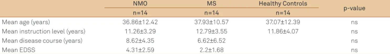
No signiicant diferences were found between the groups with respect to age (p=968) or education (p=553). Furthermore, the NMO and MS Groups did not difer in the EDSS (p=0.114) or disease duration (p=0.367). Data are shown in Table 1.
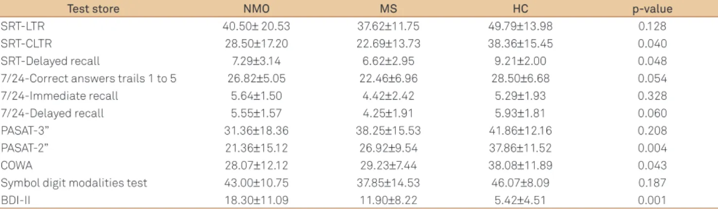
Regarding the ANOVA, there was a major signiicant ef-fect among the groups on SRT-CLTR (p=0.04), SRT delayed recall (p=0.048), PASAT-2 seconds (p=0.004), and COWA (p=0.043). Following a posteriori Tukey’s tests, the NMO pa-tients performed signiicantly worse than the controls in PASAT-2 (p=0.003). he MS Group had a worse performance than the controls on SRT-LTR (p=0.034), SRT delayed recall
NMO MS Healthy Controls
p-value
n=14 n=14 n=14
Mean age (years) 36.86±12.42 37.93±10.57 37.07±12.39 ns
Mean instruction level (years) 11.26±3.29 12.79±3.55 11.86±4.07 ns
Mean disease course (years) 8.62±4.35 6.62±6.52 ns
Mean EDSS 4.31±2.59 2.2±1.68 ns
Table 1. Demographic and clinical data of neuromyelitis optica and multiple sclerosis patients and healthy controls.
(p=0.048), and 7/24 Correct Answers Trails 1 to 5 (p=0.048). However, no diferences were found between the NMO and MS Groups, which can be seen in Table 2.
When comparing the performance of NMO patients with local normative scores, it was found that 35.71% of them showed abnormal performance in verbal memory, 9.09% had some problem in visual memory, 64.28% had in attention, and 48.85% presented abnormal performance in verbal lu-ency. In the MS Group, 61.53% of the participants showed ab-normal performance in verbal memory, 61.53% had problem in visual memory, 33.33% in attention, and 38.46% showed abnormal performance in verbal luency.
Characteristic of the Neuromyelitis optica Group
Considering the amount of impaired tests (35.71%), the sum of these percentages exhibited no alteration (7.14%), had one impaired test (28.57%), had two impaired tests, and (28.57%), had three or more impaired tests.
Regarding association between MRI and the amount of impaired tests, ive out of eight patients with a normal MRI
and four out of six with lesions had at least one cognitive im-paired test. Comparison of cognition among patients with and without lesions using MRI revealed a signiicant difer-ence only in one test, namely SRT-CLTR (p=0.016), therefore patients without lesions (n=7) performed better than those with such thing. Concerning the levels of depression, eight subjects showed score within the normal range (Grade 1), while six revealed signs of depression; one showed mild de-pression (Grade 2), three presented moderate dede-pression (Grade 3), whereas the remaining three had severe depres-sion (Grade 4). he data are summarized in Table 3.
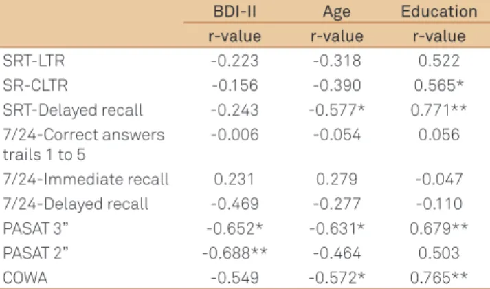
Correlations between the neuropsychological mea-sures and BDI-II and demographics variables are presented in Table 4. BDI-II correlated signiicantly and negatively to PASAT-3 (r=-0.652, p<0.05) and PASAT-2 (r=-0.688, p<0.01). However, the BDI-II scores did not correlate signiicantly with EDSS or disease duration. Furthermore, scores on SRT delay recall and PASAT-3 test were signiicantly correlated to age and education. However, cognitive tests did not correlate to evolution disease and EDSS (data not shown).
Test store NMO MS HC p-value
SRT-LTR 40.50± 20.53 37.62±11.75 49.79±13.98 0.128
SRT-CLTR 28.50±17.20 22.69±13.73 38.36±15.45 0.040
SRT-Delayed recall 7.29±3.14 6.62±2.95 9.21±2.00 0.048
7/24-Correct answers trails 1 to 5 26.82±5.05 22.46±6.96 28.50±6.68 0.054
7/24-Immediate recall 5.64±1.50 4.42±2.42 5.29±1.93 0.328
7/24-Delayed recall 5.55±1.57 4.25±1.91 5.93±1.81 0.060
PASAT-3” 31.36±18.36 38.25±15.53 41.86±12.16 0.208
PASAT-2” 21.36±15.12 26.92±9.54 37.86±11.52 0.004
COWA 28.07±12.12 29.23±7.44 38.08±11.89 0.043
Symbol digit modalities test 43.00±10.75 37.85±14.53 46.07±8.09 0.187
BDI-II 18.30±11.09 11.90±8.22 5.42±4.51 0.001
Table 2. Neuropsychological test results for the neuromyelitis optica and multiple sclerosis patients and healthy controls.
NMO: neuromyelitis optica; MS: multiple sclerosis; HC: healthy controls; SRT: selective reminding test; LTR: long-term storage; CLTR: consistent long-term retrieval; PASAT: paced auditory serial addition test; COWA: controlled oral word association; BDI-II: Beck depression inventory II.
Patient number Brain MRI BDI-II Cognitive domains
Verbal memory Visual memory Verbal fluency Attention
1 Normal 1 0 0 0 1
2 Unspecific lesions 3 1 0 1 1
3 Normal 1 0 1 0 1
4 Normal 4 0 0 1 1
5 Typical lesions AQP4 2 0 0 0 0
6 Unspecific lesions 3 1 0 0 1
7 Normal 1 0 0 0 0
8 Typical lesions AQP4 1 1 0 1 1
9 Unspecific lesions 1 0 0 0 0
10 Normal 1 0 0 0 0
11 Normal 1 1 NA 1 1
12 Normal 1 0 0 0 0
13 Normal 4 0 NA 1 1
14 Unspecific lesions 4 1 NA 1 1
MRI: magnetic resonance imaging; BDI: Beck depression inventory; AQP4: Aquaporin 4; cognitive domains=0: within normal range, 1: below the fifth percentile, NA: not applied visual alterations; BDI II=grades 1: 0 to 13 (without depression); 2: 14 to 19 (mild); 3: 20 to 28 (moderate); 4: 29 to 63 (severe).
1. Wingerchuk DM, Hogancamp WF, O’Brien PC, Weinshenker BG. The clinical course of neuromyelitis optica (Devic’s syndrome). Neurology 1999;53:1107-1114.
2. Lennon VA, Wingerchuk DM, Kryzer TJ, et al. A serum autoantibody marker of neuromyelitis optica: distinction from multiple sclerosis. Lancet Neurol 2004;364:2106-2112.
3. Blanc F, Zephir H, Lebrun C, et al. Cognitive functions in neuromyelitis optica. Arch Neurol 2008;65:84-88.
4. Nilsson P, Rorsman I, Larsson EM, Norrving B, Sandberg-Wollheim M. Cognitive dysfunction 24-31 years after isolated optic neuritis. Mult Scler 2008;14:913-918.
5. He D, Wu Q, Chen X, et al. Cognitive impairment and whole brain diffusion in patients with neuromyelistis optica after acute relapse. Brain Cogn 2011;77:80-88.
6. He D, Chen X, Zhao D, Zhou H. Cognitive function, depression, fatigue, and activities of daily living in patients with neuromyelitis optica after acute relapse. Int J Neurosci 2011;121:677-683.
7. Benedict RHB, Cookfair D, Gavett R, et al. Validity of the minimal assessment of cognitive function in multiple sclerosis (MACFIMS). J Int Neuropsychol Soc 2006;12:549-558.
8. Sepulcre J, Vanotti S, Hernandez, et al. Cognitive impairment in patients with multiple sclerosis using the Brief Repeatable Battery-Neuropsychology test. Mult Scler 2006;12:187-195.
References
Table 4. Correlations between neuropsychological measures, depression, age, and education in neuromyelitis optica patients.
BDI-II Age Education r-value r-value r-value
SRT-LTR -0.223 -0.318 0.522
SR-CLTR -0.156 -0.390 0.565*
SRT-Delayed recall -0.243 -0.577* 0.771**
7/24-Correct answers trails 1 to 5
-0.006 -0.054 0.056
7/24-Immediate recall 0.231 0.279 -0.047
7/24-Delayed recall -0.469 -0.277 -0.110
PASAT 3” -0.652* -0.631* 0.679**
PASAT 2” -0.688** -0.464 0.503
COWA -0.549 -0.572* 0.765**
SRT: selective reminding test; LTR: term storage; CLTR: consistent long-term retrieval; PASAT: paced auditory serial addition test; COWA: controlled oral word association; BDI-II: Beck depression inventory II; *p<0.05 level; **p<0.01 level.
DISCUSSION
NMO patients presented a signiicantly lower neuro-psychological performance than healthy controls of similar age and education. Surprisingly, 57.14% of the NMO Group showed CI. Similarly, Blanc et al.3 found 56.7% of CI in their
sample and He et al.5 also reported it.
Regarding cognitive performance, the few descriptions found in the literature on patients with NMO accounted a pattern of subcortical cognitive impairment, characterized by reduced speed of information processing, changes in ex-ecutive functions, attention, and memory. In this research, the NMO Group showed more dysfunction in attention and verbal luency than in memory. Blanc et al.3 described
alter-ation of long-term memory and executive functions, while He et al.5 found memory deicit and decreased speed of
in-formation processing and attention, after an acute relapse. his stage of the disease might not be the most appropriate for the evaluation of patients, because many factors involved in the pathogenesis of a relapse go beyond those involved in that of the disease itself.
Cognitive performance of NMO patients was found to be similar to that of MS ones coinciding with Blanc et al.3.
However, when compared with the published local norms, a higher proportion of MS patients showed visual memory impairment and a higher proportion of the NMO Group ev-idenced attention issues. Furthermore, CI in NMO patients could be considered surprising, since NMO is thought to be restricted to damage of optic nerves and spinal cord.
At the early stage of the disease, MRI brain lesions are al-most inexistent, with a few exceptions of little subcortical white matter lesions. However, recent MRI and pathological anatomy indings suggest that tissue damage in NMO is more extensive and includes the compromise of other areas, such as brain stem, cerebellum, and cortex16-18. Although this study
was not meant to examine the relationship between cogni-tion and MRI indings, cognitive alteracogni-tions were found both in patients with and without lesions in MRI. He et al.5 reported
evidence of microscopic brain lesions using difusion tensor imaging, which could explain the presence of cognitive altera-tions in those without lesions in conventional MRI measures.
A high number of patients (6 out of 14) manifested signs of depression, ranging from mild to severe, coincid-ing with the indcoincid-ings of Chanson et al.19, who found
inlu-ence of depression on the quality of life of patients with NMO. Moreover, He et al.6 reported a signiicant
correla-tion between cognicorrela-tion, depression, and routine activities in patients assessed after an acute relapse of the disease. Depression was related to attention measures, but not with physical disability or disease durations. he negative inlu-ence of depression in attentional functions has already been reported in MS20. In NMO, Blanc et al.3 did not observe an
association between cognition and depression.
Cognition is not associated with physical disability and disease duration, this is consistent with the indings of Blanc et al.3. he limitations of this study include the
9. Cáceres F, Vanotti S, Rao S, The Reconem Workgroup. Epidemiological characteristics of cognitive impairment of multiple sclerosis patients in a Latin American country. J Clin Exp Neuropsychol 2011;33:1094-1098.
10. Wingerchuk DM, Lennon VA, Pittock SJ, Lucchinetti CF, Weinshenker, BG. Revised diagnostic criteria for neuromyelitis optica. Neurology 2006;66:1485-1489.
11. Kurtzke JF. Rating neurological impairment in multiple sclerosis: an expanded disability status scale (EDSS). Neurology 1983;33:1444-1452.
12. Rao SM, Leo GJ, Bernardin L, Unverzagt F. Cognitive dysfunction in multiple sclerosis. I. Frequency, patterns, and prediction. Neurology 1991;41:685-691.
13. Smith A. Symbol Digits Modalities Test, Los Angeles: Western Psychological Services; 1982.
14. Beck AT, Steer RA, Brown GK. Manual for the Beck Depression Inventory-II. San Antonio, TX: Psychological Corporation; 1996.
15. Simon JH, Lib D, Traboulseec A, et al. Standardized MR Imaging Protocol for Multiple Sclerosis: Consortium of MS Centers Consensus Guidelines. Am J Neuroradiol 2006;27:455-461.
16. Rocca MA, Agosta F, Mezzapesa DM, et al. Magnetization transfer and diffusion tensor MRI show gray matter damage in neuromyelitis optica. Neurology 2004;62:476-478.
17. Rocca MA, Agosta F, Mezzapesa DM, et al. A functional MRI study of movement-associated cortical changes in patients with Devic’s neuromyelitis optica. Neuroimage 2004;21:1061-1068.
18. Yu CS, Lin FC, Li KC, et al. Diffusion tensor imaging in the assessment of normal-appearing brain tissue damage in relapsing neuromyelitis optica. Am J Neuroradiol 2006;27:1009-1015.
19. Chanson JB, Zephir H, Collongues N, et al. Evaluation of health-related quality of life, fatigue and depression in neuromyelitis optica. Eur J Neurol 2011;18:836-841.