167
CLINICS 2006;61(2):167-70
a Department of Radiology, Children’s Institute, Hospital das Clínicas, São
Paulo University Medical School - São Paulo/SP, Brazil.
b Pediatrics Surgery Division, Hospital das Clínicas, São Paulo University
Medical School - São Paulo/SP, Brazil. Email: [email protected]
LETTER TO THE EDITOR
IMPORTANCE OF ANGIOGRAPHIC STUDY IN
PREOPERATIVE PLANNING OF CONJOINED TWINS:
CASE REPORT
Francisco Cesar Carnevalea, Marcus Vinicius Borgesa, Breno Boueri Affonsoa, Ricardo Augusto de Paula Pintoa, Uenis
Tannurib, and João Gilberto Maksoudb
The occurrence of conjoined twins is rare. Its actual prevalence is unknown, but it is estimated to range from 1:50,000 to 1:200,0001–4 with a higher level of incidence
in Southwest Asia and Africa where an occurrence of 1:14,000 to 1:25,0005 is observed, with a female
predomi-nance ratio of 3:1.4
Its etiology is unknown, but an incomplete division of the zygote between 13th and 15th days after fertilization
probably occurs.3,5–7 About 40% to 60% of the conjoined
twins are born alive, and almost 35% of these live-born neonates die within 24 hours.4
Usually, the surgery for separation of twins is not per-formed soon after the birth unless some anomaly puts in risk the life of one or of both children. Therefore, in most cases it is recommended to wait for the twins to grow, mak-ing the operative procedure technically easier.
The importance of a detailed preoperative assessment of the various organs leading to precise anatomical knowl-edge and therefore to a comprehensive preoperative plan-ning is quite well known. Nevertheless, there is not an es-tablished algorithm for these evaluations that provides for the diversified anatomical variations existing from case to case.
With the development of noninvasive imaging methods, there was a decrease in the use of angiography as a preoperative assessment method in the hepatic resections. Most of the reports found in the literature emphasize the use of computed tomography (CT) scanning in preoperative planning, attributing to the other diagnostic methods a sec-ondary role with succinct descriptions.
The aim of this article is to present a case of ischiopa-gus twins whose surgical separation without the sacrifice of one of them seemed impracticable after helical CT and
ultrasonography (US) evaluations and whose successful sur-gical separation became possible after a more detailed angiographic vascular study.
CASE REPORT
Female ischiopagus tripus twins, of 15 months of age, were evaluated for the possibility of separation. They were joined below the xiphoid process at an angle of approxi-mately 90 degrees. The twins shared a single pelvis, and each child exhibited a normal anterior lower limb and a conjugated posterior lower one. One of the twins was vis-ibly bigger than the other.
The preoperative radiological evaluation showed the following:
A – Bone structure – plain radiography and helical CT scan:
Completely separated vertebral columns and sacra, without visible alterations. The region anterior to the pel-vic bones formed a single ring, with a normal pubic sym-physis and 2 acetabula articulating the normal lower limbs. In the posterior region of the pelvis, the iliac bones articu-lated an atrophied limb with a single femur and tibia and a bifid foot.
B – Cardiovascular system – plain radiography, echocardiogram and helical CT:
The larger child presented the heart directed towards the left and the aortic arch coursing to the right, with an existing inferior vena cava (IVC) and agenesis of the su-perior vena cava (SVC).
The smaller child exhibited the heart directed to the right and the aortic arch turned to the left with IVC agen-esis.
C – Urogenital system – Intravenous Pyelogram and helical CT:
168
CLINICS 2006;61(2):167-70 Importance of angiographic study in preoperative planning of conjoined twins
Carnevale FC et al.
twin had only 1 kidney and 2 ureters (with 1 of them drain-ing into the sister’s bladder), a bladder and urethra. Each infant presented independent vaginal vestibule, vagina, and uterus.
D- Gastrointestinal system – contrasted radiological study of the digestive tract (esophagus, stomach, duode-num, small bowel and colon):
Each twin presented a separate stomach, duodenum, and small bowel, sharing, however, 1 single colon and rectum. The smaller twin presented an imperforate anal canal.
E- Liver and spleen – abdomen ultrasonography (US) and helical CT:
Presence of a fusioned liver with 2 complete and inde-pendent hila and 2 gallbladders.
In the bigger twin, the parenchyma drainage was carried out through the hepatic veins to its own IVC, while in the smaller infant, the parenchyma drainage was accomplished by means of venous stems into her sister’s IVC, since she did not present an IVC of her own. There was only 1 spleen,
and it belonged to the smaller twin.
Since the hepatic venous draining of the smaller child coursed into the IVC of the bigger twin, their separating operation was contraindicated.
Subsequently, angiographic study was performed under general anesthesia, after the parents had signed an informed consent. The procedure was carried out in the angiography suite through catheterization of the right internal jugular vein and left femoral vein of the smaller infant and through the femoral arteries of both children employing a JB1 cath-eter (Cordis®, Johnson and Johnson®) and hydrophilic guide
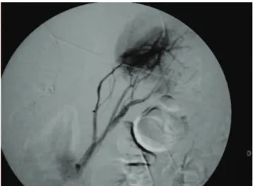
wire (Terumo 0.032", Boston Scientific®). Celiac and
me-senteric angiographic study (arterial and venous return phase) demonstrated a normal hepatic arterial supply for the smaller twin (Figures 1, 2). The study confirmed agen-esis of the IVC in the smaller twin, but it also showed in this child that the hepatic parenchyma drained into her own right atrium through small hepatic veins, thus allowing the twins to be successfully separated (Figures 3, 4).
Figure 1 - Thoracoabdominal aortography with identification of the celiac
trunk and hepatic, gastroduodenal, and superior mesenteric arteries of the smaller twin
Figure 2 - Venous return phase of the superior mesenteric artery of the smaller
twin, displaying the intra-and extra-hepatic portal vein
Figure 3 - Selective catheterization through the jugular access of the smaller
twin, displaying communication of her hepatic vein with the inferior vena cava of the bigger twin
Figure 4 - Hepatic venography of the smaller twin displaying the drainage
169
CLINICS 2006;61(2):167-70 Importance of angiographic study in preoperative planning of conjoined twins Carnevale FC et al.
DISCUSSION
Conjoined twins are classified according to the most prominent site of conjunction: thorax (thoracopagus); ab-domen (onphalopagus); sacrum (pygopagus); pelvis (ischio-pagus); skull (cephalopagus), and back (rachipagus). The most common types are: thoracopagus (73%) followed by pygopagus (19%) and finally by ischiopagus (6%).8,9
The phenomenon of conjoined twins with its rare oc-currence and the vast diversity of anatomical variation from case to case poses a unique challenge for pediatricians, pediatric surgeons, and radiologists.
The radiological assessment of the different organs for surgical preoperative planning of conjoined twins is of para-mount importance. However, the creation of a precise al-gorithm designed for such evaluation is impractical due to the vast number of anatomical variations existing in such cases.
Great emphasis is given to US and helical CT evalua-tions in most of the publicaevalua-tions, which attribute a second-ary position to the other methods of imaging diagnostics,
providing only brief and succinct descriptions. However, in this case, due to the inconclusive evaluation possible from the abdominal CT, the option of visceral angiogra-phy was adopted to accurately define the hepatic arterial and venous vascular anatomy, allowing the hepatic section without complications.
In this particular case, the initial evaluation by US and helical CT contraindicated the separation surgery of these conjoined twins, since the hepatic parenchyma drainage of one of the twins was not adequately demonstrated.
Thus, detailed angiographic assessment of the twins ac-quired a fundamental role in the preoperative study, allow-ing the successful surgical separation of the children.
Therefore, it can asserted that in the preoperative as-sessment of conjoined twins, no imaging method is supe-rior to another and that the integrated multidisciplinary evaluation for conduct definition is cardinal. Despite be-ing an invasive method, angiographic study should be in-dicated as a preoperative diagnostic complementation in cases in which the vital anatomic vascular structures are not identified by other means.
REFERENCES
1. Hanson JW. Incidence of conjoined twinning (letter). Lancet. 1975;2:1257.
2. Spitz L. Conjoined twins. Br J Surg. 1996;83:1028-30.
3. Barth RA, Filly RA, Goldberg JD, Moore P., Silverman NH. Conjoined twins: prenatal diagnosis and assessment of associated malformations. Radiology. 1990;177:201-7.
4. Edmonds LD, Layde PM. Conjoined twins in the United States, 1970-1977. Teratology. 1982;25:301-8.
5. Zimmermann AA. Embryological and anatomic considerations of conjoined twins. Birth Defects. 1967;3:18-27.
6. Jones KL. Smith’s recognizable patterns of human malformation. 2nd
170
CLINICS 2006;61(2):167-70 Importance of angiographic study in preoperative planning of conjoined twins
Carnevale FC et al.
7. Rudolph AJ, Michaels JP, Nichols BL. Obstetric management of conjoined twins. Birth Defects. 1967;3:28-37.
8. Schnaufer L. Conjoined twins. In: Raffensperger JG, ed. Swenson’s Pediatric Surgery. 4th ed. New York: Appleton Century-Crofts; 1980. p. 910-20.