online | memorias.ioc.fiocruz.br
Diagnostic reliability of an immunochromatographic test for
Chagas disease screening at a primary health
care centre in a rural endemic area
Diego Mendicino1/+, Mariana Stafuza1, Carlina Colussi1,
Mónica del Barco1, Mirtha Streiger1, Edgardo Moretti2
1Centre for Research on National Endemics, Faculty of Biochemistry and Biological Sciences, National University of Litoral, Santa Fe, Argentina 2National Coordination for Vector Control, Faculty of Medical Sciences, National University of Cordoba, Cordoba, Argentina
Many patients with Chagas disease live in remote communities that lack both equipment and trained personnel to perform a diagnosis by conventional serology (CS). Thus, reliable tests suitable for use under difficult conditions are required. In this study, we evaluated the ability of personnel with and without laboratory skills to perform im-munochromatographic (IC) tests to detect Chagas disease at a primary health care centre (PHCC). We examined whole blood samples from 241 patients and serum samples from 238 patients. Then, we calculated the percentage of overall agreement (POA) between the two groups of operators for the sensitivity (S), specificity (Sp) and positive (PPV) and negative (NPV) predictive values of IC tests compared to CS tests. We also evaluated the level of agree-ment between ELISAs and indirect haemagglutination (IHA) tests. The readings of the IC test results showed 100% agreement (POA = 1). The IC test on whole blood showed the following values: S = 87.3%; Sp = 98.8%; PPV = 96.9% and NPV = 95.9%. Additionally, the IC test on serum displayed the following results: S = 95.7%; Sp = 100%; PPV = 100% and NPV = 98.2%. Using whole blood, the agreement with ELISA was 96.3% and the agreement with IHA was 94.1%. Using serum, the agreement with ELISA was 97.8% and the agreement with IHA was 96.6%. The IC test performance with serum samples was excellent and demonstrated its usefulness in a PHCC with minimal equipment. If the IC test S value and NPV with whole blood are improved, then this test could also be used in areas lacking laboratories or specialised personnel.
Key words: Chagas disease - serological test - immunochromatographic assay
Chagas disease is a widespread zoonosis in Latin America caused by the flagellate protozoan Trypano-soma cruzi. The transmission route of Chagas disease is primarily vector borne through haematophagous triatomine insects. Triatoma infestans represents the greatest risk due to its anthropophilic habits. Other transmission routes of quantitatively lower public health significance include transfusion, connatal and digestive routes. These forms of inter-human transmission are re-sponsible for the urbanisation and globalisation of this disease (Schmuñis & Yadón 2010). The insect vectors of Chagas disease persist in dwellings of scattered and hard-to-reach rural populations where the prevalence of infection by this route is greater due to both environmen-tal and social reasons (Briceño-León & Galván 2007).
T. cruzi infection is often asymptomatic or oligo- symptomatic during the acute period. The chronic stage of infection is also latent until after 20 years or more when cardiac (most common) and/or gastrointestinal pa-thologies appear in approximately one-third of patients.
doi: 10.1590/0074-0276140153 Financial support: CAI+D/UNL
+ Corresponding author: [email protected] Received 4 May 2014
Accepted 6 November 2014
T. cruzi infection is confirmed during the chronic phase by demonstrating the host immune response against this parasite. At least two standardised serologi-cal tests with different principles or that detect antibod-ies against different specific antigens must be used for confirmation (Rassi Jr et al. 2010). To diagnose Chagas disease, the results of the two tests must be coinciden-tally positive. In cases of discrepancy (a positive and a negative test), a third test should be conducted to con-firm or rule out infection.
The most commonly used serological tests [conven-tional serology (CS)] are ELISAs and indirect haemag-glutination (IHA) tests. Currently, various serological diagnostic techniques are being evaluated, with each technique using different detection principles (Afonso et al. 2012). However, conducting these tests requires equipped laboratories and human resources trained in biochemical analysis.
Vector transmission occurs most often in rural popula-tions far from urban centres. In these areas, access to health coverage is reduced and travelling to locations where more complex services are available is extremely difficult.
risk involved in the delay caused by the transfer of the patient and the risk of using blood that has not been screened for Chagas.
Some authors have proposed collecting blood sam-ples on filter paper (Holguín et al. 2013) or storing blood samples in glycerin (Arrieta et al. 2004) for clinical and population studies in rural areas due to the lack of avail-ability of laboratories with the necessary equipment to diagnose this infection in these areas. The analysis of biological samples obtained by non-invasive methods, such as oral mucosal transudate specimens, has also been suggested (Moretti et al. 2004). Invariably, the samples must subsequently be taken to a laboratory for processing. However, this approach requires revisiting patients with positive results to confirm the infection us-ing a newly collected biological sample.
Because of these difficulties, techniques that can be used in primary health care centres (PHCCs) without high-ly complex requirements, such as specialised personnel and instruments, are required. However, such techniques must be able to deliver results quickly and reliably.
An immunochromatographic (IC) test is a rapid vi-sually read test that is widely used for the diagnosis of physiological conditions (e.g., pregnancy tests) and of in-fectious and non-inin-fectious diseases. Several IC tests can be performed on whole blood. The reagents for these tests are stored at room temperature (RT); thus, these tests do not require cold storage and the data can be easily ac-quired and interpreted. These characteristics will make new techniques suitable for use in low-resource settings (López-Chejade et al. 2010). For use as screening tech-niques, assays must also have extremely high sensitivity (S) and negative predictive values (NPV) because exclud-ing patients who seem to be seronegative, but who are actually positive is problematic. Positive samples should be sent to more complex laboratories for confirmation by other techniques. The techniques should have high speci-ficity (Sp) and positive predictive values (PPV) to avoid excessive labour use and economic burdens on the health care system. Incorrect test results also cause an emotion-al burden on the patients and their families.
We compared the results obtained with this test in the field using whole blood and serum samples with results from CS to study the performance and the usefulness of a commercial IC test for the diagnosis of Chagas infec-tion. The results of the IC tests were read by staff both with and without training in laboratory work.
SUBJECTS, MATERIALS AND METHODS
Patient selection and sample collection - Trained laboratory technicians obtained blood samples by ve-nipuncture using disposable syringes and needles from 241 patients attending clinical PHCC in the rural area of the province of Santa Fe, Argentina. The prevalence and risk of Chagas in the study area are similar to those found in the Gran Chaco ecoregion, although the vector situation is under control in Santa Fe (Mendicino et al. 2013). The study was “single-blind” because the tested samples were obtained from a serological survey of pa-tients at a greater risk of being infected due to their epi-demiological history (unsafe dwelling conditions and/or
a mother with Chagas disease). The single-blind design was chosen because of the low prevalence of this disease and because a double-blind design would have required an excessively high number of samples.
The samples were collected in disposable tubes with separator gel. No plasma samples were used so that the samples could be transported in the primary tubes without haemolysis.
Processing the whole blood IC test in the field - The whole blood samples were tested using WL Check Cha-gas test kits (Wiener Lab SAIC, Argentina). The WL Check Chagas test is a lateral flow IC test that detects antibodies specific to T. cruzi. These kits do not include any disposable items (e.g., capillary tubes or Pasteur pi-pettes) and the only laboratory instrument required to perform this test is an automatic micropipette. Each test kit consists of individual tests that can be stored at RT; the test results should be read after 25-35 min. Each in-dividual cassette has an internal quality control. If the sample contains antibodies specific to T. cruzi, then the complex bound to the antigens on the membrane pro-duces a pink and/or purple line. If the sample does not contain antibodies to T. cruzi, then the complex remains unbound and no line appears.
To conduct the IC tests, we collected 40 μL of whole
blood with an automatic micropipette before a clot formed and then followed the manufacturer’s instruc-tions for the IC test. After the required waiting time, the results were read in situ by two different operators, who were a qualified health professional (biochemist, lab technician, nurse or doctor) and an unqualified vol-unteer (driver or administrative staff). The results were recorded in spreadsheets in a single-blind manner.
The sera were separated by centrifugation at the PHCC. The samples were transported to the reference hospital in their primary tubes and stored in compliance with the cor-responding biosafety and conservation regulations.
Processing serum IC and CS samples in the labora-tory - All samples were subjected to ELISA (Chagatest ELISA, Wiener Lab SAIC) and IHA (IHA Chagas Poly-chaco, Lemos Laboratory SRL, Argentina) in the labo-ratory of the Centre for Research on National Endemic Diseases of the National University of Litoral (UNL) us-ing commercial reagents approved by the National Ad-ministration of Drugs, Food and Medical Technology. When a discrepancy between the two tests occurred, we performed indirect immunofluorescence with com-mercial conjugates and smears were prepared in our laboratory with formalin-fixed T. cruzi epimastigotes of the Tulahuen strain (Streiger et al. 1980). A sample was considered positive or negative according to the agree-ment of two of these three CS tests. The CS results were delivered to the individual patients.
Serum could not be obtained from three of the 241 pa-tients because the volume after centrifugation was too small. IC tests were performed on 238 serum samples from the same patients following the manufacturer’s instructions.
The percentage of overall agreement (POA) was de-termined as a measure of the variation in readings be-tween observers with (O1) and observers without (O2) training in laboratory work. The POA was calculated as a+d/a+b+c+d, where a = positive readings for O1 and O2, b = a positive reading for O1 and a negative reading for O2, c = a negative reading for O1 and a positive read-ing for O2 and d = negative readread-ings for O1 and O2.
We calculated the S, Sp, PPV and NPV by comparing the IC test results on whole blood and serum with the CS results. We considered the results as true positive (TP) when the IC and CS were positive for the sample and true negative (TN) when the IC and CS results were negative. A false positive (FP) was defined as a positive IC result and a negative CS result. A false negative (FN) was de-fined as a negative IC result and a positive CS result.
S is defined as the proportion of study participants considered positive by CS who were correctly identified as positive by IC test. Sp is defined as the proportion of study participants without Chagas infection according to CS who were correctly identified by IC test. We calcu-lated S and Sp using the following equations: S = TP/ TP+FN and Sp = TN/TN+FP.
PPV is defined as the proportion of study partici-pants who were reactive to the IC test (TP+FP) and who were correctly considered positive by the IC test (TP). Therefore, PPV represents the percentage of patients who actually had the disease and who also tested posi-tive. The NPV is defined as the proportion of patients who were non-reactive for this test (TN+FN) and who were correctly considered negative by the IC test (TN). The NPV represents the percentage of patients without the disease who also tested negative. We calculated these values using the following equations: PPV = TP/FP+TP and NPV = TN/FN+TN.
We also determined the agreement of the IC results with the IHA and ELISA results. This agreement was de-fined as the proportion of the IC results (positive and neg-ative) that coincided with the IHA and ELISA results.
Ethics - This study was reviewed and approved by the Advisory Committee on Research Ethics and Safety of the Faculty of Biochemistry and Biological Sciences of the UNL.
Before collecting the blood samples, the scope of the study was explained to the patients and all questions were addressed. We included all patients who accepted being subjected to analysis for Chagas by signing an in-formed consent form. The CS results were given to each patient individually and privately. The seropositive pa-tients were connected with the provincial program for Chagas control, monitoring and treatment in accordance with the National Standards for Care.
RESULTS
Variation in the readings between observers - The agreement (POA = 1) between the readings of the results of the IC tests on whole blood was 100% when assessed by different operators (personnel with and without train-ing in laboratory work).
S, Sp and PPV and NPV - As shown in Table I, the results of the IC tests on whole blood had an S of 87.3% and an Sp of 98.8% compared with CS. The PPV was 96.9% and the NPV was 95.9%.
When the results of the IC tests on serum samples were compared with the results of CS, S was 95.7%, Sp was 100%, PPV was 100% and NPV 98.2% (Table II).
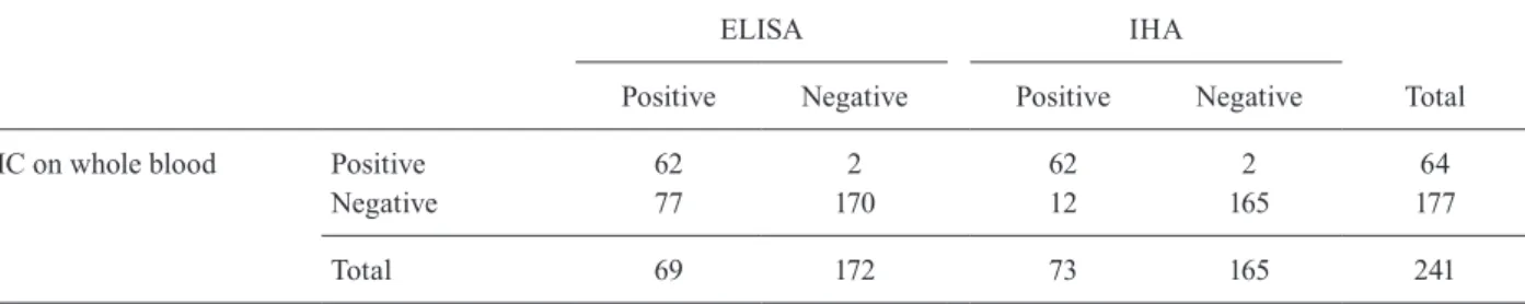
Agreement with ELISA and IHA - A comparison of the results of the IC tests on whole blood and the results of the ELISA and IHA tests are presented in Table III. The result agreement was 96.3% with ELISA and 94.1% with IHA.
The results of the IC tests on serum and the results of the ELISA and IHA tests are summarised in Table IV. The agreement between the IC and ELISA results was 97.8% and that between the IC and IHA results was 96.6%.
DISCUSSION
IC tests are advantageous for the diagnosis of Cha-gas disease in low-resource settings such as rural areas because these tests are easy to use and can be performed with both whole blood and serum. Despite these advan-tages, only a limited number of studies regarding the use of these tests with whole blood in the field have been published (Roddy et al. 2008, Chippaux et al. 2009). Several studies have evaluated the performance of these tests with serum samples in the laboratory (Ponce et al. 2005, Reithinger et al. 2010). In a recent multicentre TABLE I
Results of immunochromatography (IC) with whole blood vs conventional serology (CS)
CS
Positive Negative Total
IC on whole blood Positive 62 2 64 Negative 9 168 177
Total 71 170 241
negative predictive value: 95.9%; positive predictive value: 96.9%; sensitivity: 87.3%; specificity: 98.8%.
TABLE II
Results of immunochromatography (IC) on serum vs conventional serology (CS)
CS
Positive Negative Total
IC on serum Positive 67 0 67 Negative 3 168 171
Total 70 168 238
study, the performances of 11 commercially available IC tests for Chagas disease (including the WL Check Chagas test) were compared using samples from refer-ence laboratory serum banks (Sánchez-Camargo et al. 2014). That comparison was performed under labora-tory conditions and the authors concluded that further studies should be conducted in either the field or clinical settings in both endemic and non-endemic areas using whole blood samples. The present study was performed in a low-resource setting with serum and whole-blood samples and the results were read by trained and un-trained personnel. The behaviour of the tests has not been previously compared using both types of samples or using the variability in readings obtained from trained and untrained personnel.
The IC test evaluated in this study was easy to imple-ment. Although this test was performed by personnel un-trained in laboratory work, their readings agreed 100% with those made by health personnel trained in conduct-ing laboratory tests. Sánchez-Camargo et al. (2014) also reported a high score in the ease-of-use questionnaire for the same test when performed under laboratory con-ditions. A previous study with another commercial IC test showed 99.9% agreement among two non-laboratory health workers (Roddy et al. 2008). Although diagnostic tests should be performed by laboratory personnel, these tests can be performed in places where health workers are scarce or during acute emergencies, such as natural disasters, when emergency transfusions are required and when blood banks are not accessible.
A high level of agreement was observed among the results obtained in the laboratory with serum samples us-ing IC, ELISA and IHA tests. The use of ELISA and IHA is widespread in biochemical diagnosis centres. However, IHA has a lower S and a lower percentage of agreement compared with the other tests (Remesar et al. 2009).
The values of S, Sp, PPV and NPV were acceptable when the results of the tests with serum were compared with those of the CS results and were similar to the results obtained with the same commercial test in the study led by Sánchez-Camargo et al. (2014). The per-formances of IC tests in the laboratory were similar to those of conventional techniques (Remesar et al. 2009, Flores-Chávez et al. 2010, Afonso et al. 2012, Pereira et al. 2012), with the advantage that IC tests require only a centrifuge to separate the serum.
The results obtained with the kit used in this study and whole blood samples showed S values lower than those obtained with other IC tests for Chagas in both field (Roddy et al. 2008, Chippaux et al. 2009) and lab-oratory studies (Ponce et al. 2005). The Sp value was similar to the Sp values observed in those studies. The agreement with validated techniques (ELISA and IHA) was lower than when using serum samples.
The performance of the test evaluated in the present study is acceptable for use in screening American try-panosomiasis if performed with serum and confirmed with other reference techniques. This test requires only basic equipment to separate the serum, the performance TABLE IV
Results of immunochromatography (IC) on serum vs ELISA and indirect haemagglutination (IHA)
ELISA IHA
Total Positive Negative Positive Negative
IC on serum Positive 65 2 66 1 67
Negative 3 168 7 164 171
Total 68 170 73 165 238
agreement with ELISA: 97.8% [(65+168)/238]; agreement with IHA: 96.6% [(66+164)/238]. TABLE III
Results of immunochromatography (IC) on whole blood vs ELISA and indirect haemagglutination (IHA)
ELISA IHA
Positive Negative Positive Negative Total
IC on whole blood Positive 62 2 62 2 64
Negative 77 170 12 165 177
Total 69 172 73 165 241
and interpretation of the test are simple and the reading time is short. Because of these characteristics, this test is suitable for use in both low-complexity laboratories and PHCCs that are often more accessible to popula-tions in vector risk areas.
Each cassette of the kit has an internal quality con-trol so that the determinations can be made individually without processing a batch of samples from different pa-tients. Individual determination is one advantage of this test over IHA and ELISA, which both require positive and negative controls with each processing batch. Thus, IC tests are convenient for use in laboratories in which an analysis for Chagas is not required frequently.
When performed with whole blood samples, we be-lieve that this IC test does not have the necessary S and NPV to be used as a screening technique in low-resource settings and would lead to incorrectly identifying some patients as non-infected. If these parameters are im-proved, then this test could be useful for diagnosis in the field and in population studies. This test could also be useful in pre-transfusion screening in emergencies and in situations with no health facilities or with health fa-cilities with limited access to the population.
Evaluating the performance of the IC test on alterna-tive samples, such as oral mucosal transudate specimens, which are collected using a non-invasive method and are advantageous in terms of biosafety and acceptance by the study population, will also be valuable.
ACKNOWLEDGEMENTS
To the communities that worked with us and to the staff of the PHCC, for allowing us to conduct the study, and to the Pro-vincial Chagas Program of the Province of Santa Fe and its di-rector, Marcelo Nepote, for logistical support in the field trips.
REFERENCES
Afonso AM, Ebell MH, Tarleton RL 2012. A systematic review of high quality diagnostic tests for Chagas disease. PLoS Negl Trop Dis 6: e1881.
Arrieta R, Daquino B, Rosso N, Ferreras M, Juárez N 2004. Evalu-ación de una metodología de tamizaje en la enfermedad de Cha-gas en San Luis, Argentina. Salud Publica Mex46: 430-437.
Briceño-León R, Galván JM 2007. The social determinants of Chagas disease and the transformations of Latin America. Mem Inst Os-waldo Cruz 102 (Suppl. I): 109-112.
Chippaux J, Santalla J, Postigo J, Romero M, Clavijo NS, Schneider D, Brutus L 2009. Sensitivity and specificity of Chagas Stat-Pak test in Bolivia. Trop Med Int Health 14: 732-735.
Flores-Chávez M, Cruz I, Rodríguez M, Nieto J, Franco E, Gárate T, Cañavate C 2010. Comparación de técnicas serológicas conven-cionales y no convenconven-cionales para el diagnóstico de la enferme-dad de Chagas importada en España. Enferm Infecc Microbiol Clin 28: 284-293.
Holguín A, Norman F, Martín L, Mateos M, Chacón J, López-Vélez R, Pérez-Molina J 2013. Dried blood as an alternative to plasma or serum for Trypanosoma cruzi IgG detection in screening pro-grams. Clin Vaccine Immunol 20: 1197-1202.
López-Chejade P, Roca C, Posada E, Pinazo M, Gascon J, Portús M 2010. Utilidad de un test inmunocromatográfico para el cribado de la enfermedad de Chagas en asistencia primaria. Enferm In-fecc Microbiol Clin 28: 169-171.
Mendicino D, Stafuza M, del Barco M, Colussi C, Bizai M, Fabbro D, Nepote M, Streiger M 2013. Infección chagásica en niños de cua-tro distritos de riesgo de la provincia de Santa Fe. Acta Bioquím Clín Latinoam 47: 571-578.
Moretti E, Basso B, Gil P, Vaca B, Jacqueline J, Yasenzaniro P 2004. Detección de anticuerpos para Chagas y toxoplasmosis en trasu-dado mucoso oral. Acta Bioquím Clín Latinoam 38: 159-163.
Pereira GA, Lozada-Neto F, Barbosa VF, Ferreira-Silva M, de Mo-raes-Souza H 2012. Performance of six diagnostic tests to screen for Chagas disease in blood banks and prevalence of Trypano-soma cruzi infection among donors with inconclusive serology screening based on the analysis of epidemiological variables. Rev Bras Hematol Hemoter 34: 292-297.
Ponce C, Ponce E, Vinelli E, Montoya A, de Aguilar V, González A, Zingales B, Rangel-Aldao R, Levin M, Esfandiari J, Umesawa E, Luquetti A, da Silveira J 2005. Validation of a rapid and reliable test for diagnosis of Chagas disease by detection of Trypanosoma cruzi-specific antibodies in blood donors and patients in Central America. J Clin Microbiol 43: 5065-5068.
Rassi Jr A, Rassi A, Marin-Neto J 2010. Chagas disease. Lancet 375: 1388-1402.
Reithinger R, Grijalva M, Chiriboga R, de Noya BA, Torres J, Pavia-Ruz N, Manrique-Saide P, Cardinal M, Gürtler R 2010. Rapid detection of Trypanosoma cruzi in human serum by use of an immunochro-matographic Dipstick Test. J Clin Microbiol 48: 3003-3007.
Remesar M, Gamba C, Colaianni I, Puppo M, Sartor P, Murphy E, Neilands T, Ridolfi M, Leguizamón M, Kuperman S, del Pozo A 2009. Estimation of sensitivity and specificity of several Try-panosoma cruzi antibody assays in blood donors in Argentina.
Transfusion49: 2352-2358.
Roddy P, Goiri J, Flevaud L, Palma P, Morote S, Lima N, Villa L, Torrico F, Albajar-Viñas P 2008. Field evaluation of a rapid im-munochromatographic assay for detection of Trypanosoma cruzi
infection by use of whole blood. J Clin Microbiol 46: 2022-2027.
Sánchez-Camargo C, Albajar-Viñas P, Wilkins P, Nieto J, Leiby D, Paris L, Scollo K, Flórez C, Guzmán-Bracho C, Luquetti A, Calvo N, Tadokoro K, Saez-Alquezar A, Palma P, Martin M, Flevaud L 2014. Comparative evaluation of 11 commercialized diagnostic tests for detecting Trypanosoma cruzi antibodies in serum banks in endemic and non-endemic areas. J Clin Micro-biol doi: 10.1128/JCM.00144-14.
Schmuñis G, Yadón Z 2010. Chagas disease: a Latin American health problem becoming a world health problem. Acta Trop 115: 14-21.