Bhatnagar, KP.*
Department of Anatomical Sciences and Neurobiology, School of Medicine, University of Louisville
Louisville, Kentucky 40292, USA *e-mail: [email protected]
Received July 19, 2006 – Accepted February 2, 2007 – Distributed August 31, 2008 (With 27 figures)
Abstract
The vampire bat, Desmodus rotundus, is exceptionally agile and stealthy in nature. Feeding at night on cattle blood, it is a known scourge carrying rabies. It is endowed with a very high neocortical volume among bats, acute olfac-tory capabilities and an accessory olfacolfac-tory system. These characteristics have resulted into an impressive number of neuroanatomical investigations except a long due atlas on its brain. This study presents a cytoarchitectural atlas of the brain of the common vampire, Desmodus rotundus murinus, in the frontal plane, serially between the olfactory bulb and the medulla oblongata. Twenty six selected sections are presented, each separated by about 300 to 560 microns. The atlas figures show lugol fast blue-cresyl echt violet stained hemisections with their matching half in a labeled line drawing. About 595 discrete brain structures (some repeating) have been identified. This study is likely to provide the accurate localization of nuclear groups, whole structures, fiber tracts, and interconnections to facilitate future neuro-anatomical and neurophysiological investigations on the vampire brain.
Keywords: bats, brain, cytoarchitectural atlas, Desmodus rotundus, vampire.
O cérebro do morcego vampiro comum, Desmodus rotundus murinus
(Wagner, 1840): um atlas citoarquitetural
Resumo
O morcego vampiro, Desmodus rotundus, é excepcionalmente ágil e furtivo. Alimentando-se à noite do sangue de gado, é um conhecido flagelo portador da raiva. Entre os morcegos, apresenta um alto volume neocortical, capacida-des olfatórias agudas e um sistema ofatório suplementar. Estas características têm suscitado um número impressio-nante de estudos neuroanatômicos, no entanto, não foi produzido ainda um atlas do cérebro desta espécie. O presente estudo apresenta, assim, um atlas citoarquitetural do cérebro do vampiro comum, Desmodus rotundus murinus, no plano frontal, serialmente entre o bulbo ofatório e a medula oblongata. Vinte e seis seções selecionadas são aqui apresentadas, cada uma separada por em torno de 300 a 560 mícrones. As figuras do atlas mostram hemisseções manchadas com lugol violeta de cresilo, com a sua respectiva metade apresentada numa figura rotulada. Em torno de 595 estruturas cerebrais discretas (algumas repetidas) foram identificadas. Este estudo proverá a localização precisa de grupos nucleares, estruturas inteiras, tratos de fibras, e interconexões para facilitar futuras investigações neuroana-tômicas e neurofisiológicas realizadas no cérebro do morcego vampiro.
Palavras-chave: morcegos, cérebro, atlas citoarquitetural, Desmodus rotundus, vampiro.
1. Introduction
Of the three species of vampires (Desmodus, Diaemus, and Diphylla),Desmodus is the most common and best known as a pest and a scourge. Desmodus is unique among bats in having the largest neocortical vol-ume (size index 1024) amongst the nearly 276 species of bats investigated (Baron et al., 1996a; b, c). For this char-acteristic alone Desmodus is considered one of the most evolved bats studied thus far. Monographic works on
brain atlases have also been available in the literature (Table 1).
For the Latin American countries, Desmodus car-ries a special threat. It is called ‘thief of the night’, and causes substantial loss of cattle from transmitted rabies (Greenhall et al., 1983). Its importance is further en-hanced in the newly discovered use of the clot-buster, plasminogen activator (desmoteplase or DSPA) in its saliva (Hawkey, 1966) in prevention of strokes in the hu-man (Fauber, 2003). It is not an overstated conclusion that knowledge of the brain cytoarchitectonics on this species is essential to have.
The association of the vampire behavioral com-plexity with its voluminous neocortex has been estab-lished. (Baron et al., 1996a; b; c). These features make Desmodus an ideal bat species for neuroanatomical and neurophysiological research. This study was, therefore, undertaken to analyze the serial brain anatomy of the common vampire bat.
2. Materials and Methods
2.1. Specimens
AdultDesmodus rotundus were captured live from a wild colony in Veracruz, Mexico through the courtesy of
Forment. Five females and two males were transported to the author’s laboratory in Louisville and remained in good health in captivity for several years.
2.2. Brain preparation
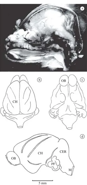
Bats were deeply anesthetized with ether and int-racardiacally perfused first with saline in nitrite solu-tion with the right atrium punctured. Perfusion was then continued using one of the three solutions: Bouin’s, 4% glutaraldehyde, or 10% buffered formalin. The skin and muscles were dissected away from the cranium and the neck. The head was severed at the neck and kept im-mersed in the fixative for several hours, after which the calvarium and the dorsal half of the upper cervical ver-tebrae were chipped away. The exposed brain and the spinal cord were further left in the fixative for several more hours. The olfactory nerves were transected and the frontal lobes were gently lifted, gradually severing other cranial nerves. The hypophysis was gently pushed out of the sella turcica. When the entire brain (Figure 1) was thus freed, it was kept in the fixative for 24-48 hours, washed, and processed in graded ethyl alcohol. Paraffin embedding as well as cellosolve (ethylene-glycol-monoethyl ether) procedures were followed for the in-dividual brains.
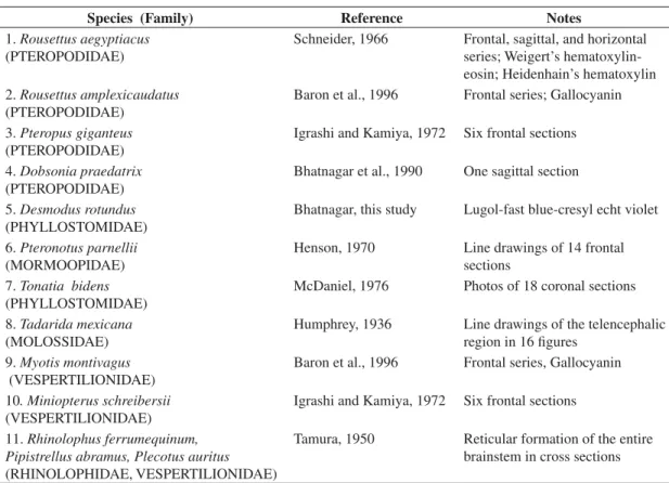
Table 1. A partial list of atlases and monographs on the brains of bats.
Species (Family) Reference Notes
1.Rousettus aegyptiacus (PTEROPODIDAE)
Schneider, 1966 Frontal, sagittal, and horizontal series; Weigert’s hematoxylin-eosin; Heidenhain’s hematoxylin 2.Rousettus amplexicaudatus
(PTEROPODIDAE)
Baron et al., 1996 Frontal series; Gallocyanin
3.Pteropus giganteus (PTEROPODIDAE)
Igrashi and Kamiya, 1972 Six frontal sections 4.Dobsonia praedatrix
(PTEROPODIDAE)
Bhatnagar et al., 1990 One sagittal section 5.Desmodus rotundus
(PHYLLOSTOMIDAE)
Bhatnagar, this study Lugol-fast blue-cresyl echt violet 6.Pteronotus parnellii
(MORMOOPIDAE)
Henson, 1970 Line drawings of 14 frontal sections
7.Tonatia bidens (PHYLLOSTOMIDAE)
McDaniel, 1976 Photos of 18 coronal sections 8.Tadarida mexicana
(MOLOSSIDAE)
Humphrey, 1936 Line drawings of the telencephalic region in 16 figures
9.Myotis montivagus (VESPERTILIONIDAE)
Baron et al., 1996 Frontal series, Gallocyanin 10. Miniopterus schreibersii
(VESPERTILIONIDAE)
Igrashi and Kamiya, 1972 Six frontal sections
11.Rhinolophus ferrumequinum, Pipistrellus abramus, Plecotus auritus (RHINOLOPHIDAE, VESPERTILIONIDAE)
Tamura, 1950 Reticular formation of the entire brainstem in cross sections
For the cellosolve infiltration, the brain was processed through four changes of cellosolve, leaving it overnight in the last change. Three changes of one hour duration each in benzene, followed by three one-hour changes in paraffin were made under vacuum at 58 °C. The infil-trated brain was then embedded in paraffin. Serial frontal (coronal) sections were cut at 20 Mm thickness, arranged on albumen-coated slides which were kept at 58 °C in an oven for drying. Staining was achieved with lugol fast blue overnight, differentiated in 70% alcohol, rinsed, and counterstained with cresyl echt violet for 5-7 minutes, cleared in xylene and mounted in permount.
decalcified heads of several specimens, perfused with Bouin’s solution, was prepared. These were stained with the one-step Gomori trichrome procedure (Bhatnagar and Kallen, 1974; Bhatnagar, 1980). A partly incomplete, 20Mm thick, stained serial series made from a female
Desmodus rotundus murinus brain was received from the late Professor William A. Wimsatt, Cornell University, Ithaca, New York.
2.3. Brain analysis
Using a macrophotography apparatus, selected sec-tions, primarily from the lugol fast blue-cresyl echt violet series, beginning with the olfactory bulb and ending with the medulla oblongata, were photographed with gaps of about 300-560 µm between sections. A total of 710 sec-tions, each 20 µm thick, was prepared. Even though over 100 sections were photographed, studied and labeled, 26 sections were finally selected for this atlas, represent-ing the entire brain, without unduly repeatrepresent-ing. In this process of selecting representative sections, a few struc-tures may be found missing. These were purposely left out to conserve space.
Desmodus brain cytoarchitecture was studied in comparison with the brain anatomy of the human (Villiger-Ludwig-Rasmussen, 1951), rat (Paxinos and Watson, 1997), muskrat (Panneton and Watson, 1991), cat, hamster, guinea pig, and other bat species (Table 1). Structures were localized and identified on a camera lu-cida drawing, an exact match control of the right half of the photographed section (Atlas Figures 2-27).
3. Results and Discussion
The primary objective of this investigation was to prepare an atlas of the brain of the vampire,Desmodus rotundusand identify the structures, such as the nuclear groups, ascending and descending fiber tracts, cranial nerve nuclei, cortical regions, and brainstem components. This species, being so commonly available and so unique in its behavioral characteristics, has long deserved detailed cytoarchitectural studies of its brain. Desmodus has the largest neocortical volume compared to 276 species of bats (Table 2; see Baron et al., 1996a; b; c). Most early studies on the vampire brain anatomy are onDesmodus ro-tundus (see Mann, 1960, 1963). Plenty of data are provid-ed in the three-volume work “Comparative Neurobiology in Chiroptera” by Baron et al. (1996a; b; c). The supe-rior olivary complex was investigated by Kuwabara and Bhatnagar (1999). They also reported that the lateral lemniscus is columnar and similar as in other echolocat-ing bats (Kuwabara and Bhatnagar, 2000). Data on the three species of vampire brains were reviewed in detail by Bhatnagar (1988a) Ultrastructural observations on the pineal gland of Desmodus were also reported (Bhatnagar, 1988b). The accessory olfactory bulb inDesmodus, though short in height, extends nearly to the full diameter of the main olfactory bulb (Cooper and Bhatnagar, 1976), an
ob-CH
OB
OB
CH CER
PF
M
5 mm
b c
d
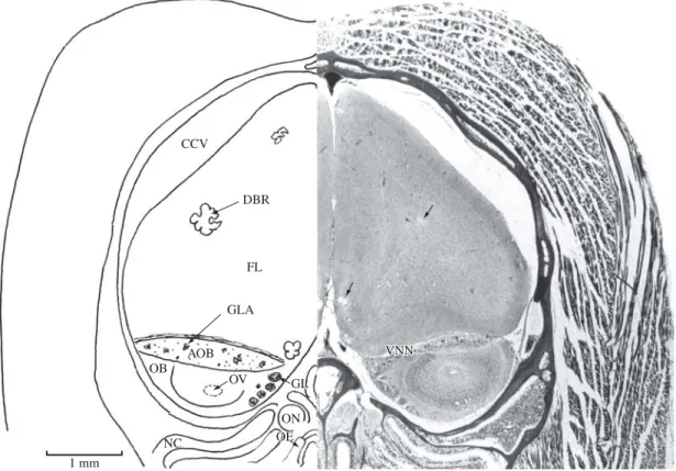
AOB OB
GL GL
ON OE OE CCV
DBR
FL
GLA
OV
NC
VNN VNN
Figure 2.This anterior-most section is from the caudal olfactory bulb region. It is from a wild caught Desmodus rotundus held captive in our laboratory. This bat’s entire brain showed ‘holes’ or degenerated regions as depicted here (arrows). Re-fer to the text. AOB, accessory olfactory bulb; CCV, cranial cavity; DBR, degenerated brain region; FL, frontal lobe; GL, glomerular layer of the main olfactory bulb; GLA, glomeruli of the accessory olfactory bulb; NC, nasal cavity; OB, main olfactory bulb; OE, olfactory epithelium; ON, olfactory nerve; OV, olfactory ventricle; VNN, vomeronasal nerve. Section No. 65, De1-201; Gomori one-step trichrome, 10Mm.
NEO CC
FM
GCC
LOT GL
OB
IOT AON 1 mm
Figure 3.AON, anterior olfactory nucleus; C, cingulum; FM, forceps minor of the corpus callosum; GCC, genu of the corpus callosum; GL, glomerular layer of the main olfactory bulb; IOT, intermediate olfactory tract; LOT, lateral olfactory tract; NEO, neocortex; OB, olfactory bulb. Section No. 70.
GCC C
STR
RCC
PC
TU PC
AON
1 mm
Figure 4. AON, anterior olfactory nucleus; C, cingulum; GCC, genu of the corpus callosum; PC, piriform (olfactory) cortex; RCC, radiation of the corpus callosum; STR, striatum (caudate-putamen). TU, olfactory tubercle. Section No. 75.
NEO CC
VHC VHC FH STR ICAP GP
AC POL NLOT
IST TS F POM
1 mm 1 mm
C C
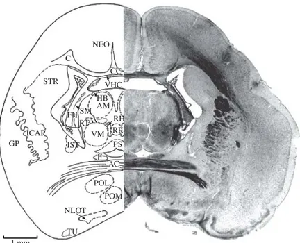
Figure 7.AC, anterior commissure; C, cingulum; CC, cor-pus callosum; F, fornix; FH, fimbria of the hippocamcor-pus; GP, globus pallidus; ICAP, internal capsule; IST, interstitial nucleus of stria terminalis; NEO, neocortex; NLOT, nucleus of the lateral olfactory tract; POL, lateral preoptic nucleus; POM, medial preoptic nucleus; STR, striatum; TS, triangu-lar nucleus of septum; VHC, ventral hippocampal commis-sure. Section No. 105, 96-5, 10880Mm.
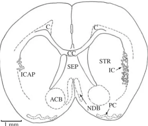
ICAP
SEP CC
C C
STR IC
PC NDB ACB
1 mm
Figure 5.ACB, accumbens nucleus; C, cingulum; CC, corpus callosum; ICAP, internal capsule; NDB, nucleus of diagonal band (tract); PC, piriform (olfactory) cortex; SEP, septal nucleus; STR, striatum (caudate-putamen). Section No. 81.
STR C
CC CC MSEP
CF
LSEP F IST
NDB POM
AC ACB
ICAP
1 mm
Figure 6.AC, anterior commissure; ACB, accumbens nu-cleus; C, cingulum; CC, corpus callosum; CF, column of fornix; F, fornix; ICAP, internal capsule; IST, interstitial nucleus of stria terminalis; LSEP, lateral septal nucleus; MSEP, medial septal nucleus; NDB, nucleus of diagonal band (tract); POM, medial preoptic nucleus; STR, striatum (caudate-putamen). Section No. 89, 99-4, 11180 µm.
servation that has not been made in other mammalian ol-factory bulbs, including those of bats. There is an olol-factory ventricle. In one of the brains, prepared from a wild caught
Desmodus (see Figure 2), isolated telencephalic regions were devoid of brain tissue, as though it had been punched out. It would be interesting to investigate if this strange morphological finding bears any relationship to the rabies virus carrying characteristics of the vampire.
TheDesmodus brain is short, lissencephalic (smooth; sulci lacking on the outer surface), with high hemi-spheres (Figure 2), and a rostral sulcus. Parafloculus is unexposed. The pons is large, and the pyramidal
tracts are huge until they cross. Accurately detailed and painstakingly measured volumes on the vampire brains (Desmodus rotundus, and including another vampire species,Diphylla ecaudata) are provided in Baron et al. (1996a; b; c ). These are summarized in Table 3. For the sake of space, those specifically not included in Table 3 are the volumes of subdivisions and parts of medulla oblongata, mesencephalic components, cerebellar nu-clei, and lateral geniculate body. For the data on these structures and others, the reader is directed to Baron et al. (1996a; b; c).
Even though major senses (vision, hearing, echolo-cation and olfaction) are well developed in the vampire, it is not practical for the bat to use phyllostomid type of sonar for locating its prey. Statements have been made in the literature that vampires use olfaction to a greater extent in feeding (Mann, 1963). If greater sizes or vol-umes are related to greater sense of acuity, then the size of the olfactory and the vomeronasal organs are indica-tive of an olfactory sense acuter than in most bat species. Even by cursory examination, of the three vampires,
Diaemus appears to have larger volumes dedicated to the olfactory and the vomeronasal (accessory) systems than
Desmodus. Diphylla appears to occupy the last position amongst vampires in this regard.
pale-NEO C
CC CC VHC
AM HB
RH RE RE VM
PS F F
AC
POL
POM POM
NLOT
TU IST GP
ICAP ICAP
STR
FH FH
AV AV RT SM
1 mm
Figure 8.AC, anterior commissure; AM, anteromedial thalamic nucleus; AV, anteroventral thalamic nucleus; C, cingulum; CC, corpus callosum; F, fornix; FH, fimbria of hippocampus; GP, globus pallidus; HB, habenular nucleus; ICAP, internal capsule; IST, interstitial nucleus of stria terminalis; NEO, neocortex; NLOT, nucleus of the lateral olfactory tract; POL, lateral preoptic nucleus; POM, medial preoptic nucleus; PS, periventricular stellatocellular nucleus; RE, reuniens thalamic nucleus; RH, rhomboid thalamic nucleus; RT, reticular thalamic nucleus; SM, striamedullaris of the thalamus; STR, striatum; TU, olfactory tubercle; VHC, ventral hippocampal commissure; VM, ventromedial nucleus of the thalamus. Section No. 118, 93-3, 10540Mm.
Figure 9.AC, anterior commissure; AD, anterodorsal thalamic nucleus; AH, anterior nucleus of hypothalamus; AM, antero-medial thalamic nucleus; AV, anteroventral thalamic nucleus; C, cingulum; CC, corpus callosum; F, fornix; FH, fimbria of hippocampus; GP, globus pallidus; HB, habenular nucleus; ICAP, internal capsule; ITR, inferior thalamic radiation; LH, lateral nucleus of hypothalamus; LPO, lateral preoptic nucleus; LT, lateral thalamic nucleus; MPO, medial preoptic nucleus; NEO, neocortex; NLOT, nucleus of the lateral olfactory tract; PS, periventricular stellatocellular nucleus; PT, paratenial thalamic nu-cleus; PVH, periventricular nucleus of the hypothalamus; RE, reuniens thalamic nunu-cleus; RH, rhomboid thalamic nunu-cleus; RT, reticular thalamic nucleus; SM, stria medullaris of the thalamus; STR, striatum; VHC, ventral hippocampal commissure; VM, ventromedial nucleus of the thalamus; VT, ventral nucleus of the thalamus. Section No. 133, 90-3, 10240Mm.
NEO CC VHC C
HB HB SM
PT AM
PS PS AD LT
AV VM RE
ITR PS PVH PVH LH
AH MPO LPO
NLOT
NLOT AC
F RTVT FH STR
ICAP GP
RH RH
CC C
VHC
PS PS MTN AM
MTL
RE RH RH
PVH PNM
AH LNH RT
VN
RIC
AMY
PNSP PNSP AD
AV SM SM
LHB MHB MHB
ICAP FH LT STR
LV
1 mm
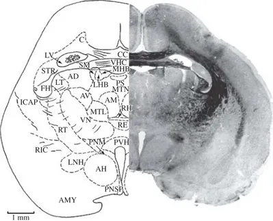
Figure 10.AD, anterodorsal thalamic nucleus; AH, anterior nucleus of hypothalamus; AM, anteromedial nucleus of thalamus; AMY, amygdala; AV, anteroventral thalamic nucleus; C, cingulum; CC, corpus callosum; FH, fimbria of hippocampus; ICAP, internal capsule; LHB, lateral nucleus of habenula; LNH, lateral nucleus of hypothalamus; LT, lateral thalamic nucleus; LV, lateral ventricle; MHB, medial nucleus of habenula; MTL, median nucleus of thalamus, lateral portion; MTN, median thalamic nucleus; PNM, paraventricular nucleus, magnocellular part; PNSP, paraventricular nucleus, parvicellular part; PS, periventricu-lar stellatocelluperiventricu-lar part; PVH, periventricuperiventricu-lar nucleus of hypothalamus; RE, reuniens thalamic nucleus; RH, rhomboid thalamic nucleus; RIC, retrolenticular internal capsule; RT, reticular thalamic nucleus; SM, stria medullaris of thalamus; STR, striatum; VHC, ventral hippocampal commissure; VN, ventral nucleus of thalamus. Section No. 151, 86-1, 9880Mm.
CC CC DHC DHC C
RCC
H H ADN ADNRFRFHBHB
SM
LT FH STR
VT CM
AV
ITR MTL MTLMTNMTN
AM AM VM
RE RE PVH
AH LH
OT
AMY SO
NII OC SCH
PS PS RH RH ICAP
RT RT TRM
ICRL ICRL
1 mm
NEO
C RCC
H
LT
DHC DHC SM SM ADN HBHB
RF RF
MT LMT AV
VNTD MTRI
VN
ICAP CP
LH
OT
AMY
VH DH
RE RH CM RT
FH STR
ICAP
ITR
ML VMVM ZI
SCH SCH
OC
1 mm 1 mm
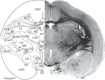
Figure 12.AD, anterodorsal thalamic nucleus; AMY, amygdala; AV, anteroventral nucleus of thalamus; C, cingulum; CM, centromedian thalamic nucleus; CP, cerebral peduncle; DH, dorsal nucleus of hypothalamus; DHC, dorsal hippocampal com-missure; FH, fimbria of hippocampus; H, hippocampus; HB, habenular nucleus; ICAP, internal capsule; ITR, inferior thalamic radiation; LH, lateral nucleus of the hypothalamus; LMT, lateral medial nucleus of thalamus; LT, lateral thalamic nucleus; ML, medial lemniscus; MT, median nucleus of thalamus; MTRI, intermediate thalamic radiation; NEO, neocortex; OC, optic chiasm; OT, optic tract; RE, reuniens thalamic nucleus; RCC, radiation of corpus callosum; RF, retroflex fasciculus; RH, rhomboid tha-lamic nucleus; RT, reticular thatha-lamic nucleus; SCH, suprachiasmatic nucleus; SM, stria medullaris of thalamus; STR, striatum (caudate-putamen) nucleus; VH, ventral nucleus of the hypothalamus; VM, ventromedian nucleus of thalamus; VN, ventral nucleus of thalamus; VNTD, ventral nucleus of thalamus, dorsal portion; ZI, zona inserta. Section No. 194, 79-5, 9220Mm.
PMN PFN VNTD VNTD
VH LGB
DG
H NOT CPL
V3
RF RF PNT LT
OT CP
FH
ML MTG
DH
VH LHZI MPF
SOPC AMY
SRT SRT
PCOM PCOM PTN
IMN HB
1 mm
MGB LGB
VNTVNTD PNT DG BSC
LNPPTNPTNCSCCSC PB PB NOT
PCOM PCOM IMN
RF RF
ML MTF
PNH PNH H2H1 CP
H HN OT STRASTRA
PFNPFN
AN I
AMY MPF
ZI
DPN DPN
VPN
NEO SNGSNG
1 mm
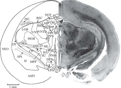
Figure 14.AMY, amygdala; AN, arcuate nucleus; BSC, brachium of superior colliculus; CP, cerebral peduncle; CSC, com-missure of superior colliculus; DG, dentate gyrus; DPN, dorsal premammillary nucleus; H, hippocampus; HN, hippocampal nucleus; H1, H2, Forel’s fields; I, infundibulum; IMN, interstitial magnocellular nucleus of posterior commissure; LGB, lateral geniculate body; LNP, lateral nucleus of thalamus (posterior portion); MGB, medial geniculate body; ML, medial lemniscus; MPF, medial prosencephalic nucleus; MTF, mamillotegmental fasciculus; NEO, neocortex; NOT, nucleus of optic tract; OT, optic tract; PB, pineal body (epiphysis); PCOM, posterior commissure; PFN, parafascicular nucleus; PNH, posterior nucleus of hypothalamus; PNT, posterior nucleus of thalamus; PTN, pretectal nucleus; RF, retroflex fasciculus; SNG, substantia nigra; STRA, superior thalamic radiation; VNT, ventral nucleus of thalamus; VNTD, ventral nucleus of thalamus, dorsal portion; VPN, ventral premamillary nucleus; ZI, zona incerta. Section No. 225, 72-5, 8560 Mm.
BSC LN
SC PB
CSC
PNT
PCOM PCOM ND ND
RFRF VNTD VNT MGB LGB DN DN
ML
SNG SNGZI BIC
STRA
DG
HN CP
MTF
MPF
PNH AN
HYP I H
1 mm
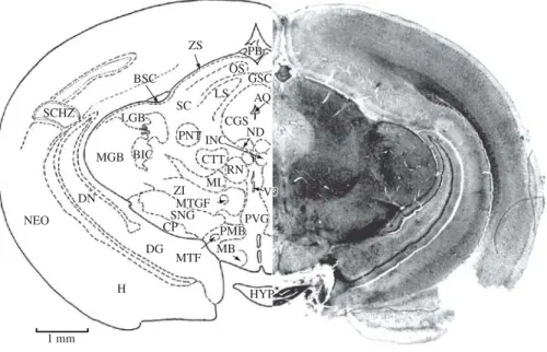
ZS
BSC
SC LGB LGB
LS OS
GSC
AQ
ND CGS
RN CTT PNT
MGB BICBIC
DN
NEO
H DG
CP SNG
ZI ML MTGF MTGF
PVG PVG PMB PMB MB MTF
HYP HYP INC INC
V3 V3 SCHZ
PB PB
1 mm
Figure 16.AQ, aqueduct, cerebral; BIC, brachium of inferior colliculus; BSC, brachium of superior colliculus; CGS, central gray substance; CP, cerebral peduncle; CSC, commissure of superior colliculus; CTT, central tegmental tract; DG, dentate gyrus; DN, dentate nucleus; H, hippocampus; HYP, hypophysis; INC, interstitial nucleus of Cajal; LGB, lateral geniculate body; LS, leminiscal stratum; MB, mamillary body; MGB, medial geniculate body; ML, medial lemniscus; MTGF, mamil-lotegmental fasciculus; MTF, mamillothalamic fasciculus; ND, nucleus of Darkschewitsch (one of the accessory oculomotor nuclei); NEO, neocortex; OS, optic stratum; PB, pineal body; PMB, peduncle of mamillary body; PNT, posterior nucleus of thalamus; PVG, periventricular gray; RN, red nucleus; SC, superior colliculus; SCHZ, schizocortex; SNG, substantia nigra; ZI, zona incerta; ZS, zonal stratum; V 3, third ventricle. Section No. 251, 66-1, 7980Mm.
NEO
OS ZS SC
LS
RS CTT
CGS
RN NON MLF
NIV
IPN SNG
CP DG
H
DN
HYP MGB BICBIC
ML
RSTD NIII
1 mm
NEO
BIC
SC OS
LS CGS
CSC
DLLL
CTT MGB
RST SNG
CPML P IPN
HYP NIII
DBC DTST MLF
OCN OCN
1 mm
Figure 18.BIC, brachium of inferior colliculus; CGS, central gray substance; CP, cerebral peduncle; CSC, commissure of superior colliculus; CTT, central tegmental tract; DBC, decussation of middle cerebral peduncle; DLLL, dorsal loop of lateral lemniscus; DTST, decussation of tectospinal tract; HYP, hypophysis; IPN, interpeduncular nucleus; LS, lemniscal stratum; MGB, medial geniculate body; ML, medial lemniscus; MLF, medial longitudinal fasciculus; NEO, neocortex; N III, oculomotor nerve; OCN, oculomotor nucleus; OS, optic stratum; P, pons; RST, rubrospinal tract; SC, superior colliculus; SNG, substantia nigra. Section No. 295, 58-1-5, 7380Mm.
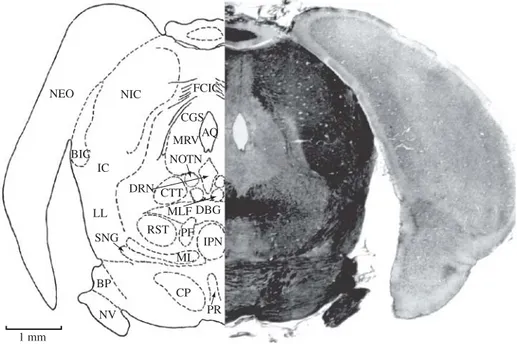
NEO
IC NIC
BIC
LL
SNG RST PF IPN ML
CP PR NV
BP
CTT DBG NOTN
AQ FCIC
CGS MRV
DRN MLF
1 mm
CER
IC NEO
CGS CGS MTNV MTNV
RST
IPN ML LL
PR
CP LPN NV
BP
PF PF BIC MLF
DBC DRN
1 mm
Figure 20. BIC, brachium of inferior colliculus; BP, brachium of middle cerebral peduncle; CER, cerebellum; CGS, central gray substance; CP, cerebral peduncle; DBC, decussation of brachium of inferior colliculus; DRN, dorsal raphe nuclei; IC, inferior colliculus; IPN, interpeduncular nucleus; LL, lateral lemniscus; LPN, lateral pontine nuclei; ML, medial lemniscus; MLF, medial longitudinal fasciculus; MTNV, mesencephalic tract of trigeminal nerve; NEO, neocortex; NV, trigeminal nerve; PF, predorsal fasciculus; PR, pontine raphe and nuclei; RST, rubrospinal tract. Section No. 329, 51-1-4, 6640 Mm.
CER
IC
LL
MRV
BC BC
MLF NDT NDT CGS V4
RST RETF
TST TSTDRN
ML
MRN TB NTB LL NLL NLL NV
BP VCN
NV NV PFL
DLF DLF
PT PT PT PT 1 mm
TB SOC
NVII VCN PFL
DN
DLF MLFMLF
TST
RPN RPN
ML ML
NTB NTB TBML TBML RST NVII RB BP BP
PNNV PNNV
MNNV MNNV NV STNV STNV
1 mm 1 mm
BIC
PT
(CSTA) (CSTA)
Figure 22.BIC, brachium of inferior colliculus; BP, brachium pontis; DLF, dorsal longitudinal fasciculus (mesencephalic); DN, dentate nucleus; ML, medial lemniscus; MLF, medial longitudinal fasciculus; MNNV, motor nucleus of trigeminal nerve; NTB, nucleus of trapezoid body; NV, trigeminal nerve; N VII, facial nerve; PFL, paraflocculus; PNNV, principal nucleus of trigeminal nerve; PT, pyramidal tract (corticospinal), anterior; RB, restiform body; RPN, raphe nucleus; RST, rubrospinal tract; SOC, superior olivary nucleus (complex); STNV, spinal tract of trigeminal nerve; TB, trapezoid body; TBML, trapezoid body and medial lemniscus; TST, tectospinal tract; VCN, ventral cochlear nucleus. Section No. 402, 35-1-2, 5360Mm.
CER
GEN
DN
FN FN
V4
PFL PFL
VTCN STNV
TN LVN
SOC NOFN
RPN ML TST
GFN GFN NVI
MLF BIC
BIC SVN SVN RB
NCFBT NCFBT NCFBT
RST RST
VCN VCN
NVIII
PFI
DPT DPT 1 mm
LAF AT
CP
RS
SN NOFN NOFN NIXNGN
DLF ML
TST
ION 1 mm
V4 V4 MVN SNSTVN
STNV
RB
VCN VCN
MLF MLF
VSCT VSCT
NPHN NPHN
) PT (CSTL) PT (CSTL) PT PT (C(CSTTLTL)))L)
Figure 24.AT, acoustic tuberculum; CP, choroid plexus; DLF, dorsal longitudinal fasciculus; ION, inferior olivary nucleus; LAF, lateral aperture, fourth ventricle; ML, medial lemniscus; MLF, medial longitudinal fasciculus; MVN, medial vestibular nucleus; NGN, nucleus of glossopharyngeal nerve; NOFN, nucleus of origin of facial nerve; NPHN, nucleus prepositus of hypoglossal nerve; N IX, glossopharyngeal nerve; PT (CSTL), pyramidal (corticospinal tract); RB, restiform body; RS, reticular substance; SN, salivatory nucleus; SNSTVN, spinal nucleus and spinal tract of vestibular nerve; STNV, spinal tract of trigeminal nerve; TST, tectospinal tract; VCN, ventral (anterior) cochlear nucleus; N VIII, vestibulocochlear nerve; VSCT, ventral spinocerebellar tract; V 4, fourth ventricle. Section No. 464, 25-2-4, 4300 Mm.
CER MOL
GNL
PL
FL
RB
STNV
LRN
NSTNV
PT (CSTL) VSCT
ION RS NX AFAF
MLF HNO HNO DFLDFL SPV4 SPV4 V4 NFG NFG NFC
FSNFS FSNFS
NDOVN NDOVN
TST TST
ION
NXII VCST DAON DAON
1 mm
FG FC
NX
DSCT
LST LST VSCT
SOT
LVST TST VCST VCST RF
MLF LCST
RST
RST NDOANNDOAN
NVOAN NSTNV
NSTNV
STNV
0.5 mm
DAON, dorsal (posterior) accessory olivary nucleus; DLF, dorsal longitudinal fasciculus; DSCT, dorsal (poste-rior) spinocerebellar tract; ENFC, external nucleus of fasciculus cuneatus; FC, fasciculus cuneatus; FG, fasciculus gracilis; FSNFS, fasciculus solitarius and nucleus of fasciculus solitarius; HNO, hypoglossal nucleus of origin; IAF, internal arcuate fibers; ION, caudal inferior olivary nucleus; LCST, lateral corticospinal tract; LSTT, lateral spinothalamic tract; LVST, lateral vestibulospinal tract; ML, medial lemniscus; MLF, medial longitudinal fasciculus; NDOVN, nucleus of dorsal origin of vagus nerve; NFC, nucleus of fasciculus cuneatus; NFG, nucleus of fasciculus gracilis; NVOVN, nucleus of ventral origin of vagus nerve; N X, vagus nerve; N XII, hypoglossal nerve; RB, restiform body (inferior cerebellar peduncle); RS, reticular substance; SOT, spinoolivary tract (end point); STNNV, spinal tract and nucleus of trigeminal nerve; TST, tectospinal tract; VCST, ventral corticospinal tract; VSCT, ventral spinocerebellar tract. Section No. 565, 15-2-5, 1900Mm.
Figure 27.DSCT, dorsal spinocerebellar tract; FC, fasciculus cuneatus; FG, fasciculus gracilis; LCST, lateral (pyramidal) corticospinal tract; LST, lateral spinothalamic tract; LVST, lateral vestibulospinal tract; MLF, medial longitudinal fasciculus; NDOAN, nucleus of dorsal origin of accessory nerve; NSTNV, nucleus of spinal tract of trigeminal nerve; NVOAN, nucleus of ventral origin of accessory nerve; RF, reticular formation; RST, rubrospinal tract; SOT, spinoolivary tract; STNV, spinal tract of trigeminal nerve; TST, tectospinal tract; VCST, ventral corticospinal tract; VSCT, ventral spinocerebellar tract. Section No. 675, 4-2-10, 760Mm.
FC RB ENFC
DSCT
STNNV
NX SOT
ION
NXII
TST ML MLF MLF
VCST RS IAF
HNO HNO NDOVN
NVOVN NVOVN
LCST
LCST LCST LSTT LSTT VSCT
LVST
NFC FG
NFG
DAON DAON FSNFS
DLF
teran species including the Desmodontidae. Data on Desmodus and Diphylla have been calculated and shown separately. The values for human brain are provided for comparison. (After Baron et al., 1996a, p. 89).
Megachiroptera % (Range)
Microchiroptera % (Range)
Desmodus (%)
Diphylla (%)
Human (%) Medulla oblongata 7.1 (5.3-8.9) 13.1 (7.8-19.7) 8.5 8.3 0.8 Cerebellum 13.5 (11.5-16) 19.5 (15.3-28.9) 17.6 17.4 11.0 Mesencephalon 5.4 (4.2-6.9) 9.8 (5.5-14.6) 6.2 7.0 0.6 Diencephalon 8.6 (7.9-9.4) 8.0 (6.7-10.6) 7.9 8.4 2.7 Telencephalon 65.3 (60.1-70.2) 49.6 (37.7-63.4) 59.8 58.9 85.0
Table 3. Neurobiological data on Desmodus rotundus (from Baron et al., 1996a; b; c).
Brain length 16.80 mm
Standard brain volume 964 mm3
Net brain volume 944 mm3
Brain weight 999 mg
Body weight 36.3 g
Volumes of brain parts, mm3
Telecephalon 564.8
Neocortex 312.5
main olf bulb 33.0
acc. olf bulb 0.355
Diencephalon 74.9
Mesencephalon 58.3
Cerebellum 166.4
Medulla oblongata 80.0
opallium and the synaptic cascades of its sectors, cortical involvement of instinctive action patterns and responses based on neocortical associations are the remaining cir-cuitary. The pursuit of the main and the accessory olfac-tory systems in Desmodus is likely to provide the neuro-anatomical explanation of its behavioral complexities.
For neurological information on Desmodus, the read-er is directed to Greenhall et al. (1983, species account), Mann (1960, neurobiology), Escobar et al. (1968, infe-rior olivary nucleus), Yamamoto et al. (1955, abducens nucleus, pyramidal tract and the vestibular nucleus), Palacios Prü and Briceno (1972, glial cell and neuro-nal dendrite ultrastructural relationship in the olfactory bulb), and Palacios Prü (1970, ultrastructure of mossy fibers).
The cytoarchitectural organization of the Desmodus brain deserves to be compared in parallel with that of other vampire species, bats of other families (especially megachiroptera), and some other representative mam-mals. When nuclear groups, fiber tracts, and their inter-connections are thus compared and contrasted, the neu-roanatomical basis of behavior in the vampire is more likely to become apparent. This atlas has been presented with exactly that future goal in mind.
Acknowledgements — A serial section series of the vampire brain prepared by Lois Copeland was kindly gifted to the author by the late Professor William Abel Wimsatt of Cornell University, Ithaca, New York. Without his help and encouragement, not only this atlas, but the author’s other studies in the field of bat biology and neuroanatomy would not have become a reality. As a tribute to a great mentor and comparative morpho- physiologist, this work is dedicated to Professor William Wimsatt. Cathy Caple at Louisville spent long hours in the dark room to create the best photographic images from the serial sections. Lucinda Schultz helped with histological preparations. Grateful thanks are due to the Editor-in-Chief, Dr Takako Matsumara-Tundisi, and Dr. Paula Matvienko-Sikar, of the Brazilian Journal of Biology®,
for encouragement and many courtesies. Dr. Wilson Uieda and Dr. Jader MarinhoFilho kindly provided the Scholarly support.
References
BARON, G., STEPHAN, H. and FRAHM, H., 1996a. Comparative Neurobiology in Chiroptera: macromorphology, brain structures, tables and atlases. Birkhäuser Verlag: Basel. vol. 1, p. 1-529.
olivary complex of the vampire bat, Desmodus rotundus (Chiroptera: Phyllostomidae). Acta Chiropterologica, vol. 1, no. 1, p. 81-92.
-, 2000. The nuclei of the lateral leminiscus of the vampire bat, Desmodus rotundus. Association for Research in Otolaryngology, vol. 23, p. 180. (abstract).
McDANIEL, VR., 1976. Brain anatomy. In Baker, RJ., Jones, JK. and Carter, DC. (Eds.). Biology of Bats of the New World Family Phyllostomatidae. Part I. Lubbock: Mus. Texas Tech Univ. Spec. Publ. vol. 10, p. 147-200.
MANN, G., 1960. Neurobiologia de Desmodus rotundus. Invest. Zool. Chil., vol. 6, p. 79-99.
-, 1963. The rhinencephalon of Chiroptera. Invest. Zool. Chil., vol. 9, p. 1-93.
PALACIOS-Prü, EL., 1970. Aspectos estructurales sobre fibras musgosas: studio comparative Cavia cobaya y Desmodus rotundus. Método de Golgi y microscopía electrónica. Acta Cient. Venezolana, vol. 21, p. 226-233.
PALACIOS-Prü, EL. and BRICENO, RVM., 1972. An unusual relationship between glial cells and neuronal dendrites in olfactory bulbs of Desmodus rotundus. Brain Res., vol. 36, no. 2, p. 404-408.
PANNETON, WM. and WATSON, BJ., 1991. Stereotaxic atlas of the brainstem of the muskrat, Ondatra zibethicus. Brain Res. Bull., vol. 26, no. 4, p. 479-509.
PAXINOS, G. and WATSON, C., 1997. The rat brain in stereotaxic coordinates. New York: Academic.
SCHNEIDER, R., 1957. Morphologische Untersuchungen am Gehirn der Chiropteren (Mammalia). Abh. Senckenb. Naturforsch. Ges., vol. 495, p. 1-92.
-, 1966. Das Gehirn von Rousettus aegyptiacus (E. Geoffroy 1810) (Megachiroptera, Chiroptera, Mammalia). Ein mit Hilfe mehrerer Schnittserien ersteller Atlas. Abh. Senckenb. Naturforsch. Ges. vol. 513, p. 1-160.
TAMURA, I., 1950. Comparative anatomical studies on the brain stem, with special references to the reticular formation and its relating nuclei of Chiroptera. Acta Anat. Niigata, Ensia Niigata,vol. 50, p. 65-98.
WAGNER, JA., 1840. Die Säugethiere in Abbildungen nach der Natur. Suppl. 1. Abt. Die Affen und Flederthiere, München. 558 p.
VILLIGER, E., LUDWIG, E. and RASMUSSEN, AT., 1951. Atlas of the cross section anatomy of the brain. New York: Blakiston.
YAMAMOTO, S., SHIMODA, B., MOMMA, R. and SAI, H., 1955. Studies on brain stem. IX. Comparative anatomical study of brain stems of some species of bats. Tohoku J. Exp. Med., vol. 61, no. 4, p. 339-344.
haracteristics in functional systems, echoethological adaptation, adaptive radiation and evolution. Birkhäuser Verlag: Basel. vol. 3, p. 1075-1596.
BHATNAGAR, KP., 1980. The chiropteran vomeronasal organ: its relevance to the phylogeny of bats. In Wilson, DE. and Gardner, AL. (Eds.). Proceedings of the Fifth International Bat Research Conference. Lubbock: Texas Tech Press. p. 289-315. -, 1988a. Anatomy. In Greenhall, AM. and Schmidt, U. (Eds.). Natural History of Vampire Bats. Boca Raton, Florida: CRC Press. p. 41-70.
-, 1988b. Ultrastructure of the pineal body of the common vampire bat, Desmodus rotundus. Am. J. Anat., vol. 181, no. 2, p. 163-178.
BHATNAGAR, KP., and KALLEN, FC., 1974. Morphology of the nasal cavities and associated structures in Artibeus jamaicensisandMyotis lucifugus. Am. J. Anat., vol. 139, no. 2, p. 167-190.
BHATNAGAR, KP., FRAHM, HD. and STEPHAN, H., 1990. The megachiropteran pineal organ: a comparative morphological and volumetric investigation with special emphasis on the remarkably large pineal of Dobsonia praedatrix. J. Anat. (Lond.),vol. 168, p. 143-166.
BRAUER, K., and SCHOBER, W., 1970. Catalogue of Mammalian Brains. Part I. Jena: Fischer.
-, 1976. Catalogue of Mammalian Brains. Part II. Jena: Fischer.
COOPER, JG. and BHATNAGAR, KP., 1976. Comparative anatomy of the vomeronasal complex in bats. J. Anat. (Lond.), vol. 122, no. 3, p 571-601.
ESCOBAR, A., SAMPEDRO, ED. and DOW, RS., 1968. Qualitative data on the inferior olivary nuclei in man, cat and vampire bat. J. Comp. Neur., vol. 132, p. 397-404.
FAUBER, J., 2003. Vampire bats’ saliva may help in treating blood clots, stroke. Milwaukee Journal Sentinel, January 10. GREENHALL, AM., JOERMANN, G., and SCHMIDT, U., 1983. Desmodus rotundus. Mammalian Species, vol. 202, p. 1-6.
HAWKEY, CM., 1966. Plasminogen activator in the saliva of the vampire bat, Desmodus rotundus. Nature, vol. 211, p. 434-435. HENSON, OW., 1970. The central nervous system. In Wimsatt, WA. (Ed.). Biology of Bats. vol. II. New York: Academic. p. 57-152.
HUMPHREY, T., 1936. The telecephalon of the bat. I. The non-cortical nuclear masses and certain pertinent fiber connections. J. Comp. Neur., vol. 65, no. 1, p. 603-711.