ISSN 0100-879X
BIOMEDICAL SCIENCES
AND
CLINICAL INVESTIGATION
www.bjournal.com.br
www.bjournal.com.br
Volume 45 (6) 473-564 June 2012
Braz J Med Biol Res, May 2012, Volume 45(6) 482-487
doi: 10.1590/S0100-879X2012007500040
Chi-square analysis of the reduction of ATP levels in L-02
hepatocytes by hexavalent chromium
Yang Yuan, Li Peng, Hu Gong-Hua, Dai Lu, Zhong Xia-Li1, Zhou Yu and Zhong Cai-Gao
Institutional Sponsors
The Brazilian Journal of Medical and Biological Research is partially financed by
Faculdade de Medicina de Ribeirão Preto Campus
Ribeirão Preto
Explore High - Performance MS Orbitrap Technology In Proteomics & Metabolomics
Chi-square analysis of the reduction of
ATP levels in L-02 hepatocytes
by hexavalent chromium
Yang Yuan
1,2, Li Peng
1, Hu Gong-Hua
1, Dai Lu
1, Zhong Xia-Li
1, Zhou Yu
1and Zhong Cai-Gao
11School of Public Health, Central South University, Changsha Hunan, China 2Department of Clinical Laboratory, Huaihua Medical College, Huaihua Hunan, China
Abstract
This study explored the reduction of adenosine triphosphate (ATP) levels in L-02 hepatocytes by hexavalent chromium (Cr(VI)) using chi-square analysis. Cells were treated with 2, 4, 8, 16, or 32 μM Cr(VI) for 12, 24, or 36 h. Methyl thiazolyl tetrazolium (MTT) experiments and measurements of intracellular ATP levels were performed by spectrophotometry or bioluminescence assays following Cr(VI) treatment. The chi-square test was used to determine the difference between cell survival rate and ATP levels. For the chi-square analysis, the results of the MTT or ATP experiments were transformed into a relative ratio with respect to the control (%). The relative ATP levels increased at 12 h, decreased at 24 h, and increased slightly again at 36 h following 4, 8, 16, 32 μM Cr(VI) treatment, corresponding to a “V-shaped” curve. Furthermore, the results of the chi-square analysis demonstrated a significant difference of the ATP level in the 32-μM Cr(VI) group (P < 0.05). The results suggest that the chi-square test can be applied to analyze the interference effects of Cr(VI) on ATP levels in L-02 hepatocytes. The decreased ATP levels at 24 h indicated disruption of mitochondrial energy metabolism and the slight increase of ATP levels at 36 h indicated partial recovery of mitochondrial function or activated glycolysis in L-02 hepatocytes.
Key words: Hexavalent chromium; ATP level; Interference effect; Chi-squaretest
Introduction
Correspondence: Zhong Cai-Gao, School of Public Health, Central South University, Changsha Hunan, 410078 China. E-mail: [email protected]
Received May 21, 2011. Accepted March 9, 2012. Available online March 23, 2012. Published June 4, 2012. Hexavalent chromium, Cr(VI), is a well-documented
human carcinogen and is widely found in human living environments as the result of industrial production or discharge (1). Recently, Cr(VI) pollution has been found in crops due to the uptake of Cr(VI) from the soil and in rivers in some regions of China (2,3). The toxicity caused by oral Cr(VI) ingestion is thought to be due to toxicity to the liver, which is the main organ of biological metabolism, and liver damage (or hepatotoxicity) from Cr(VI) exposure
has been confirmed in animal experiments and in cultured
L-02 hepatocytes, which showed hepatocyte ultrastructure disruption, mitochondrial damage and apoptosis (4-6).
Mitochondria are the main site of ATP synthesis, which is produced by the tricarboxylic acid cycle (TCA) and oxida-tive phosphorylation (OXPHOS) in the inner mitochondrial membrane (7). At present, the detailed interference effect of Cr(VI) on the cellular ATP levels is not known. Gener-ally, Cr(VI) can induce apoptosis and lead to a decrease in
cell survival or cell number, and differences in cell number result in the different cellular ATP levels. And in this case manual adjustment of cell number is commonly applied to balance differences in cell number in each group. Small sample t-tests or one-way ANOVA were applied to compare the differences between the Cr(VI) treatment groups and control. However, in our view, adjusting the cell number is not the only way to analyze the toxic effects of Cr(VI)
in vitro. The chi-square statistical test (χ2) can also be
used to analyze the physiological or toxicological effects of Cr(VI) in vitro.
The χ2 test is a commonly used statistical method
and consists of the Pearson chi-square, linear-by-linear chi-square, McNemar and Mantel-Haenszel tests, among
others. Currently, χ2 analysis is widely applied to compare
the difference of a relative ratio existing between two or
Reduction of ATP levels in L-02 hepatocytes by Cr(VI) 483
factors, to assess risk, and to predict trends of disease
development. However, the χ2 test is rarely applied in the
field of in vitro cytotoxicity. For this reason, after cultured
L-02 hepatocytes were exposed to 0, 2, 4, 8, 16, and 32 μM Cr(VI) for 12, 24, or 36 h, a χ2 test was applied to analyze
the interference effect by comparing the difference between cell survival rate and intracellular ATP levels to establish a novel method of analyzing the cytotoxicity induced by toxic chemicals in vitro.
Material and Methods
Material
Potassium dichromate [K2Cr2O7, abbreviated as Cr(VI)]
was purchased from Sigma (USA). Roswell Park Memorial Institute (RPMI-1640) culture medium was purchased from Solarbio (USA), and newborn calf serum was purchased from Shi Ji Qing (China). A methyl thiazolyl tetrazolium (MTT) cell death kit was purchased from Amresco (USA). The ATP assay kit-S0026 was purchased from Beyotime (China). The human embryonic liver cell line L-02 (L-02 hepatocytes) was obtained from the Shang Hai Center of Cell Culture of Chinese Academy of Sciences. A 0.1 M Cr(VI) stock solution was prepared by adding 29.418 g K2Cr2O7
to 1000 mL ddH2O. The stock was then diluted in culture
medium to 2, 4, 8, 16, 32 µM.
L-02 hepatocyte culture and Cr(VI) exposure
L-02 hepatocytes were cultured on a six-well plate with RPMI-1640 medium containing 15% newborn calf serum at 37°C in a 5% CO2 atmosphere. The culture medium was
changed every 1-2 days. When the cell density reached 60%
confluence, the cells were exposed to Cr(VI) for different
periods of time (12, 24, or 36 h) at 37°C. Untreated cultures were used as a control group. Cell survival was analyzed by the MTT method according to manufacturer instructions.
MTT reduction assay
The MTT assay was performed according to manufac-turer instructions. The growing cells were collected by 0.25% Trypsin digestion, centrifugation, and supernatant removal. Two milliliters of RPMI-1640 culture medium containing 15% newborn calf serum was added to resuspend the cells as a single cell suspension. The cell suspension was then inoculated in a 96-well culture plate at a density of 1.0 x 10⁴ cells/well. The following day, the cells were grown in medium containing 0, 2, 4, 8, 16, or 32 µM Cr(VI) for 12, 24, or 36 h at 37°C. Following Cr(VI) treatment, MTT was
added at a volume of 10 μL/well and cultured for 4 h at 37°C, then 100 μL formazan lysate was added, and the cells were
cultured for 6 h at 37°C. Finally, the 96-well culture plate was removed from the incubator and continually shaken for 5 min on a micro-oscillator to completely dissolve the formazan. Immediately, cell vitality was analyzed by measur-ing absorbance at 492 nm with a multifunction microplate
reader (Thermo Varioskan Flash 3001, USA).
ATP bioluminescence assay
L-02 hepatocytes were seeded at a density of 2.5 x 10⁵
cells/well on three six-well plates. When the cells reached
60% confluence, they were exposed to Cr(VI) for 12, 24,
or 36 h. Following Cr(VI) treatment, intracellular ATP levels were determined using a bioluminescent ATP assay kit. The
cells were disrupted in 200 μL lysis buffer by mechanical
disruption, and centrifuged at 12,000 g to collect the cell
supernatant. Meanwhile, an aliquot (100 μL) of an ATP
detection working solution was added to each well of a black 96-well culture plate and incubated for 3 min at room
temperature. Then, four replicates of 40-μL samples of the
cell lysate from each group were added to the wells. After allowing the reaction to take place for a few seconds, the luminescence value was measured. In addition, the 96-well plates also contained serial dilutions of an ATP standard solution to generate a standard curve, and the ATP levels in L-02 hepatocytes were calculated by comparison with the ATP standard curve.
Data analysis
Data were analyzed statistically with Microsoft Office
Excel 2003 and SPSS 13.5. The results of the ATP and MTT assays are reported as means ± SD. The statistical
significance of differences between means was deter -mined by an F-test (ANOVA analysis) followed by least
significant difference (LSD) post hoc tests. The survival rate of the cultured cells (from the MTT assay) and the relative ATP levels are reported as percent (%) change
from control. Statistical significance was determined by Pearson chi-square or linear χ2 tests. For the purpose
of χ2 analysis,the compared groups were divided by the
same number to achieve a gain of less than 100%. A P
< 0.05 values (two-sided test) was accepted as statisti
-cally significant.
Results
Cell viability
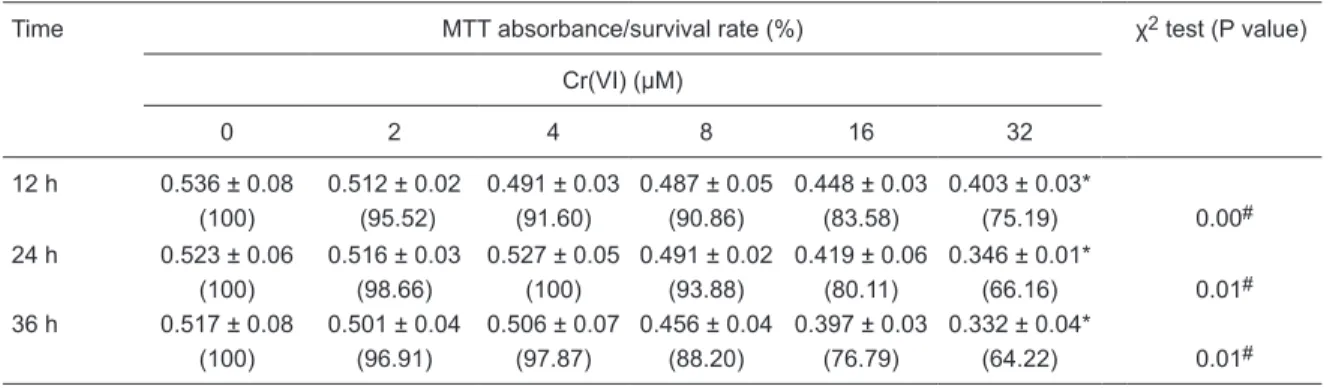
Following treatment with 2, 4, 8, 16, and 32 µM Cr(VI), L-02 hepatocyte viability decreased progressively over
12, 24, or 36 h (P < 0.05). The survival rates ranged from
88.20 to 100% after treatment with low concentrations of Cr(VI) (2, 4, and 8 µM), and the high Cr(VI) concentrations (16 and 32 µM) led to lower cell survival rates (64.22 to 83.58%). Further details from this experiment are shown in Table 1.
ATP level in L-02 hepatocytes
Following 12 h of Cr(VI) treatment, the ATP levels of L-02 hepatocytes were increased. However, after 24 h of
treatment, intracellular ATP levels decreased significantly
µM Cr(VI) group. Following 36 h of Cr(VI) treatment, the low ATP levels showed a slight up-regulation, while the ATP levels in the 16 and 32 µM Cr(VI) groups remained lower than control. The graphic change of relative ATP
levels was described as a “V-shaped” curve (Table 2,
Figure 1).
χ2 analyses comparing cell survival rate and relative
ATP levels
Following 12 h of Cr(VI) treatment, the χ2 test showed
a significant difference in ATP levels in the 8, 16, and 32 µM groups (P < 0.05). Following 24 h of Cr(VI) treatment, the χ2 test showed a significant difference in ATP levels in
the 4, 8, 16, and 32 µM groups (P < 0.05). Following 36 h of Cr(VI) treatment, the χ2 test showed a significant differ
-ence in ATP levels in the 8 and 32 µM Cr(VI) groups (P< 0.05) (Table 3).
Figure 1. Relative ATP levels in L-02 hepatocytes following hexavalent chromium (Cr(VI)) treatment.
Table 1. Effect of Cr(VI) on the viability of L-02 hepatocytes.
Time MTT absorbance/survival rate (%) χ2 test (Pvalue)
Cr(VI) (μM)
0 2 4 8 16 32
12 h 0.536 ± 0.08 0.512 ± 0.02 0.491 ± 0.03 0.487 ± 0.05 0.448 ± 0.03 0.403 ± 0.03*
(100) (95.52) (91.60) (90.86) (83.58) (75.19) 0.00#
24 h 0.523 ± 0.06 0.516 ± 0.03 0.527 ± 0.05 0.491 ± 0.02 0.419 ± 0.06 0.346 ± 0.01*
(100) (98.66) (100) (93.88) (80.11) (66.16) 0.01#
36 h 0.517 ± 0.08 0.501 ± 0.04 0.506 ± 0.07 0.456 ± 0.04 0.397 ± 0.03 0.332 ± 0.04*
(100) (96.91) (97.87) (88.20) (76.79) (64.22) 0.01#
Cr(VI) = hexavalent chromium; MTT = methyl thiazolyl tetrazolium. Data are reported as means ± SD and percent within parentheses, N = 4. *P < 0.05 (least significant difference multiple comparisons followingthe F-test); #P < 0.05 for linearity (linear χ2 test).
Table 2. ATP levels in L-02 hepatocytes following hexavalent chromium (Cr(VI)) treatment.
Time ATP content/relative level (%) χ2 test (Pvalue)
Cr(VI) (μM)
0 2 4 8 16 32
12 h 8.44 ± 0.49 10.26 ± 0.63* 9.59 ± 0.56* 11.33 ± 0.26* 10.92 ± 0.42* 11.71 ± 0.39*
(100) (121.56) (113.62) (134.24) (129.38) (138.74) 0.03#
24 h 14.77 ± 0.68 17.22 ± 0.83* 10.10 ± 0.23* 8.10 ± 0.22* 5.32 ± 0.54* 1.62 ± 0.09*
(100) (116.58) (68.38) (54.84) (36.02) (10.96) 0.00#
36 h 19.27 ± 0.72 18.49 ± 2.04 21.05 ± 1.87 23.79 ± 0.52* 16.07 ± 1.61* 6.72 ± 0.09*
(100) (95.96) (109.24) (123.46) (83.40) (34.88) 0.15
Reduction of ATP levels in L-02 hepatocytes by Cr(VI) 485
Discussion
Cr(VI) is a common environmental pollutant that is widely
used in electroplating, metal refining, printing, dyeing, tan -ning, and other industrial and agricultural processes, and its carcinogenicity has been documented by the Interna-tional Research Agency of Cancer (IRAC) (1). In China, epidemiological studies suggested that occupational Cr(VI) exposure led to chronic damage of liver, lung, nasal mucosa, skin and other organs, and an increased risk of cancer inci-dence (8-10). Furthermore, the study of Cr(VI) cytotoxicity revealed that Cr(VI) could readily cross the cell membrane
through nonspecific anion channels, resulting in excessive
generation of reactive oxygen species. Consequently, induced oxidative stress, genetic damage, mitochondrial dysfunction, activation of apoptosis-related caspases, and mitochondrial-mediated apoptosis were observed (11-13), while chromium-induced genotoxicity and apoptosis were closely associated with Cr(VI) carcinogenesis (14).
Mitochondria are the main site of ATP synthesis, which is produced mainly through the TCA cycle and OXPHOS, named as mitochondrial aerobic respiration (7). Under nor-mal physiological conditions, mitochondrial aerobic
respira-tion is the main way of energy provision, while glycolysis in the cytoplasm is negligible due to the low effectiveness of ATP production (7). Interestingly, the glycolysis metabolism is activated as a compensatory means of energy produc-tion existing in many cancer cells (15-17). At present, it is unclear whether toxic chemicals cause also the activation of glycolysis in the process of toxicity. In an adverse environ-ment of exposure to toxic chemicals, several studies have shown that the disorder of energy metabolism induced by toxic chemicals was closely associated with mitochondrial dysfunction. For example, acute ethanol exposure led to suppression of mitochondrial ATP generation and fatty acid oxidation and decreased respiration and accessibility of mi-tochondrial adenylate kinase in permeabilized hepatocytes (18,19). Exposureto 5 and 10 µM Pb reduced decreased cellular ATP levels in the neuronal cell lines PC-12 and SH-SY5Y, which correlated with voltage-dependent anion channel (VDAC) transcription and expression (20). VDAC is an important protein located on the outer mitochondrial membrane, which controls mitochondrial life and death (21). At present, the effect of Cr(VI) hepatotoxicity on cellular ATP levels remains ambiguous; therefore, it is important to elucidate the interference effect of Cr(VI) on ATP levels Table 3. χ2 analysis of ATP level and cell viability of L-02 hepatocytes after treatment with hexavalent
chro-mium (Cr(VI)) for 12, 24, and 36 h.
Concentration of
Cr(VI) (μM) Cell survival rate (%) Relative level of ATP in cells (%)
12 h 100.00 121.56 113.62 134.24 129.38 138.74
0 100.00 1.000
2 95.52 0.065
4 91.60 0.120
8 90.86 0.002*
16 83.58 0.001*
32 75.19 0.000*
24 h 100.00 116.58 68.38 54.84 36.02 10.96
0 100.00 1.000
2 98.66 0.202
4 100.00 0.022*
8 93.88 0.003*
16 80.11 0.000*
32 66.16 0.000*
36 h 100.00 95.96 109.24 123.46 83.40 34.88
0 100.00 1.000
2 96.91 0.887
4 97.87 0.396
8 88.20 0.011*
16 76.79 0.564
32 64.22 0.014*
in L-02 hepatocytes.
Different doses of Cr(VI) can lead to differences in cell survival rates and cell number from control and consequently alter intracellular ATP levels. Therefore, it was interesting
to scientifically evaluate the interference effect of Cr(VI) on ATP level in cells. For the first time, a chi-square test was
used to analyze experimental data on the toxicity of Cr(VI), which is a novel method of analysis of the toxicological ef-fects induced by Cr(VI). Chi-square testing was applied to compare differences between cell survival rates and ATP
levels. If there were significant differences between the
variables, this would indicate that the change in intracel-lular ATP levels is not related to changes in celintracel-lular survival rates, which could indicate that Cr(VI) interferes with ATP synthesis in L-02 hepatocytes.
The experimental results showed that Cr(VI) led to a gradual decrease of cell survival rate in L-02 hepatocytes at 12, 24, or 36 h of exposure, and the 32 µM Cr(VI)
treat-ment was able to significantly decrease the cell survival rate.
Meanwhile, the relative ATP level showed a pattern of Cr(VI) interference with ATP levels described as an increase at 12 h, a decrease at 24 h, and a new slight increase at 36 h,
looking like a “V-shaped” curve. Furthermore, the results of the Pearson χ2 test showed that doses of 8, 16, and 32 µM
Cr(VI) induced a significant increase of ATP levels at 12 h, while 4, 8, 16, and 32 µM Cr(VI) doses induced a significant
decrease of ATP at 24 h. However, after Cr(VI) treatment for 36 h, the ATP levels increassed slightly again, but the
ATP levels in the 16 and 32 µM Cr(VI) groups were still lower than control. In our view, the increase of ATP level at 12 h indicated activation of mitochondrial aerobic respira-tion, the decreased ATP levels at 24 h indicated disruption of mitochondrial energy metabolism and, interestingly, the slight increase of ATP levels at 36 h indicated partial recovery of mitochondrial function or activated glycolysis in L-02 hepatocytes.
In summary, the χ2 test enabled us to distinguish the
confounding effects of decreased cell survival rate from
changes in intracellular ATP content. This study is the first
to demonstrate that exposure to 32 µM Cr(VI) leads to a
significant increase in cellular ATP at 12 h, a decrease at
24 h, and a slight increase again at 36 h. Furthermore, in
future studies, the χ2 statistical test could also be considered
as a reference for exploring cytotoxicity or pharmacological mechanisms of other chemicals. It would be interesting to further explore the molecular mechanism of mitochondrial
energy metabolism- or glycolysis-related genes by the χ2
method during Cr(VI) toxicity.
Acknowledgments
Research supported by the Natural Science Founda-tion of China (#30972511) and the Science InnovaFounda-tion Project from the Educational Committee of Hunan Province (#CX2010B098).
References
1. International Agency for Researchon Cancer. [Chromium, nickel and welding. IARC Monographs on the evaluation of the carcinogenic risks to humans]. IARC 1990; 51: 49-256. 2. Wang Wei, Liu Dong-hua, Jiang Wu-sheng, Hou Wen-qiang.
[Effects of plant growth in the soil polluted by chromium].
Agro-Environ Protection 2002; 21: 257-259.
3. Zhang Dong, Zhu Li-xia, Yin Guo-xun. [Investigation on hexavalent chromium pollution of karst groundwater in Ji-aozuo, China]. Earth Environ 2009; 37: 237-242.
4. Xiao-Ling Zhou, Han Ying-Shi, Ceng Qing-Shan, Wang Xiang-Pu. [Study of mouse liver tissue cytochemistry and morphology induced by acute chromium trioxide exposure].
J Toxicol 1990; 4: 92-94.
5. Zeng Ming, Wang Xiang-Pu, An Fei-Yun, Gao Ze-Xuan, Wang An. [An experimental study on the toxic liver and renal damage induced by chromium]. China Public Health 1999; 15: 869-870.
6. Xiao JW, Zhong CG, Li B. [Study of L-02 hepatocyte apop-tosis induced by hexavalent chromium associated with mitochondria function damage]. Wei Sheng Yan Jiu 2006; 35: 416-418.
7. Voet D, Voet JG, Pratt CW. Fundamentals of biochemistry. 2nd edn. Hoboken: John Wiley and Sons, Inc.; 2006. 8. Cai Shi-xiong, Huang Mei-yuan, Luo Yu-mei, Fu Zhen-ying,
Chui Yu-zhen, Chen Fu-ming. [Survey of malignant tumor
incidence in chromate salt production workers]. Chinese J Industr Hygiene Occupational Dis 1986; 4: 210-213. 9. Liao Yongling, Zhou Xu, He Chunlan, Tang Yaoyuan, Zou
Lunling, Chen ZhiLian, et al. [Survey on occupational dis-eases of skin and nasopharynx of 233 workers exposed to chromium]. Occup Health 2001; 17: 2-4.
10. Li Deng-jiu, Qian Sheng-feng, Li Liang, Guo Ping, Wang Xiao-fang. [Health survey of chromate salt production work-ers]. China Occup Safety Health Manag System Certification 2001; 21: 37-39.
11. Kasprzak KS. Oxidative DNA and protein damage in metal-induced toxicity and carcinogenesis. Free Radic Biol Med
2002; 32: 958-967.
12. Valko M, Rhodes CJ, Moncol J, Izakovic M, Mazur M. Free radicals, metals and antioxidants in oxidative stress-induced cancer. Chem Biol Interact 2006; 160: 1-40.
13. Son YO, Hitron JA, Wang X, Chang Q, Pan J, Zhang Z, et al. Cr(VI) induces mitochondrial-mediated and caspase-depen-dent apoptosis through reactive oxygen species-mediated p53 activation in JB6 Cl41 cells. Toxicol Appl Pharmacol
2010; 245: 226-235.
Reduction of ATP levels in L-02 hepatocytes by Cr(VI) 487
15. Gatenby RA, Gillies RJ. Why do cancers have high aerobic glycolysis? Nat Rev Cancer 2004; 4: 891-899.
16. Elstrom RL, Bauer DE, Buzzai M, Karnauskas R, Harris MH, Plas DR, et al. Akt stimulates aerobic glycolysis in cancer cells. Cancer Res 2004; 64: 3892-3899.
17. Shi DY, Xie FZ, Zhai C, Stern JS, Liu Y, Liu SL. The role of cellular oxidative stress in regulating glycolysis energy metabolism in hepatoma cells. Mol Cancer 2009; 8: 32. 18. Hoek JB, Cahill A, Pastorino JG. Alcohol and mitochondria:
a dysfunctional relationship. Gastroenterology 2002; 122: 2049-2063.
19. Holmuhamedov E, Lemasters JJ. Ethanol exposure
de-creases mitochondrial outer membrane permeability in cultured rat hepatocytes. Arch Biochem Biophys 2009; 481: 226-233.
20. Prins JM, Park S, Lurie DI. Decreased expression of the voltage-dependent anion channel in differentiated PC-12 and SH-SY5Y cells following low-level Pb exposure. Toxicol Sci 2010; 113: 169-176.