UNIVERSIDADE FEDERAL DO CEARÁ
FACULDADE DE FARMÁCIA, ODONTOLOGIA E ENFERMAGEM PROGRAMA DE PÓS-GRADUAÇÃO EM ODONTOLOGIA
FABIANNI MAGALHÃES APOLONIO
ANÁLISE DA ATIVIDADE PROTEOLÍTICA DAS MMPS -2 E -9 EM DENTINA HUMANA SADIA E SUA INTERAÇÃO NA INTERFACE ADESIVA
FABIANNI MAGALHÃES APOLONIO
ANÁLISE DA ATIVIDADE PROTEOLÍTICA DAS MMPS -2 E -9 EM DENTINA HUMANA SADIA E SUA INTERAÇÃO NA INTERFACE ADESIVA
Tese apresentada ao Programa de Pós-Graduação em Odontologia da Faculdade de Farmácia, Odontologia e Enfermagem da Universidade Federal do Ceará, como requisito parcial para obtenção do Título de Doutor em Odontologia.
Área de Concentração: Clínica Odontológica Orientador: Prof. Dr. Vicente de Paulo Aragão Saboia
Dados Internacionais de Catalogação na Publicação Universidade Federal do Ceará
Biblioteca de Ciências da Saúde
A654a Apolonio, Fabianni Magalhães.
Análise da atividade proteolítica das mmps -2 e -9 em dentina humana sadia e sua interação na interface adesiva / Fabianni Magalhães Apolonio. – 2014.
94 f. : il.
Tese (Doutorado) – Universidade Federal do Ceará, Faculdade de Farmácia, Odontologia e Enfermagem, Departamento de Clínica Odontológica, Programa de Pós-Graduação em Odontologia, Doutorado em Odontologia, Fortaleza, 2014.
Área de Concentração: Clínica Odontológica.
Orientação: Prof. Dr. Vicente de Paulo Aragão Saboia.
1. Ácido Cítrico. 2. Adesivos Dentinários. 3. Colágeno. 4. Dentina. I. Título.
FABIANNI MAGALHÃES APOLONIO
ANÁLISE DA ATIVIDADE PROTEOLÍTICA DAS MMPS -2 E -9 EM DENTINA HUMANA SADIA E SUA INTERAÇÃO NA INTERFACE ADESIVA
Tese apresentada ao Programa de Pós-Graduação em Odontologia da Faculdade de Farmácia, Odontologia e Enfermagem da Universidade Federal do Ceará, como requisito parcial para obtenção do Título de Doutor em Odontologia.
Aprovada em: ___/___/___
BANCA EXAMINADORA
_______________________________________________ Prof. Dr. Vicente de Paulo Aragão Saboia (Orientador)
Universidade Federal do Ceará – UFC
_______________________________________________ Prof. Dr. Sérgio Lima Santiago
Universidade Federal do Ceará – UFC
_______________________________________________ Prof. Dr. Mário Áureo Gomes Moreira
Universidade Federal do Ceará – UFC
_______________________________________________ Prof. Dra. Polliana Mendes Candia Scaffa
Universidade Anhanguera de São Paulo
_______________________________________________ Prof. Dra. Vanara Florêncio Passos
AGRADECIMENTOS ESPECIAIS
À DEUS, por ter me dado a vida e por tê-la mantido sempre com tantas alegrias e repleta de pessoas especiais.
À minha FAMÍLIA, pelo apoio, exemplo e carinho. E em especial aos meus pais, MARIA VALDECLEIDE MAGALHÃES APOLONIO e FRANCISCO FABIO APOLONIO, meu marido, LUCIANO MOREIRA DE SOUSA NETO e ao meu irmão, MATHEUS MAGALHÃES APOLONIO por serem meu alicerce e fortaleza em todos os momentos da vida.
Ao meu professor orientador, Dr. VICENTE DE PAULO ARAGÃO SABOIA, por ter contribuído para minha formação intelectual, científica e profissional durante o mestrado e doutorado. Agradeço pela paciência, ensinamentos, conselhos, inteligência e honestidade em todo esse período. Durante estes anos, você não nos ensinou somente a sermos pesquisadores melhores, mas a sermos pessoas melhores e íntegras. Obrigada pela confiança e apoio dedicados a mim.
Aos professores Dr. LORENZO BRESCHI e Dra. ANNALISA MAZZONI, pela valiosa contribuição à minha formação e orientação durante estágio doutoral no exterior realizado no Laboratório do Instituto de Pesquisas Rizzoli – Universidades de Bologna e Trieste – Itália. Aprendi muito como pesquisadora nesse período. Muito obrigada pelo carinho e apoio.
Aos professores SÉRGIO LIMA SANTIAGO, LIDIANY KARLA AZEVEDO RODRIGUES, MONICA YAMAUTI, CARLOS AUGUSTO OLIVEIRA FERNANDES e demais professores da pós-graduação pela convivência e ensinamentos durante o curso de Doutorado.
MELO SAMPAIO. Obrigada pelo carinho, pelo companheirismo, pelo apoio e compreensão nos momentos estressantes.
Aos colegas estrangeiros que fiz durante a participação no estágio doutoral no exterior: ROSA CURCI, POLLIANA SCAFFA, RAFAEL VITTI, MARÍLIA RIPPIE, RODA SESEOGULLARI-DIRIHAN, VALÉRIA ANGELONI e VITTORIO CHECHI. Muito obrigada pelos momentos de descontração e aprendizado.
Às funcionárias da Pós-Graduação em Odontologia da UFC, LÚCIA RIBEIRO MARQUES LUSTOSA E JANAINE MARQUES LEAL, pelo auxílio e disponibilidade.
AGRADECIMENTOS
À Universidade Federal do Ceará, na pessoa do reitor Prof. Dr. JESUALDO PEREIRA FARIAS.
À Faculdade de Farmácia, Odontologia e Enfermagem (FFOE/UFC), na pessoa de sua diretora Profa. Dra. MARIA GORETTI RODRIGUES DE QUEIROZ.
Ao Curso de Odontologia, na pessoa do seu coordenador, Prof. Dr. FABRÍCIO BITU SOUSA.
Ao Programa de Pós-graduação em Odontologia – PPGO, na pessoa da sua coordenadora, Prof. Dra. LIDIANY KARLA AZEVEDO RODRIGUES.
RESUMO
As metaloproteinases são enzimas capazes de degradar o colágeno exposto na interface dentina/resina provocando sérios danos à manutenção da integridade da interface adesiva. Este estudo está dividido em três capítulos, cujos objetivos foram: 1) Avaliar o efeito da desmineralização através de diferentes ácidos na ativação de MMP-2 em dentina humana (Capítulo 1); 2) Avaliar o efeito de um sistema adesivo de passo único na ativação de MMP-2 e -9 dentinária usando zimografia in situ e teste de atividade enzimática; 3) Avaliar a capacidade do agente de ligação cruzada 1-etil-3-(3-dimetilaminopropril) carbodiamida (EDC) na inibição da atividade das MMPs em dentina (Capítulo 3). Como abordagens metodológicas foram realizados 3 estudos in vitro com avaliação enzimática em dentina humana. Proteína dentinária foi extraída após desmineralização com os ácidos fosfórico a 1% e a 10% e cítrico a 10% por 1, 5 e 10 min, e submetida a zimografia gelatinosa e teste de análise enzimática especifica, para a avaliação da atividade de MMP-2 (Capítulo 1). Dentina em pó/blocos foi tratada com o adesivo de passo único Adper Easy Bond (3M ESPE) e a atividade de MMP-2 e -9 foi avaliada através de zimografia in situ e quantificada através do ELISA (Capítulo 2). Dentina em pó/blocos foi tratada com os sistemas adesivos Optibond FL ou Scotchbond 1XT com ou sem tratamento prévio com EDC. A atividade enzimática foi analisada através de zimografia gelatinosa e in situ (Capítulo 3). A atividade de MMP-2 esteve presente em todos os grupos testados e aumentou após desmineralização com ácido fosfórico 10% e ácido cítrico 10% (Capítulo 1). Após tratamento com o adesivo de passo único, houve aumento da atividade enzimática dentinária. A análise in situ mostrou que a ação das MMPs está associada ao colágeno exposto e não protegido pelos monômeros adesivos (Capítulo 2). Zimograma revelou um aumento na expressão de MMP-2 e -9 após a exposição aos sistemas adesivos, enquanto o uso de EDC 0.3M como pré-tratamento, inativou as gelatinases dentinárias (Capítulo 3). Pode-se concluir que tanto soluções ácidas (Capítulo 1), quanto os adesivos autocondicionantes são capazes de ativar as MMPs dentinárias (Capítulo 2) e que algumas substâncias, como o agente de ligação cruzada EDC, são capazes de inibir a ativação destas enzimas (Capítulo 3).
ABSTRACT
MMPs are enzymes that can degrade exposed collagen in dentin/resin interface causing serious damage to maintaining the integrity of the adhesive interface. The present study is divided in three chapters, whose aims: 1) To evaluate the effect of demineralization by different acid solutions in MMP-2 activation on human dentin (Chapter 1); 2) To evaluate the effect of a one-step adhesive system on dentinal MMP-2 and -9 activation using in situ zymography and an enzymatic activity assay (Chapter 2); and 3) To evaluate the ability of MMPs inhibition by 1-ethyl-3-(3-dimethylaminopropryl) carbodiimide (EDC) EDC cross-linker on dentin (Chapter 3). As for the methodology approaches, 3 in vitro studies were performed to evaluate enzymatic expression of human dentin. Dentin protein was extracted after demineralization by 1% phosphoric acid, 10% phosphoric acid and 10% citric acid for 1, 5 and 10 minutes, and subjected to gelatin zymographic and activity assay (Chapter 1). Dentin powder/slabs were treated with one-step adhesive Adper Easy Bond (3M ESPE) and MMP-2 and -9 activities were evaluated using in situ zymography and quantified by means of an specific enzymatic assay (Chapter 2). Dentin powder/slabs were treated with one of those adhesive systems: Optibond FL or Scotchbond 1XT with or without pre-treatment using EDC. The enzymatic activity was analyzed using gelatin zymography and in situ zymography (Chapter 3). MMP-2 activity was present in all tested groups and increased after demineralization by 10% phosphoric acid and 10% citric acid (Chapter 1). After treated with one-step adhesive, enzymatic activity increased. In situ evaluation showed that MMPs action is associated with the exposed and unprotected collagen promoted by adhesive monomers (Chapter 2). Zymograms revealed increased expression of dentin endogenous MMP-2 and -9 after adhesives systems application, while the use of 0.3M EDC as a primer, inactivated dentin gelatinizes (Chapter 3). It can be concluded that both acid solutions (Chapter 1) and self-etch adhesives (Chapter 2) are able to activate dentin MMPs and that solutions like EDC cross-linker can inhibit the activation of those enzymes (Chapter 3).
SUMÁRIO
1 INTRODUÇÃO ………. 10
2 PROPOSIÇÃO ……….……… 13
3 CAPÍTULOS ……….……… 14
3.1 CAPÍTULO 1 ………... 15
Does different acid etching induces activation of MMP-2? 3.2 CAPÍTULO 2 ……….……. 27
Effects of a one-step adhesive on dentin MMPs activity 3.2 CAPÍTULO 3 ……….………. 40
Carbodiimide Inactivation of MMPs and Effect on Dentin Bonding 4 DISCUSSÃO GERAL ...………..……... 56
5 CONCLUSÃO GERAL ………..….…... 59
REFERÊNCIAS GERAIS ………..…... 60
Introdução
1- INTRODUÇÃO GERAL
A união à dentina está fundamentada no mecanismo de hibridização que
envolve a capacidade de difusão dos monômeros resinosos na matriz de colágeno
desmineralizada (Nakabayashi et al., 1982). No entanto, esses monômeros não são
capazes de penetrar e preencher completamente a dentina desmineralizada
(Hashimoto et al., 2003; Carvalho et al., 2005; Chiaraputt et al., 2011; Hass et al.,
2012), deixando a matriz orgânica dentinária exposta e susceptível à degradação
(Sano et al., 1999; Hashimoto et al., 2003).
As evidências científicas permitem especular que o comprometimento da
interface de união dentina/compósito é provavelmente o resultado de um efeito
combinado da degradação de seus componentes resinosos, que gradualmente
absorvem água tornando-se cada vez mais permeáveis e susceptíveis à eluição
(Malacarne et al., 2006; Reis et al., 2007) e da degradação da matriz dentinária
exposta pelo condicionamento ácido e não protegida pela resina adesiva (Hashimoto
et al., 2003; Pashley et al., 2004). A hipótese de que a desestruturação do colágeno
constituinte da camada híbrida possa ocorrer em função de um mecanismo
proteolítico endógeno vem sendo paulatinamente verificada por estudos conduzidos
em condições in vitro e in vivo (Pashley et al., 2004; Hebling et al., 2005; Armstrong
et al., 2006; Mazzoni et al., 2006; Nishitani et al., 2006; Carrilho et al., 2007a,b) que
atestam a presença de atividade colagenolítica intrínseca na dentina (Tjäderhane et
al., 1998; Sulkala et al., 2002, 2007).
Na estrutura dental, a participação das metaloproteinases de matriz
(MMPs) na degradação da matriz orgânica dentinária passou a ser fortemente
considerada quando, em 2004, Pashley e colaboradores, mostraram que a
desestruturação do colágeno exposto e parcialmente infiltrado pelo sistema adesivo
poderia ocorrer em função de um mecanismo proteolítico endógeno, sugerindo uma
explicação à prematura degradação das restaurações adesivas. Os resultados
desse trabalho chamaram a atenção para a importância do estudo de tais enzimas
As MMPs constituem um grupo de vinte e quatro enzimas que são
responsáveis pela degradação de componentes da matriz extra-celular e membrana
basal (Visse & Nagase, 2003), incluindo vários tipos de colágeno. Estudos
demonstraram que diferentes MMPs podem ser secretadas por odontoblastos
(Palosaari et al., 2000, 2003), sendo que a presença dessas enzimas na dentina
sadia também foi reportada (Martin-De Las Heras et al., 2000; Sulkala et al., 2007;
Mazzoni et al., 2009). Já no tecido cariado, a expressão das MMPs -2, -8 e -9 foi
identificada nas formas ativa e inativa, bem como foram verificadas suas atividades
gelatinolíticas (Tjäderhane et al., 1998). Essas proteases são capazes de degradar
diversos componentes da matriz extracelular em pH neutro, incluindo o colágeno em
sua forma nativa ou desnaturado (Visse & Nagasse, 2003).
Além das MMPs, outra classe de proteases capazes de degradar
componentes da matriz extracelular que recentemente passaram a ser estudadas na
estrutura dental são as cisteíno-catepsinas (CTs). A expressão gênica para
diferentes CTs foi demonstrada por Tersariol et al. (2010) em tecido pulpar e em
odontoblastos, sendo que os mesmos autores mostraram a presença de CT-B em
dentina sadia por imuno-histoquímica. Ainda nesse estudo, foi observada uma
correlação positiva entre a atividade de MMPs e CTs, ambas extraídas da dentina
sadia (Tersariol et al., 2010).
O emprego do ácido fosfórico pode ser responsável pela reativação de
enzimas colagenolíticas antes inativadas pela deposição dos cristais de
hidroxiapatita (Hashimoto et al., 2003; Pashley et al., 2004; Mazzoni et al., 2006;
Carrilho et al., 2009). Alguns monômeros constituintes dos sistemas adesivos sejam
eles de condicionamento total e ouauto-condicionantes, por sua característica ácida
também tem sido considerados promotores da ativação de enzimas endógenas da
matriz dentinária responsáveis pela degradação do colágeno (Mazzoni et al., 2006;
Nishitani et al., 2006; Moon et al., 2010). Estas enzimas podem trazer
conseqüências para a integridade do colágeno da matriz em decorrência da ação de
enzimas da própria dentina (Carrilho et al., 2009; Mazzoni et al., 2011a,b; Mazzoni et
al., 2012). Se por um lado o condicionamento ácido aumenta a resistência da
interface adesiva de forma imediata, por outro, parece contribuir para a redução de
sua longevidade pela reativação de enzimas capazes de degradar o colágeno.
O estudo das enzimas presentes na dentina é geralmente feito por
métodos imuno-histoquímicos, que permitem avaliar a presença e localização das
enzimas no complexo dentinopulpar assim como avaliar a atividade dessas enzimas
e a degradação da matriz orgânica dentinária (Shimada et al., 2009;
Tezvergil-Mutluay et al., 2010; Mazzoni et al., 2011a). No entanto, o estudo da presença e
atividade de proteases em tecidos mineralizados pode ser feito a partir da extração
dessas enzimas do tecido, o que normalmente envolve a desmineralização de tal
estrutura. Existem diferentes protocolos de extração que permitem a remoção ou
extração das proteases da dentina envolvendo diferentes soluções para
desmineralização e diferentes condições de pH (Martin-De Las Heras et al., 2000;
Breschi et al., 2010, Mazzoni et al., 2007). Porém, a efetividade desses métodos
para extração de proteínas da dentina, assim como a sua influência sobre a
atividade das enzimas extraídas e das enzimas remanescentes na dentina é
desconhecida.
Sabendo que os efeitos da atividade destas enzimas na degradação do
colágeno pode comprometer a longevidade das restaurações adesivas, foram
desenvolvidos alguns inibidores sintéticos das MMPs (Gendron et al., 1999; Sorsa et
al., 2006; Carrilho et al., 2007a,b; Cova et al., 2011; Mazzoni et al., 2013). Dentre
estas soluções, o uso recente de agentes de ligação cruzada vem ganhando
popularidade (Liu et al., 2011; Tjäderhane et al., 2012) após estudos que mostraram
um aumento da força mecânica da rede de colágeno e da resistência à degradação
enzimática (Al-Ammar et al., 2009; Macedo et al., 2009; Cova et al., 2011; Mazzoni
et al., 2013). A eficácia comprovada dos agentes de ligação cruzada na estabilidade
do colágeno dentinário fez com que, recentemente, o composto 1-etil-3 -
(3-dimetilaminopropil) carbodiamida (EDC) seja proposto como um agente de proteção
do colágeno capaz de preservar a resistência de união ao longo do tempo e inibir a
ação das MMPs (Bedran-Russo et al., 2010; Tezvergil-Mutluay et al., 2012; Mazzoni
et al., 2013).
Sendo assim, diante desses novos achados, o objetivo deste estudo foi
verificar o efeito potencial de diferentes componentes (composições ácidas e
sistemas adesivos) na ativação das metaloproteinases 2 e 9, além de investigar o
Proposição
2- PROPOSIÇÃO
Essa tese de doutorado será apresentada em capítulos, tendo como
objetivos:
Capítulo 1: Avaliar o efeito da desmineralização por de diferentes ácidos na ativação de MMP-2 em dentina usando as técnicas de zimografia e teste de
atividade específico.
Capítulo 2: Investigar os efeitos de um sistema adesivo autocondicionante de passo único na atividade das MMP-2 e -9 correlacionando as
técnicas de zimografia in situ e teste de atividade específico.
Capítulo 3: Avaliar a capacidade do agente de ligação cruzada EDC na inibição da atividade das MMPs usando as técnicas de zimografia e zimografia in
Capítulos
3- CAPÍTULOS
REGIMENTO INTERNO
Esta tese está baseada no Artigo 46 do Regimento Interno do Programa
de Pós-graduação em Odontologia da Universidade Federal do Ceará, que
regulamenta o formato alternativo para dissertações de Mestrado e teses de
Doutorado, e permite a inserção de artigos científicos de autoria ou co-autoria do
candidato. Desta forma, esta tese é composta de três capítulos contendo artigos a
serem submetidos para publicação em revistas científicas, conforme descrito abaixo:
Capítulo 1
“Does different acid etching induces activation of MMP-2?” Apolonio FM,
Saboia VPA, Breschi L, Curci R, Mazzoni A. Este artigo será submetido à publicação
no periódico Journal of Adhesive Dentistry.
Capítulo 2
“Effects of a one-step adhesive on dentin MMPs activity” Apolonio FM,
Mazzoni A, Saboia VPA, Santi S, Angeloni V, Curci R, Di Leonarda R, Tay FR,
Pashley DH, Breschi L. Este artigo será submetido à publicação no periódico
European Journal of Oral Sciences.
Capítulo 3
“Carbodiimide Inactivation of MMPs and Effect on Dentin Bonding”
Mazzoni A, Apolonio FM, Saboia VPA, Santi S, Angeloni V, Checchi V, Curci R, Di
Leonarda R, Tay FR, Pashley DH, Breschi L. Este artigo foi submetido e está
Capítulo 1
3.1 CAPÍTULO 1
Does different acid etching induces activation of MMP-2?
F.M. Apolonio1, V.P.A. Saboia1, L. Breschi2, R. Curci3, A. Mazzoni4
1 Department of Restorative Dentistry, Federal University of Ceará,
Fortaleza, Brazil;
2 Department of Biomedical and Neuromotor Sciences, DIBINEM,
University of Bologna and IGM-CNR, Unit of Bologna, Italy
3
Laboratory of Musculoskeletal Cell Biology, RAMSES, IOR, Bologna,
Italy;
4
Department of Biomedicine, Unit of Dental Sciences and Biomaterials,
University of Trieste, Trieste, Italy;
For: Journal of Adhesive Dentistry
(To be submitted in January 2015)
Correspondence: Prof. Lorenzo Breschi, Department of Biomedical and Neuromotor Sciences, DIBINEM, University of Bologna - Alma Mater Studiorum, Via San Vitale
59, 40125, Bologna, Italy, Tel: +39-051-2088139; Fax: +39-051-225208; email:
lorenzo.breschi@unibo.it
ABSTRACT
Purpose: This study aimed to investigate the effect of different acid treatments on MMP-2 dentin activation. The hypothesis tested was that acid composition,
concentration or time exposed could change the MMP-2 dentin activity. Materials and Methods: Aliquots of 100mg of dentin powder were treated as follow: mineralized dentin (G1); demineralized using: 1% Phosphoric acid for 1 (G2), 5 (G3)
or 10 (G4) minutes; 10% Phosphoric acid for 1 (G5), 5 (G6) or 10 (G7) minutes; and
10% Citric acid for 1 (G8), 5 (G9) or 10 (G10) minutes. Dentin protein was extracted
and MMP-2 activity was evaluated by means of an activity assay and gelatin
zymography. Results: Both zymography analysis and activity assay revealed that MMP-2 activity was present in all tested groups (G1-10) and increased when dentin
was demineralized using 10% Phosphoric acid (G5-7) and 10% Citric acid (G8-10),
accepting the hypothesis. Conclusion: It may be concluded that low pH solutions can activate MMP-2 but the intensity depends on the concentration of the solution.
Key words: biochemical assays, citric acid, collagen, phosphoric acid, zymography.
INTRODUCTION
Enzymatic degradation of the collagen matrix by host-derived enzymes
plays a significant role in the destruction of the bonded interface2, 3 ,25. Several matrix
metalloproteinases (MMPs) have been identified in dentin, and suggested to be
responsible for the digestion of collagen fibrils exposed at the adhesive interface12.
Mature human odontoblasts synthesize at least gelatinases MMP-2 and -9,
collagenases MMP-8 and -13, and enamelysin MMP-2023,24,28,29,31. MMP-2 is
probably the most abundant MMP in human mineralized dentin matrix16 identified
throughout the entire depth of dentin1 and within the hybrid layer (HL) created by a
two-step etch-and-rinse adhesive system14. This protein has been shown to be
extremely robust, resisting acidic and thermal denaturation30. It was speculated that
MMP-2 is known to activate other proforms of MMPs20.
In mineralized dentin, MMPs become covered with apatitic nanocrystals,
making them immobile and non-functional22. However, during restorative procedures,
resin components, those enzymes can be exposed and activated by cleavage of their
propeptide17,18. Although a small fraction of these proteases may be extracted by
acids5,15 most remain bound to the matrix in their active forms, where they can slowly
hydrolyze the collagen matrix26.
Traditionally, investigators have extracted proteases from the dentin matrix
for identification and evaluation of their functional activity by zymography2,15 and
more recently by an specific enzymatic assay17,23. To investigate the activity of
proteases in mineralized dentin one way generally need to demineralize this
substrate. There are many enzyme extraction protocols involving different solutions
for demineralization in different pH conditions2,12,15 .
Previous papers had used phosphoric acid4,13,14,16, citric acid15 and
EDTA11,12,30 for the demineralization step in proteases evaluation. However, no
previous studies showed if differences in acid composition, concentration or time
exposed could change enzymatic activity on dentin. This is a very important data
since those changes could underestimate or superestimate enzyme activity.
The purpose of this study was to investigate the effects of different acid
treatments, concentration and time exposure, on MMP-2 dentin activation, by means
of a correlative analysis based on gelatin zymography and an specific enzymatic
assay. The tested hypothesis was that acid composition, concentration or time
exposed change the MMP-2 dentin activity.
MATERIALS AND METHODS
Reagents were purchased from Sigma Chemical Co. (St. Louis, MO, USA)
unless otherwise specified.
Zymography analysis
Freshly extracted non-carious human third molars were used in this
study, which was approved by Ethical Committee of the University of Trieste, Italy.
Zymographic analysis was performed in accordance with Mazzoni et al., 201213. In
brief, mineralized dentin powder was obtained from eight human third molars by
freezing the dentin in liquid nitrogen and triturating it using Retsch miller (Model
were separated. One of them was used as control G1 as mineralized dentin
(untreated group), the other nine were demineralized using: 1% phosphoric acid (PA)
for 1 (G2), 5 (G3) or 10 (G4) minutes; 10% phosphoric acid for 1 (G5), 5 (G6) or 10
(G7) minutes; and 10% citric acid (CA) for 1 (G8), 5 (G9) or 10 (G10) minutes.
Demineralized dentin was treated with 100 µL of the respective acid and neutralized
with 70 µl of 5N NaOH (Mazzoni et al., 2012).
After the previously described treatment dentin powder aliquots were
re-suspended in extraction buffer (50 mM Tris-HCl, pH 6, containing 5 mM CaCl2, 100
mM NaCl, 0.1% Triton X-100, 0.1% non-ionic detergent P-40, 0.1 mM ZnCl2, 0.02%
NaN3) for 24 hrs at 4°C as previously described (Breschi et al., 2010). The
specimens were sonicated for 10 min (at ≈ 30 pulses) and centrifuged for 20 min at
4°C (20,800 g) and the supernatant was removed. The protein content was further
concentrated using a Vivaspin centrifugal concentrator (10 kDa cut-off) for 30 min at
4ºC (15,000 g, 3 times). Total protein concentrations of dentin extracts were
determined by the Bradford assay.
Dentin protein aliquots (60 µg of protein) were diluted in Laemmli sample
buffer at a 4:1 ratio and subjected to electrophoresis under non-reducing conditions
in 10% sodium dodecyl sulfate-polyacrylamide gel (SDS-PAGE) containing 1 mg/mL
gelatin which had been fluorescently labeled with MDPF. Pre-stained low range
molecular-weight SDS-PAGE standards (Bio-Rad) were used as molecular-weight
markers. After electrophoresis, the gels were washed for 1h in 2% Triton X-100 and
were then incubated in activation solution (50 mmol/L Tris-HCl, 5 mmol/L CaCl2, pH
7.4) for 48h. After that, the gels were photographed under UV illumination with
long-wavelength UV (Gel Doc XR System, Bio-Rad). Gelatinases (MMP-2) in the samples
were analyzed in duplicate by gelatin zymography.
Assay to determine MMP-2 activity
The enzymatic activity of MMP-2 was determined with the Biotrak™
activity assay system (GE Healthcare, Buckinghamshire, UK). Protein extraction from
treated dentin powder was performed in 50 mM Tris-HCl buffer, pH 7.4. Standard
curves were prepared, and samples were incubated in the supplier-provided assay
Italy). Assays were performed in triplicate and completed according to the
manufacturer’s instructions.
The statistical analysis was performed with all acid treatment, and since
values were normally distributed (Kolmogorov-Smirnov test), data were analyzed with
a one-way analysis of variance (ANOVA) and Tukey’s post hoc test (p < 0.05).
RESULTS
Zymographic Analysis
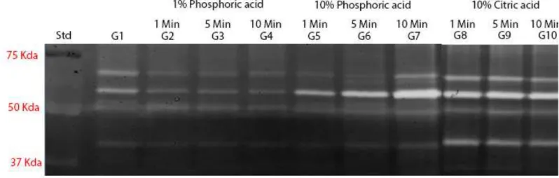
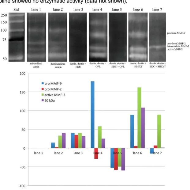
Zymograms of gelatinolytic activity are shown in Fig. 1. Zymograms of all
tested groups (G1-10) showed MMP-2 pro- and active-form (72- and 66-kDa,
respectively).
Time exposure of demineralization agent changed MMP-2 activity only for
10% phosphoric acid (G5-7). For those groups, a crescent activity could be seen
from G5 to G7. For all other groups, no difference between the three times of
application was found.
In comparison with control group (mineralized dentin), 10% phosphoric
acid (G5-7) and 10% citric acid (G8-10) showed increasing while 1% phosphoric acid
(G2-4) showed reduction in MMP-2 activity.
Control zymograms incubated with 5 mM EDTA and 2 mM
1,10-phenanthroline showed no enzymatic activity (data not shown).
Figure 1: Zymographic analysis of dentin powder treated with different demineralizing agents and application times. MMP-2 pro- and active-form (72- and
are reported in the standard lane (Std). Lane 1: Proteins extracted from dentin
powder mineralized (G1 – untreated dentin) showing the presence of MMP-2 pro-
and active-form. Lane 2, 3 and 4: demineralized dentin with 1% phosphoric acid after
1min (G2), 5min (G3) and 10min (G4) respectively, showing a lower expression of
MMP-2 pro- and active-form in a lower expression when compared to the mineralized
group. Lane 5, 6 and 7: demineralized dentin with 10% phosphoric acid after 1min
(G5), 5min (G6) and 10min (G7) respectively, showing a crescent activity of MMP-2
pro- and active-form in comparison with the mineralized and 1% phosphoric acid
groups. Lane 8, 9 and 10: demineralized dentin with 10% citric acid after 1min (G8),
5min (G9) and 10min (G10) respectively, showing a great activity of MMP-2 pro- and
active-form in comparison with G1 to G6.
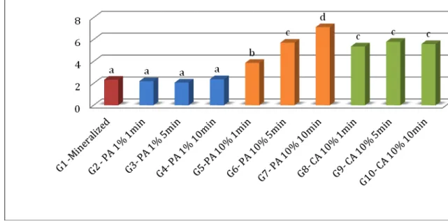
Assay for the Activities of MMP-2
Statistical analysis of data showed normal distribution and equal
variances. The MMP-2 activity (ng/mL) recorded in untreated mineralized and treated
with different acids and application times are summarized in Fig. 2. The differences
among the 10 groups were significant (p < 0.01).
Figure 2. Expression of MMP-2 activity (ng/mL) obtained with the Biotrak™ activity assay system. Bar 1, control mineralized dentin powder (G1); Bar 2, 3 and 4, dentin
Bar 5, 6 and 7, dentin powder etched with 10% phosphoric acid for 1, 5 and 10
minutes respectively (G5-7); Bar 8, 9 and 10, dentin powder etched with 10% citric
acid for 1, 5 and 10 minutes respectively (G8-10).
DISCUSSION
Dentin demineralization is an important step in dentin bonding procedures
that exposes the collagen fibril meshwork for micromechanical retention of adhesive
resins20,21. It is known that exposing dentin to low pH solutions activates MMPs that
can slowly degrade the collagen matrix26. However, none investigated the effect of
different acid solutions on MMP-2 activation and its effect regarding the time
exposure. The tested hypothesis of this study was that different acids, concentration
or time exposure could change the MMP-2 dentin activity. This was accepted since
acids showed different degrees of enzyme activation and 10% phosphoric acid
showed differences concerning time exposure.
MMP-2 may be one of the most relevant dentin-bound MMPs involved in
hybrid layer degradation14. This enzyme was found to be entrapped within the hybrid
layer created by a two-step ecth-and-rinse adhesive system and aged in artificial
saliva for 12 months. The presented results showed the presence of both MMP-2
active and pro-form in all tested groups. The amount of MMP-2 available for
detection changed in function of dentin treatment,14 showing that acid formulation is
able to change enzymatic expression.
Gelatin zymography is an extremely sensitive technique mainly used for
the detection of MMP-2 and -9 activities14,15,19 including gelatinase activities in
adhesive-treated dentin2,5. The specific enzymatic assay is an innovative approach
with the ability to quantify the activities of a tested specific dentinal MMP. This study
correlates those two enzymatic assays, an specific and a non-specific (gelatin
zymography). Both, zymography and the specific enzymatic assay, results showed
an increase of MMP-2 activity for 10% phosphoric acid and 10% citric acid in
comparison with mineralized groups. Analysis of these data provides direct evidence
of the gelatinase activities in acid-etched dentin in disagreement with previous
The majority of previous studies used 1% or 10% PA showing activity of
MMP-2 and -94,14,13,16. Just a few of them used 37% PA9,26. Although this is the
concentration used in clinical practice, the lower pH showed a reduction on enzyme
activity7,26. The main reason of this study was to evaluate the acid composition,
concentration and time of exposure that showed the highest activity of MMP-2 to
potentiate the enzyme extraction.
However, controversy exists over the interaction of PA with MMPs. It has
been reported that PA increases dentin MMP activity in a concentration of 1%2,
whereas other studies reported that 10% PA decreased dentin MMP activity under
different experimental conditions12. The present study showed that the concentration
of PA is crucial to the result. 10% PA was the unique acid that showed a continuous
increase in MMP-2 activity over time and the one that exhibited higher MMP-2
activity. 1% PA showed no difference in MMP-2 activity in ELISA and zymography
test, showing that a low concentration of this acid could not enlarge MMP-2
expression. These results are in accordance with DeVito-Moraes who evaluated
MMPs expression after 1%, 10% and 37% PA exposure, by means of western blot,
gelatin zymography, in situ zymography and hydroxyproline assays, showing a great
MMP expression of 10% PA in comparison with 1% and 37% PA7.
Citric acid is usually present in the daily diet, medications and
industrialized food. In dental practice, CA is often used to remove smear layer on root
dentin before using resin cements8,27. The present results showed an increase on
MMP-2 activity in comparison with mineralized dentin independent on time exposure.
This is most likely due to its great capacity for demineralization6 that can even
expose collagen fibrils and active MMPs. This is in accordance with Mazzoni et al.
(2007)15 and Kato et al. (2011)11 studies that reported intense gelatinolitic activity of
MMP-2 after exposition to citric acid 0.87 M.
1% phosphoric acid groups showed a reduction of MMP-2 activity. This
result may be either assigned to the zymography protocol of protein extraction. After
exposed to acid agents, dentin powder is neutralized using NaOH and the
supernatant is removed. We believe that this supernatant contains one part of total
protein and it is unvalued, reducing real protein expression. As all acids tested have
protein/enzymes within the dentine matrix and causing profound changes, especially
in secondary and tertiary structure of protein16. This may happen with all acid groups
but it was more evident with those that showed lower MMP activity.
For all acids tested, excluding 10% PA, time exposition does not show
difference on activation of this enzyme. This suggests that, in low concentration, acid
suddenly became neutralized by Calcium and Phosphate present on dentin and the
increase in time exposition could not change MMPs activity. Examining the buffering
capacity of dentin powder exposed to those acids, it can be observed that the
baseline pH of acids changed from 1.64 to 2.0, before etching, to 5.0 to 7.4 after
mixed with dentin powder. After neutralization of dentin powder with NaOH, it could
be observed a dense precipitate in eppendorf. The authors believe that such
precipitates are reaction products of the interaction of acids with dentin matrix apatite
that mask the collagen fibrils from matrix proteases. This hypothesis was either
suggested by Iwasa10 who showed a “dense, insoluble precipitate” that obscured the
fibrillar nature of the underlying collagen matrix on the surfaces of dentin exposed to
single step adhesives.
Based on the results of the present study, it can be concluded that low pH
solutions can activate dentin MMP-2 in different magnitude. So we suggest that 10%
PA and 10% CA are the best ones to be used on enzymatic tests since those acids
increased MMP-2 activity.
AKNOWLEDGMENTS
The authors declared no potential conflicts of interest with respect to the
authorship and/or publication of this article. The study was supported by CAPES
Foundation (Process 9000-11-9 FM Apolonio).
Clinical relevance
Dentin demineralization promoted by acid solutions can activate dentin MMPs
REFERENCES
1. Boushell LW, Kaku M, Mochida Y, Bagnell R, Yamauchi M. Immunohistochemical
localization of matrixmetalloproteinase-2 in human coronal dentin. Arch Oral Biol
2008;53:109–16
2. Breschi L, Mazzoni A, Nato F, Carrilho M, Visintini E, Tjäderhane L et al.
Chlorhexidine stabilizes the adhesive interface: a 2-year in vitro study. Dent Mater
2010;26: 320–325
3. Breschi L, Mazzoni A, Ruggeri A, Cadenaro M, Di Lenarda R, De Stefano Dorigo E.
Dental adhesion review: aging and stability of the bonded interface. Dent Mater
2008;24:90–101.
4. Carrilho MR, Tay FR, Donnelly AM, Agee KA, Tjäderhane L, Mazzoni A, et al.
Host-derived loss of dentin matrix stiffness associated with solubilization of collagen. J
Biomed Mater Res B Appl Biomater 2009;90:373-380.
5. De Munck J, Van den Steen PE, Mine A, Van Landuyt KL, Poitevin A, Opdenakker
G, et al. Inhibition of enzymatic degradation of adhesive-dentin interfaces. J Dent
Res 2009;88:1101-1106.
6. De-Deus G, Paciornik S, Pinho Mauricio MH, Prioli R. Real-time atomic force
microscopy of root dentine during demineralization when subjected to chelating
agents. Int Endod J. 2006;39:683-92
7. DeVito-Moraes, André Guaraci. Estudo da influência das soluções
desmineralizadoras na atividade proteolítica da dentina humana sadia / André
Guaraci De Vito Moraes; orientador Carlos Eduardo Francci; co-orientadora Marcela
Rocha Oliveira Carrilho. - Tese (Doutorado) -Programa de Pós-Graduação em
Odontologia. Área de Concentração: Materiais Dentários. - Faculdade de
Odontologia da Universidade de São Paulo.São Paulo, 2012.
8. Ebert J, Leyer A, Günther O, Lohbauer U, Petschelt A, Frankenberger R, Roggendorf
MJ. Bond strength of adhesive cements to root canal dentin tested with a novel
pull-out approach. J Endod. 2011;37:1558-61.
9. Hashimoto M, Tay FR, Ohno H, Sano H, Kaga M, Yiu C, Kumagai H, Kudou Y,
Kubota M, Oguchi H.. SEM and TEM analysis of water degradation of human
10. Iwasa M, Tsubota K, Shimamura Y, Ando S, Miyazaki M, Platt JA. PH changes upon
mixing single-step self-etching adhesives with powdered dentin. J Adhes Dent
2011;13:207-212.
11. Kato MT, Hannas AR, Leite AL, Bolanho A, Zarella BL, Santos J, Carrilho M,
Tjäderhane L, Buzalaf MA. Activity of matrix metalloproteinases in bovine versus
human dentine. Caries Res. 2011;45(5):429-34.
12. Martin-De Las Heras S, Valenzuela A, Overall CM. The matrix metalloproteinase
gelatinase A in human dentine. Arch Oral Biol 2000;45:757–65.
13. Mazzoni A, Breschi L, Carrilho M, Nascimento FD, Orsini G, Ruggeri A, et al. A
review on nature, role and functions of dentin non-collagenous proteins. Part II:
enzymes, serum proteins and growth factors. Endod Top 2012(a);21:19–40.
14. Mazzoni A, Carrilho M, Papa V, Tjäderhane L, Gobbi P, Di Lenarda R, et al. MMP-2
assay within the hybrid layer created by a two-step etch-and-rinse adhesive:
biochemical and immunohistochemical analysis. J Dent 2011(b);39:470-477.
15. Mazzoni A, Mannello F, Tay FR, Tonti GA, Papa S, Mazzotti G, et al. Zymographic
analysis and characterization of MMP-2 and -9 isoforms in human sound dentin. J
Dent Res 2007;86:436-440.
16. Mazzoni A, Papa V, Nato F, Carrilho M, Tjäderhane L, Ruggeri A Jr, et al.
Immunohistochemical and biochemical assay of MMP-3 in human dentine. J Dent
2011(a);39:231-237.
17. Mazzoni A, Nascimento FD, Carrilho M, Tersariol I, Papa V, Tjäderhane L, et al.
MMP activity in the hybrid layer detected with in situ zymography. J Dent Res
2012(b);91:467-472.
18. Mazzoni A, Pashley DH, Nishitani Y, Breschi L, Tjäderhane L, Toledano M, et al.
Reactivation of inactivated endogenous proteolytic activities of phosphoric
acid-etched dentin by etch-and-rinse adhesives. Biomater 2006;27:4470-4476.
19. Mazzoni A, Scaffa P, Carrilho M, Tjäderhane L, Di Lenarda R, Polimeni A et al.
Effects of etch-and-rinse and self-etch adhesives on dentin MMP-2 and MMP-9. J
Dent Res. 2013;92(1):82-6.
20. Nagase H. Activation mechanisms of matrix metalloproteinases. Biol Chem
1997;378:151-160.
21. Nakabayashi N & Pashley DH. Hybridization of dental hard tissues. Chicago, IL:
22. Nishitani Y, Yoshiyama M, Wadgaonkar B, Elrod D, Breschi L, Mannello F, et al.
Activation of gelatinolytic/collagenolytic activity in dentin by self-etching adhesives.
Eur J Oral Sci 2006;114:160-166.
23. Niu LN, Zhang L, Jiao K, Li F, Ding YX, Wang DY, et al. Localization of MMP-2,
MMP-9, TIMP-1, and TIMP-2 in human coronal dentine. J Dent 2011;39:536-542.
24. Palosaari H, Wahlgren J, Larmas M, Rönkä H, Sorsa T, Salo T, et al. The expression
of MMP-8 in human odontoblasts and dental pulp cells is down-regulated by
TGF-beta1. J Dent Res 2000;79:77–84.
25. Pashley DH, Tay FR, Breschi L, Tjderhane L, Carvalho RM, Carrilho M, et al. State of
the art etch-and-rinse adhesives. Dent Mater 2011;27:1–16.
26. Pashley DH, Tay FR, Yiu CKY, Hashimoto M, Breschi L, Carvalho RM, et al.
Collagen degradation by host-derived enzymes during aging. J Dent Res
2004;83:216–221.
27. Ravikumar J, Bhavana V, Thatimatla C, Gajjarapu S, Reddy SG, Reddy BR. The
effect of four different irrigating solutions on the shear bond strength of endodontic
sealer to dentin - An In-vitro study. J Int Oral Health 2014;6:85-8.
28. Sulkala M, Larmas M, Sorsa T, Salo T, Tjäderhane L. The localization of matrix
metalloproteinase-20 (MMP-20, Enamelysin) in mature human teeth. J Dent Res
2002;81:603–7.
29. Sulkala M, Pääkkönen V, Larmas M, Salo T, Tjäderhane L. Matrix
metalloproteinase-13 (MMP-metalloproteinase-13, collagenase-3) is highly expressed in human tooth pulp. Connec T Res
2004;45:231–7.
30. Sulkala M, Tervahartiala T, Sorsa T, Larmas M, Salo T, Tjäderhane L. Matrix
metalloproteinase-8 (MMP-8) is the major collagenase in human dentin. Arch Oral
Biol 2007;52:121-127
31. Tjäderhane L, Salo T, Larjava H, Larmas M, Overall CM. A novel organ culture
method to study the function of the human odontoblasts in vitro: gelatinase
expression by odontoblasts is differentially regulated by TGF-beta1. J Dent Res
1998;77:1488–98.
Capítulo 2
3.2 CAPÍTULO 2
Effect of a one-step adhesive on dentin MMPs activity
F.M. Apolonio1, A. Mazzoni2, V.P.A. Saboia1, S. Santi3, V. Angeloni2, M.
Cadenaro2, F.R. Tay4, D.H. Pashley4, L. Breschi5
1 Department of Restorative Dentistry, Federal University of Ceará,
Fortaleza, Brazil;
2 Department of Biomedicine, Unit of Dental Sciences and Biomaterials,
University of Trieste, Trieste, Italy;
3
Institute of Molecular Genetics, CNR-National Research Council of Italy ,
Bologna , Italy ; SC Laboratory of Musculoskeletal Cell Biology, Rizzoli Orthopedic
Institute , Bologna , Italy;
4 Department of Oral Biology, College of Dental Medicine, Georgia
Regents University, Augusta, GA, USA;
5
Department of Biomedical and Neuromotor Sciences, DIBINEM,
University of Bologna and IGM-CNR, Unit of Bologna, Italy
For: European Journal of Oral Sciences
(To be submitted in January 2015)
Correspondence: Prof. Lorenzo Breschi, Department of Biomedical and Neuromotor Sciences, DIBINEM, University of Bologna - Alma Mater Studiorum, Via San Vitale
59, 40125, Bologna, Italy, Tel: +39-051-2088139; Fax: +39-051-225208; email:
lorenzo.breschi@unibo.it
ABSTRACT
Degradation of the hybrid layer has been related to enzymes activity present in the
dentin matrix that destroys unprotected collagen fibrils. The aim of the study was to
evaluate the effect of a one-step adhesive system on dentinal MMP-2 and -9 using in
situ zymography and an enzymatic activity assay. This study tested the null
hypothesis that there are no differences in the activities of dentinal MMPs after
treatment with a one-step adhesive system. MMP-2 and -9 activities in
adhesive-treated dentin with Adper easy Bond (3M ESPE) were quantified by means of an
activity assay system. MMPs activity within the hybrid layer was additionally detected
in situ with the in situ zymography technique using the one-step self-etch adhesive
system. Enzymatic assay revealed increase on MMP-2 and -9 activities after
adhesive treatment. The in situ zymography revealed that gelatinolytic activity is
present within the hybrid layer created with the tested one-step self-etch adhesive
and the data indicate that host-derived gelatinases remain localized within the HL
and are active after bonding procedure. It can be concluded that one-step self-etch
adhesives can activate endogenous MMP-2 and -9 that could promotes collagen
degradation over time.
INTRODUCTION
Among contemporary dentin-bonding systems, self-etch adhesives have
consistently increased their popularity in the last years due to reduction of clinical
application steps1 minimizing also the technique sensitivity2,3. Recent formulations of
self-etch adhesives have shifted to one-step systems, in which all components are
combined into a single solution corresponding to a single clinical application step.
Despite their user-friendliness and low technique sensitivity, one-step adhesives
have exhibited lower immediate resin-dentin bond strengths and extensive interfacial
nanoleakage expression when compared with other bonding approaches4-6.
Bond durability is the main goal to achieve for adhesive restorations. It is
known that degradation processes occur at the resin/dentin interface7-10 and that this
mechanism is very complex and even not completely elucidated11,12. One reason that
contributes to the degradation of the hybrid layer (HL) over time is the activity related
to endogenous proteases enzymes, such as metalloproteinases (MMPs) and
hydrolization of the exposed collagen fibrils within the HL, if unprotected by adhesive
monomers16-19.
MMPs are a family of zinc- and calcium-dependent enzymes important for
tooth development20. In mineralized dentin, MMPs become covered with apatitic
nanocrystals, making them immobile and non-functional18. However, when exposed
to a low pH solution such as the pH of dentin bonding agents, these enzymes
became activated17,18,21-23 and can cause progressive degradation of exposed
collagen fibrils16-18,24, 25. Evidence of collagenolytic and gelatinolytic activities in
partially demineralized dentin treated with either etch-and-rinse or self-etch
adhesives confirmed, in fact, the potential involvement of these endoproteases in the
disruption of incompletely resin-infiltrated collagen fibrils within HLs7,8,17,18,21,26.
The presence of MMP-2 and -9 in human sound dentin powder was
assayed by gelatin zymography, Western-blot analysis17, activity assays15,21 and by
an immunohistochemical approach14. More recently Mazzoni and co-workers, using
an innovative technique, the in situ zymography technique15 based on a fluorescent
enzyme assay kit, have allowed screening of the relative proteolytic activity in
adhesive-treated dentin. The advantage of this latest technique is the possibility to
evaluate in situ how dentinal MMPs act at the dentin/resin interface using dentin
bonding agents as there are caused in clinical practice.
The purpose of this study was to investigate the effects of a one-step
adhesive systems on MMP-2 and -9 activities, by means of a correlative analysis
based on, in situ zymography and an specific enzymatic assay. The tested null
hypothesis was that there are no differences in the activities of host-derived MMP-2
and -9 after treatment with one-step adhesive systems.
MATERIALS AND METHODS
Reagents were purchased from Sigma Chemical Co. (St. Louis, MO, USA)
unless otherwise specified.
Assay for the Activities of MMP-2 and -9
Ten extracted human third molars were obtained from anonymous
individuals following their signed consent under a protocol approved by the University
of Trieste (Italy). Teeth, ground free of enamel, pulpal soft tissue, and cementum,
triturated by means of a steel mortar/ pestle (Reimiller, Reggio Emilia, Italy). The fine
mineralized dentin powder was pooled, dried, and kept frozen until use.
Aliquots of dentin powder were prepared, randomized divided and treated
as follow: G1 = mineralized dentin powder (MD); G2 = MD treated with Adper Easy
Bond (3M ESPE; AEB), in accordance with Mazzoni et al. (2013)21 by mixing the MD
powder with the all-in-one adhesive for 30 min followed by rinses in acetone to
remove the resin from the dentin powder.
The enzymatic activities of MMP-2 and -9 were determined with the
Biotrak™ activity assay system (GE Healthcare, Buckinghamshire, UK). Protein
extraction from the mineralized and adhesive-treated dentin powder was performed
in 50 mM Tris-HCl buffer, pH 7.4. Standard curves were prepared, and samples were
incubated in the supplier-provided assay buffer for 2 and 6 hrs, for MMP-2 and -9,
respectively, at 4°C. After extensive rinses, the detection reagent was added, and
absorbance was read at 405 nm (Bio-Rad, Segrate Milano, Italy). Assays were
performed in triplicate and completed according to the manufacturer’s instructions.
The statistical analysis was performed and since values were normally
distributed (Kolmogorov-Smirnov test), data were analyzed with a one-way analysis
of variance and Scheffe’s post hoc test (p < 0.05).
In situ zymography
Additional two extracted human third molars were used for in situ
zymography analysis. In detail, slabs of 1-mm-thick of middle/deep dentin were
obtained from each tooth by means of a low-speed saw (Micromet, Remet,
Casalecchio di Reno, Italy). A standardized smear layer was created by wet-polished
with #600-grit SiC paper under running water for 60s and the dentin surfaces were
treated with the tested all-in-one-adhesive in accordance with the manufacturer’s
instructions (Table 1). A 1-mm-thick flowable composite (Filtek 250 flow, 3M ESPE)
was placed on the top of the adhesive-treated dentin and properly polymerized
(Curing Light 2500, 3M ESPE). Bonded specimens were cut vertically into
1-mm-thick slabs to expose the adhesive/dentin interfaces by means of slow-speed saw
(Micromet) and each bonded dentin/composite slide glued to a glass microscope
support and ground down to obtain ca. 500-µm-thick specimens.
In situ zymography was performed with self-quenched
USA) in accordance with Mazzoni et al. (2012)27. In brief, the fluorescent gelatin
mixture was placed on top of each slab and covered with a coverslip, and the slides
were light-protected and incubated in humidified chambers at 37°C for 24h. The
hydrolysis of quenched fluorescein-conjugated gelatin substrate, indicative of
endogenous gelatinolytic enzyme activity, was assessed by examination with a
confocal laser scanning microscope [excitation (ex), 488 nm; and emission (em),
lp530 nm; Nikon A1-R, Tokyo, Japan]. The 2-D images obtained were then combined
to create 3D-like structural images to provide additional information regarding the
depth of gelatinolytic activity.
Negative control sections were incubated as described above, except that:
(1) 250 mM ethylenediaminetetraacetic acid (EDTA) was dissolved in the mixture of
quenched fluorescein-conjugated gelatin or (2) 2 mM 1,10-phenanthroline or (3)
standard non-fluorescent instead of fluorescent conjugated gelatin was used. EDTA
and 1,10-phenanthroline were used as negative controls because they are
well-known MMPs inhibitors.
Table 1: Components, compositions, and application procedure of Adper Easy Bond adhesive system (information supplied by the manufacturers).
Adhesive
System Composition Application procedure Adper Easy
Bond, 3M ESPE, Seefeld, Germany, pH: 2.4*
HEMA, Bis-GMA, Group
methacrylated phosphoric esters, 1,6
hexanediol dimethacrylate,
methacrylate functionalized
polyalkenoic acid, silica filler, ethanol,
water, camphorquinone, stabilizers.
Dry; apply 2 layers onto
surface cavity; air-thin for
approximately 5 s until the
film no longer moves;
light-cure for 10 s.
HEMA: 2-hydroxyethyl methacrylate; Bis-GMA: Bis-phenol A diglycidylmethacrylate.
*Information received from the manufacturer.
RESULTS
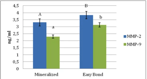
The MMP-2 and -9 activities (ng/mL) recorded in untreated mineralized,
and adhesive treated dentin powder with Adper Easy Bond are summarized in Fig. 1.
The differences among the tested groups were significant (p < 0.01). With
mineralized dentin powder as the control, the enzyme activity after treatment with
AEB increased approximately 15% for MMP-2 and 37% for MMP-9 (Fig. 1).
Figure 1: Expression of MMP-2 and -9 activities (ng/mL) obtained using Biotrak™ activity assay system. Bar 1: control mineralized dentin powder (MD); Bar
2: mineralized dentin treated with Adper Easy Bond (AEB).
In situ zymography
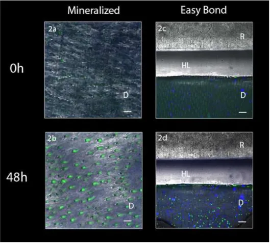
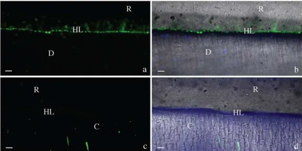
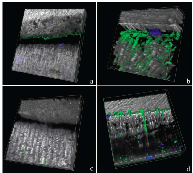
The in situ zymography performed on adhesive-dentin interfaces
created with Adper Easy Bond revealed an intense green fluorescence within the HL
after incubation, indicating that the fluorescein-conjugated gelatin was hydrolyzed at
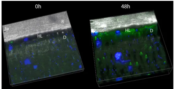
these sites (Fig. 2). The 3-D model of the acquired stacked images confirmed the
Figure 2: Resin/dentin interfaces created with Adper Easy Bond (AEB) adhesive system incubated with quenched fluorescein-labeled gelatin obtained by merging
differential interference contrast image. Image acquired in green channel. Bar = 5
µm. D = dentin; R = resin; HL = hybrid layer. Fig 2a: Representative image of
mineralized dentin at baseline showing no fluorescence. Fig 2b: Representative
image of mineralized dentin after 48h showing fluorescence. Fig. 2c: Representative
adhesive interface created by AEB at baseline showing minimal fluorescence. Fig.
2d: Representative AEB adhesive interface after gelatin incubation showing intense
Figure 3: Tri-dimensional models of the acquired images at baseline (Fig. 3a) and after incubation with gelatin showing extensive fluorescence within the hybrid layer
Fig. 3b). (R=resin composite, HL= Hybrid layer, D= Dentin).
No fluorescence was detected in negative controls, i.e., (1) EDTA-treated,
(2) 2 mM 1,10-phenanthroline-treated, or (3) specimens incubated with standard
non-fluorescent gelatin (data not shown).
DISCUSSION
This paper proposed to investigate the enzymatic activity that can
contribute to the degradation process of the adhesive/dentin interfaces created by an
all-in-one adhesive system. Results of the study showed that activities of MMP-2 and
-9 increased after the application of Adper Easy Bond, thus the hypothesis tested
that the activities of host derived dentinal MMP-2 and -9 are not influenced by the
tested dentin bonding agent should be rejected.
Current adhesive technology of one-step adhesive systems tends to
simplify bonding procedures by reducing the application steps, shortening the clinical
application time and decreasing the technique sensitivity with the attempt to improve
its standardization28. However, the degradation process of these adhesives is still not
well understood. Hydrolytic degradation has been described frequently as a major
factor affecting the stability of the hybrid layer either with self-etch or etch-and-dry
adhesives9, especially for one-step systems. Additionally, suboptimal
polymerization29, continuous etching, and phase separation30 have been shown to
the importance of enzymatic degradation of suboptimally impregnated collagen fibrils
that remain within the hybrid layer and that can be degraded following the activation
of collagen-bound MMPs8,12,21.
Self-etch adhesives have shown activation of the host-derived dentinal
MMPs17,18,21,22,26. In addition, a self-etch adhesive has been shown to increase
MMP-2 synthesis in human odontoblasts31, possibly increasing MMP-2 penetration into the
hybrid layer via dentinal fluid. MMPs can be activated in low-pH environments32,33 by
inducing the cysteine switch34. The acidic resin monomers contained either in
etch-and-rinse or self-etch adhesives may, in fact, activate latent forms of MMPs
(pro-MMPs) via the cysteine-switch mechanism that exposes the catalytic domains of
these enzymes that were blocked by pro-peptides21.
Although one-step adhesive systems rely on the same bonding
mechanism, they differ from each other in many aspects, such as acidic resin
monomer composition, water content and acidity. The tested adhesive had a pH
value of 2.4 (Table 1) and this ‘mild’ pH may explain the increase in MMP-2 and -9
activity recorded with the specific assay system employed (Fig. 1). These results are
in line with a previous study in which the effect of AEB was investigated also with
gelatin zymography showing that Adper Easy Bond increased dentinal gelatinolytic
activity of MMP-2 and 921,35.
The approach used in this study includes the possibility to check the
difference between the activation of MMPs using biochemical and morphological
analysis. Homogenization of tissues is, in fact, mandatory for activity assays, allowing
detection and quantification of enzymes in relation to the adhesive treatments,
precluding, however, the possibility to localize the MMPs at the adhesive/dentin
interface. For this reason, we additionally performed an in situ zymography technique
to obtain precise localization of the MMPs activity within the HL created by the tested
one-step adhesive system. The in situ zymography revealed that gelatinolytic activity
is present within the HL created with the tested one-step self-etch adhesive and the
data obtained clearly indicate that host-derived gelatinases remain localized within
the HL and are active after bonding procedure.
Those results contribute to confirm that self-etch adhesives can activate
MMPs in dentin since the one-step adhesive system tested improved enzymatic
Aurelio Valmori for photographical support. The study was funded by grants from
MIUR (Italy) - FIRB RBAP1095CR to L. Breschi (P.I.), PRIN 2009SAN9K5 to L.
Breschi (P.I.) and CAPES Foundation (Process 9000-11-9 FM Apolonio). The
authors declare no potential conflicts of interest with respect to the authorship and/or
publication of this article.
REFERENCES
1. Pashley DH, Tay FR, Breschi L, Tjarderhane L, Carvalho RM, Carrilho
M, Tezvergil-Mutlay A. State of the art etch-and-rinse adhesives. Dent Mater 2011;
27:1-16.
2. Van Meerbeek B, Van Landuyt K, De Munck J, Hashimoto M, Peumans
M, Lambrechts P Yoshida Y, Inoue S, Suzuki K. Technique-sensitivity of
contemporary adhesives. Dent Mater J. 2005;24:1-13.
3. Hiraishi N, Breschi L, Prati C, Ferrari M, Tagami J, King NM. Technique
sensitivity associated with air-drying of HEMA-free, single-bottle, one-step self-etch
adhesives. Dent Mater J. 2007;23:498-505.
4. Navarra CO, Cadenaro M, Codan B, Mazzoni A, Sergo V, De Stefano
Dorigo E, Breschi L. Degree of conversion and interfacial nanoleakage expression of
three one-step self-etch adhesives. Eur J Oral Sci 2009;117:463-469.
5. Amaral RC, Stanislawczuk R, Zander-Grande C, Gagler D, Reis A,
Loguercio AD. Bond strength and quality of the hybrid layer of one-step self-etch
adhesives applied with agitation on dentin. Oper Dent 2010;35:211-209.
6. Shinoda Y, Nakajima M, Hosaka K, Otsuki M, Foxton RM, Tagami J.
Effect of smear layer characteristics on dentin bonding durability of HEMA-free and
HEMA-containing one-step self-etch adhesives. Dent Mater J 2011;30:501-510. 7. Carrilho MR, Carvalho RM, de Goes MF, di Hipólito V, Geraldeli S, Tay
FR, Pashley DH, Tjäderhane L. Chlorhexidine preserves dentin bond in vitro. J Dent
Res, 2007a;86:90-4.
8. Carrilho MRO, Geraldeli S, Tay FR, de Goes MF, Carvalho RM,
Tjäderhane L, Reis AF, Hebling J, Mazzoni A, Breschi L, Pashley DH. In vivo
9. Breschi L, Mazzoni A, Ruggeri A, Cadenaro M, Di Lenarda R, De
Stefano Dorigo E. Dental adhesion review: aging and stability of the bonded
interface. Dent Mater, 2008;24:90-101.
10. Liu Y, Tjäderhane L, Breschi L, Mazzoni A, Li N, Mao J, Pashley
DH, Tay FR. Limitations in bonding to dentin and experimental strategies to prevent
bond degradation. J Dent Res, 2011;90: 953-968.
11. Donmez N, Belli S, Pashley DH, Tay FR. Ultrastructural
correlates of in vivo/in vitro bond degradation in self-etch adhesives. J Dent Res.
2005;84:355-9.
12. Scheffel D, Hebling J, Scheffel R, Agee K, Turco G, de Souza
Costa C, Pashley D. Inactivation of Matrix-bound Matrix Metalloproteinases by
Cross-linking Agents in Acid-etched Dentin. Oper Dent. 2014;39:152-8.
13. Mazzoni A, Mannello F, Tay FR, Tonti GA, Papa S, Mazzotti G,
Di Lenarda R, Pashley DH, Breschi L. Zymographic analysis and characterization of
MMP-2 and -9 isoforms in human sound dentin. J Dent Res 2007;86:436-440.
14. Mazzoni A, Pashley DH, Tay FR, Gobbi P, Orsini G, Ruggeri A,
Carrilho M, Tjäderhane L, Di Lenarda R, Breschi L. Immunohistochemical
identification of MMP-2 and MMP-9 in human dentin: correlative FEI-SEM/TEM
analysis. J Biomed Mater Res A 2009;88:697-703.
15. Mazzoni A, Papa V, Nato F, Carrilho M, Tjäderhane L, Ruggeri
A, Gobbi P, Mazzotti G, Tay FR, Pashley DH, Breschi L. Immunohistochemical and
biochemical assay of MMP-3 in human dentine. J Dent 2011;39:231-237.
16. Pashley DH, Tay FR, Yiu C, Hashimoto M, Breschi L, Carvalho
RM, Ito S.Collagen degradation by host-derived enzymes during aging. J Dent Res
2004;83:216-221.
17. Mazzoni A, Pashley DH, Nishitani Y, Breschi L, Tjäderhane L,
Toledano M, Pashley EL, Tay FR. Reactivation of inactivated endogenous proteolytic
activities of phosphoric acid-etched dentin by etch-and-rinse adhesives. Biomater
2006;27:4470-4476.
18. Nishitani Y, Yoshiyama M, Wadgaonkar B, Elrod D, Breschi L,
Mannello F, Mazzoni A, Carvalho RM, Tjäderhane L, Tay FR, Pashley DH. Activation
of gelatinolytic/collagenolytic activity in dentin by self-etching adhesives. Eur J Oral