Carvacryl acetate, a derivative of carvacrol, reduces nociceptive and
in
fl
ammatory response in mice
Samara R.B. Damasceno
a, Francisco Rodrigo A.M. Oliveira
b, Nathalia S. Carvalho
a, Camila F.C. Brito
a,
Irismara S. Silva
a, Francisca Beatriz M. Sousa
a, Renan O. Silva
a, Damião P. Sousa
c, André Luiz R. Barbosa
a,
Rivelilson M. Freitas
b, Jand-Venes R. Medeiros
a,⁎
aLaboratory of Experimental Physiopharmacology, Biotechnology and Biodiversity Center Research, Federal University of Piauí, Parnaíba, Piauí, Brazil.
bDepartment of Biochemistry and Pharmacology, Post-graduation Program in Pharmaceutics Science of Federal University of Piauí, Teresina, Piauí, Brazil
cDepartment of Physiology, Federal University of Paraíba, João Pessoa, Paraíba, Brazil
a b s t r a c t
a r t i c l e
i n f o
Article history: Received 6 July 2013 Accepted 2 November 2013
Keywords: Anti-inflammatory Anti-nociceptive Carvacryl acetate Carvacrol
Aims:The present study aimed to investigate the potential anti-inflammatory and anti-nociceptive effects of carvacryl acetate, a derivative of carvacrol, in mice.
Main methods:The anti-inflammatory activity was evaluated using various phlogistic agents that induce paw edema, peritonitis model, myeloperoxidase (MPO) activity, pro and anti-inflammatory cytokine levels. Evalua-tion of antinociceptive activity was conducted through acetic acid-induced writhing, hot plate test, formalin test, capsaicin and glutamate tests, as well as evaluation of motor performance on rotarod test.
Keyfindings:Pretreatment of mice with carvacryl acetate (75 mg/kg) significantly reduced carrageenan-induced paw edema (Pb0.05) when compared to vehicle-treated group. Likewise, carvacryl acetate (75 mg/kg) strongly inhibited edema induced by histamine, serotonin, prostaglandin E2and compound 48/80. In the peritonitis model, carvacryl acetate significantly decreased total and differential leukocyte counts, and reduced levels of myeloperoxidase and interleukin-1 beta (IL-1β) in the peritoneal exudate. The levels of IL-10, an anti-inflammatory cytokine, were enhanced by carvacryl acetate. Pretreatment with carvacryl acetate also decreased the number of acetic acid-induced writhing, increased the latency time of the animals on the hot plate and decreased paw licking time in the formalin, capsaicin and glutamate tests. The pretreatment with naloxone did not reverse the carvacryl acetate-mediated nociceptive effect.
Significance:In conclusion, the current study demonstrated that carvacryl acetate exhibited anti-inflammatory activ-ity in mice by reducing inflammatory mediators, neutrophil migration and cytokine concentration, and anti-nociceptive activity due to the involvement of capsaicin and glutamate pathways.
Crown Copyright © 2014 Published by Elsevier Inc. All rights reserved.
Introduction
Inflammation is a dynamic and complex process that arises in re-sponse towards cellular injury. It has an important role in tissue repair, yet in some cases it can cause undesirable effects such as tissue damage and loss of function. Inflammation process is characterized by the pro-duction of a cascade of mediators that regulate important factors of the inflammatory response, as the increase in vascular permeability and recruitment of leukocytes in the blood (Rodriguez-Vita and Law-rence, 2010). Once released, these inflammatory mediators may acti-vate or sensitize nociceptors adjacent to the injured tissue resulting in pain sensation (Andrade et al., 2012). Currently several analgesics and anti-inflammatory drugs are associated with important side effects,
low efficacy and specificity. For this reason, studies are conducted to identify novel therapeutic options to develop and introduce new drugs with greater safety and efficacy.
Medicinal plants are natural products known to be a significant source of new chemical substances with potential therapeutic effects (Calixto, 2005). Among natural products, use of essential oils is a prom-ising option because of proven therapeutic action and for being also commonly added in food to obtain a specific taste (Ipek et al., 2005). In this context, we can include the carvacrol (5-isopropyl-2-methylphenol), a monoterpenic phenol present in essential oils of nu-merous aromatic plants of the family Laminaceae (Vincenzi et al., 2004). Previous studies have demonstrated antimicrobial (Klein et al., 2013), antioxidant (Yanishlieva et al., 1999; Beena et al., 2013), anti-nociceptive (Melo et al., 2012) and anti-inflammatory (Landa et al., 2009) properties of this compound.
In this study, we decided to investigate a semisynthetic derivative of carvacrol, carvacryl acetate, a compound unexplored as their pharmaco-logical properties. Since carvacrol, as well as other phenols, such as
–
⁎ Corresponding author at: Av. São Sebastião, 2819, CEP: 64202-020, Parnaíba, PI, Brazil. Tel.: +55 86 99862374; fax: +55 86 33235406.
E-mail address:[email protected](J.-V.R. Medeiros).
0024-3205/$–see front matter. Crown Copyright © 2014 Published by Elsevier Inc. All rights reserved. http://dx.doi.org/10.1016/j.lfs.2013.11.001
Contents lists available atScienceDirect
Life Sciences
thymol, is more toxic than many esters, the synthesis of carvacryl ace-tate was performed to obtain a derivative of carvacrol with improved pharmacological profile and less toxicity. Thus we suggest that the pres-ence of an ester group, instead of the hydroxyl group of carvacrol, may confer different safety and efficacy features to this compound compared to monortepene carvacryl.
Considering the potential use of natural products in the develop-ment of new drugs, the aim of the present study was to investigate the anti-inflammatory and antinociceptive effects of carvacryl acetate, a derivative of carvacrol, in experimental models.
Materials and methods
Drugs and reagents
λ-Carrageenan, indomethacin, serotonin, histamine, compound 48/80, prostaglandin E2(PGE2), acetic acid, formaldehyde and dimethyl sulfoxide (DMSO) were purchased from Sigma Chemical (Saint Louis, MO, USA). Heparin and morphine were provided by Merck, Sao Paulo, Brazil. All drugs were dissolved in sterile 0.9% (w/v) NaCl (saline). The carvacryl acetate was dissolved in 2% DMSO. All other chemicals were of analytical grade and obtained from standard commercial suppliers.
Compounds
Carvacryl acetate (98% purity;Fig. 1) was obtained by acetylation of carvacrol, using acetic anhydride as acylating agent and pyridine as cat-alyst. As described below,first in a 50 mlflask, equipped with magnetic stirrer, coupled to a Friedrich condenser and an inert atmosphere was added carvacrol (5 g, 0.033 mol), pyridine (7.5 ml) and acetic anhy-dride (12.5 ml). Then subjected to magnetic stirring and under constant reflux for 24 h. Continuing the procedure for preparing the reaction mixture was poured into ice water (60 ml) and extracted from reaction product in a separator funnel using chloroform as the solvent (three times 60 ml). The chloroform phases were combined and washed with saturated copper sulphate (three times 60 ml). The chloroform phase was washed with water (three times 60 ml) and dried with anhydrous Na2SO4. Subsequently, the solvent was evaporated on a rotary evaporator. The reaction product was subjected to column chromatography using silica gel as stationary phase and a mixture of hexane/ethyl acetate (95:5) as mobile phase. There was obtained 4.779 g (0.025 mol) of carvacryl acetate and 76% yield (Vogel et al., 1996; Moraes et al., 2013). The structural identification of carvacryl acetate was performed by analysis of1H and13C NMR, IR and com-pared with literature data.
Animals
Male Swiss mice (25–30 g) were housed at a temperature of 25 ± 2 °C under a 12/12-h light/dark cycle with food and water ad libitum. All experiments were performed in accordance with theGuide for Care and Use of Laboratory Animals(National Institute of Health, Be-thesda, MD, USA) and were approved by the Ethics Committee in Re-search of the Federal University of Piauí (protocol no. 0066/10).
Experimental protocol
Carrageenan-induced paw edema in mice
The animals were randomly divided into six groups (n= 5), and edema was induced by injection of 50μl of a suspension of carrageenan (500μg/paw) in 0.9% sterile saline into the right hind paw (group I) ac-cordingSilva et al. (2013). Mice were pretreated intraperitoneally (i.p.) with either 2% DMSO (group II untreated control); indomethacin 10 mg/kg (group III reference control); or carvacryl acetate, 25, 50 or 75 mg/kg (groups IV, V, and VI, respectively). Paw volume was measured immediately before (V0), and at 1, 2, 3, and 4 h after carrageenan treat-ment (Vt), using a plethysmometer (Panlab, Barcelona, Spain). The effect of pretreatment was calculated as percent inhibition of edema relative to the paw volume of the DMSO-treated controls by using the following for-mula (Winter et al., 1962).
%inhibition of edema¼ðVt−V0ÞControl−ðVt−V0ÞTreated Vt−V0
ð ÞControl 100
Paw edema induced by different phlogistic agents
To induce edema, the animals were administered 50μl injections of serotonin (1% w/v), histamine (100μg/paw), prostaglandin E2 (3 nmol/paw), or compound 48/80 (12μg/paw) into the right hind paw (Silva et al., 2013; Chaves et al., 2013; Claudino et al., 2006). The contralateral paw received 50μl of 2% DMSO and served as an untreated control. In the experiment, the animals were pretreated with carvacryl acetate (75 mg/kg, i.p.) or indomethacin (10 mg/kg, i.p.; reference con-trol) 30 min before these intraplantar injections of phlogistic agents.
Carrageenan-induced peritonitis
For the determination of neutrophil migration into the peritoneal cavity, mice were injected intraperitoneally with 2.0% DMSO, indo-methacin 10 mg/kg or carvacryl acetate (75 mg/kg, i.p.). Thirty minutes later, the animals were injected with carrageenan (250μl; 500 μg/cavi-ty), as adapted from de reports ofChaves et al. (2013). Mice were eutha-nized 4 h later and the peritoneal cavity was washed with 1.5 ml of heparinized phosphate buffered saline (PBS) to harvest peritoneal cells. The volumes recovered were similar in all experimental groups and were equivalent to ~95% of the injected volume. Total cell counts were performed in a Neubauer chamber, and differential cell counts (100 cells total) were carried out on cytocentrifuge slides stained with hematoxylin and eosin. The results are presented as the number of neu-trophils per milliliter of peritoneal exudate. Aliquots of the peritoneal exudates were stored at−70 °C for later analysis of cytokine and myeloperoxidase (MPO) content.
Myeloperoxidase activity assay
MPO assay was based on the method ofBradley et al. (1982)and partly modified. Briefly, 400μl of the peritoneal exudates was centri-fuged at 40,000 ×gfor 7 min at 4 °C. After, 10μl of the supernatants were collected and MPO activity was assayed by measuring the change in absorbance at 450 nm usingo-dianisidin edihydrochloride and 1% hydrogen peroxide. The results were expressed in units/ml. A unit of MPO activity was defined as that converting 1μmol of hydrogen perox-ide to water in 1 min at 22 °C.
Evaluation of TNF-α, IL-1β and IL-10 levels in carrageenan-induced peritonitis
the test samples and each standard at various dilutions were added in duplicate and incubated at 4 °C for 24 h. The plates were washed three times with buffer. After washing the plates, 50μl of biotinylated sheep polyclonal anti-TNF-α, anti-IL-1βor IL-10 (diluted 1:1000 with assay buffer 1% BSA) were added to the wells. After further incubation at room temperature for 1 h, the plates were washed and 50μl of streptavidin-HRP diluted 1:5000 were added to all wells. The reagent o-phenylenediamine dihydrocloride (50μl) was added 15 min later, and the plates were incubated in the dark at 37 °C for 15–20 min. After color development, the reaction was stopped with the addition of sulfuric acid (1 M) and absorbance was measured at 490 nm. The re-sults are expressed as pg/mg protein and reported as mean ± SEM.
Acetic acid-induced abdominal writhing
Acetic acid administration causes irritation, resulting in painful con-tortions, followed by hind limb extension. Each experimental group was pretreated with DMSO, carvacryl acetate (75 mg/kg, i.p.) or morphine (5 mg/kg, s.c., reference control). After 30 min, 0.6% acetic acid (10 ml/kg body weight, i.p.) was administered. After waiting for 10 min, the number of constrictions, including abdominal muscle con-tractions and hind paw extension, were recorded over 20 min, as de-scribed byKoster et al. (1959).
Hot plate test
Mice were treated according to the method described byEddy and Leimbach (1953). Each mouse was dropped twice on a heated plate (55 ± 1 °C), separated by a 30 min interval. Thefirst trial familiarized the animal with the test procedure and the second served as the control reaction time (licking of a paw or jumping), recorded as the response la-tency on a hot plate (Insight, Ribeirao Preto, São Paulo, Brazil; model EFF-361). Animals with baseline latencies of more than 20 s were ex-cluded from the study. Mice were treated with carvacryl acetate (75 mg/kg, i.p.) or morphine (5 mg/kg, s.c.; reference drug) 30 min be-fore the test, and the control group received the same volume of 2% DMSO. Measurements were performed before (zero time) and 30, 60 and 90 min after treatment, with a cutoff time of 45 s to prevent devel-opment of paw lesion.
Formalin-induced licking
Mice were pretreated with either 2% DMSO, carvacryl acetate, (75 mg/kg, i.p.), or morphine (5 mg/kg, s.c.; reference control). Thirty minutes after administration, 2.8% formalin (20μl) was administered subcutaneously (s.c.) into the right hind paw. Licking time was recorded from 0 to 5 min (phase 1, corresponding to a direct chemical stimula-tion of nociceptors) and 20–25 min after formalin injection (phase 2, in-volving release of inflammatory mediators) (Fasmer et al., 1986).
Evaluation of the opioid pathway
To examine the involvement of opioid receptors in the antinociceptive activity of carvacryl acetate for formalin-induced pain, mice (n= 6) were pretreated with 2% DMSO or naloxone (3 mg/kg, sc; opioid antagonist). After 15 min, the animals were treated with carvacryl acetate (75 mg/kg, i.p.) or morphine (5 mg/kg, s.c.; opioid ag-onist). Thirty minutes after administration, 2.8% formalin (20μl) was administered subcutaneously (s.c.) into the right hind paw. Licking time was recorded from 0 to 5 min (phase 1, corresponding to a direct chemical stimulation of nociceptors) (Hunskaar et al., 1985).
Capsaicin-induced nociception
The animals were treated with vehicle (2% DMSO, i.p.), carvacryl ace-tate (75 mg/kg, i.p.) or morphine (5 mg/kg, s.c.; reference control), thirty minutes later mice received 20μl of capsaicin solution (3μg/paw) in the right hind paw. The nociceptive response (time that the animal remained licking or biting the injected paw) was timed over a period of 5 min after capsaicin administration (Sakurada et al., 2003).
Glutamate-induced nociception
The animals were treated with carvacryl acetate (75 mg/kg, i.p.) or MK-801 (0.03 mg/kg, i.p.; an NMDA receptor antagonist), thirty mi-nutes later mice received 20μl of glutamate solution (30μmol/paw) in the right hind paw. The nociceptive response (time that the animal remained licking or biting the injected paw) was timed over a period of 15 min after glutamate administration (Beirith et al., 2002).
Evaluation of the motor performance
The rota-rod test permits the detection of muscle relaxing agents or drugs that produce motor incoordination. Earlier, the animals were evaluated to select those that shown ability in walking on the revolving bar under the same conditions used in the test and these were divided into groups (n= 5). On the day of the test, animals were treated with 0.5 ml/25 g vehicle (2% DMSO—control, i.p.), carvacryl acetate (75 mg/kg, i.p.) or diazepam (5 mg/kg, v.o., positive control for motor impairment). After 60 min, the animals were placed on the horizontal rotating bar (12 rpm), which was a non-slip plastic rod located 28 cm over the base, for 1 min. The number of falls was counted, with a max-imum of three replacements on the bar (Dunham and Miya, 1957).
Statistical analysis
Results are expressed as means ± SEM of at leastfive animals per group, and statistical analysis was performed using one-way analysis of variance (ANOVA) followed by the Newman–Keuls post hoc test, when appropriate. Statistical significance was set atPb0.05.
Results
Carrageenan-induced paw edema
Subplantar injection of carrageenan promoted an increase in paw volume dependent on the time; this increase was maintained until the fourth hour, getting the maximum value at the third hour (Table 1). Paw edema was significantly decreased by indomethacin (Pb0.05) throughout the experimental period, with a maximal inhibition of 81.3%, at the third hour. The carvacryl acetate were evaluated at the doses of 25, 50 and 75 mg/kg, where was observed significant inhibition of edema at a dose of 75 mg/kg at all times evaluated, granting the max-imum of inhibition at the third and fourth hour, 88.8% (Pb0.001) and 97% (Pb0.001) respectively, when compared to the carrageenan group. Because the dose of 75 mg/kg afforded most of the protection against the inflammatory effects caused by carrageenan, this dose was selected for the study of the carvacryl acetate-mediated anti-inflammatory and anti-nociceptive effects.
Paw edema induced by different phlogistic agents
Carvacryl acetate (75 mg/kg, i.p.) significantly decreased the paw edema elicited by histamine, prostaglandin E2, compound 48/80 and se-rotonin (Fig. 2). The paw edema was most pronounced in thefirst thirty minutes after the subplantar injection of phlogistic agents. The peak in-hibitory effects observed at the time of half hour with the dose of 75 mg/kg of carvacryl acetate in the edemas of histamine (Fig. 2a), prostaglandin E2(PGE2)(Fig. 2b), compound 48/80 (Fig. 2c) and seroto-nin (Fig. 2d) were 34.14% (Pb0.001), 96.48% (Pb0.001), 83.33% (Pb0.001) and 37.50% (Pb0.01), respectively. The reference drug, in-domethacin (10 mg/kg, i.p.), inhibited paw edema induced by all these inflammatory mediators significantly.
Carrageenan-induced peritonitis
Peritoneal inflammation was induced and the number of cells re-cruited into the peritoneal cavity was measured as an indication of the inflammation degree. After the induction of inflammation by carrageen-an, the total leukocyte count in the peritoneal cavity in carrageenan
group was 13220 × 103± 2610 × 103cells/ml. The pre-treatment with the carvacryl acetate (75 mg/kg) decreased the number of leuko-cytes total count to 2720 × 103± 681.3 × 10³cells/ml (79.42% inhibi-tion,Pb0.001) (Fig. 3a). The carvacryl acetate (1805 × 103± 400.7 × 10³cells/ml; 84.18% inhibition,P
b0.001) also significantly decreased the neutrophil migration into the peritoneal cavity, as compared to car-rageenan group (11410 × 103± 2392 × 103cells/ml) (Fig. 3b). Simi-larly, when compared to carrageenan treatment, pretreatment with indomethacin produced an 82.2% reduction (Pb0.001) in total leuko-cyte count and a 94.3% reduction (Pb0.001) in neutrophil migration to the peritoneal cavity.
Myeloperoxidase activity assay
Table 2shows that carvacryl acetate (75 mg/kg) inhibited neutro-phil infiltration, which was evident from the myeloperoxidase (MPO) activity measured in the peritoneal exudate. Pretreatment of mice with carvacryl acetate reduced MPO activity to 4.1 ± 0.6 U/ml versus
7.6 ± 0.8 U/ml of the carrageenan group, which was equivalent to a reduction of 46.05% (Pb0.05).
Evaluation of TNF-α, IL-1β and IL-10 levels in carrageenan-induced peritonitis
The intraperitoneal administration of carrageenan caused a large in-crease in levels of TNF-α and IL1-β (202.20 ± 19.97 pg/ml and 1995 ± 13.19 pg/ml, respectively) in peritoneal exudate, when com-pared to untreated animals (36.67 ± 12.57 pg/ml in levels of TNF-α and 53.78 ± 32.93 pg/ml in levels of IL1-β). Pretreatment of animals with carvacryl acetate (75 mg/kg; i.p.) significantly reduced the con-centrations of IL1-β to 951.1 ± 143.3 pg/ml (52.32% inhibition, Pb0.001), when compared to carrageenan group, but did not decrease TNF-αlevels (Table 2). On the other hand, pretreatment with carvacryl acetate (75 mg/kg; i.p.) induced a significant increase of IL-10 levels (71.46 ± 11.69 pg/ml) in peritoneal exudate when compared to con-trol group (Cg: 28.03 ± 08.18 pg/ml).
Table 1
Effect of carvacryl acetate on carrageenan-induced paw edema in mice. Values of paw edema are expressed in mean ± SEM (n = 5). Inhibition percentage of paw edema is indicated in parenthesis.
Treatment Dose (mg/kg) Paw edema (ml)
1 h 2 h 3 h 4 h
DMSO 0.002 ± 0.002 0.002 ± 0.002 0.002 ± 0.002 0.002 ± 0.002
Control (Cg) 0.058 ± 0.019 0.060 ± 0.014 0.107 ± 0.016 0.066 ± 0.006 Indomethacin 10 0.012 ± 0.007⁎(79.3) 0.014 ± 0.011⁎(76.7) 0.020 ± 0.015⁎⁎(81.3) 0.014 ± 0.011⁎⁎⁎(78.8) Carvacryl Acetate 25 0.054 ± 0.006 (06.9) 0.040 ± 0.012 (33.3) 0.054 ± 0.012⁎(49.5) 0.060 ± 0.012 (09.09)
50 0.040 ± 0.013 (31.0) 0.048 ± 0.012 (20.0) 0.046 ± 0.006⁎(57.0) 0.050 ± 0.011 (24.24) 75 0.016 ± 0.005⁎(72.4) 0.014 ± 0.006⁎(76.7) 0.012 ± 0.005⁎⁎⁎(88.8) 0.002 ± 0.002⁎⁎⁎(97.0)
⁎ pb0.05 compared with control. ⁎⁎ pb0.01 compared with control.
⁎⁎⁎ pb0.001 compared with control (one way ANOVA followed by Newman–Keuls' test).
Acetic acid-induced abdominal writhing
Fig. 4shows that the number of abdominal writhing in mice, induced by acetic acid, in the period of 20 min was 26.75 ± 8.18. The pre-treatment with the carvacryl acetate (75 mg/kg, i.p.) decreased the number of writhing to 10.33 ± 4.24, granting an inhibition of 61.39% (Pb0.05). The morphine (5 mg/kg, s.c.), an opioid receptor agonist, re-duced in 94.77% (Pb0.05) the number of writhing, when compared with the acetic acid group, as expected.
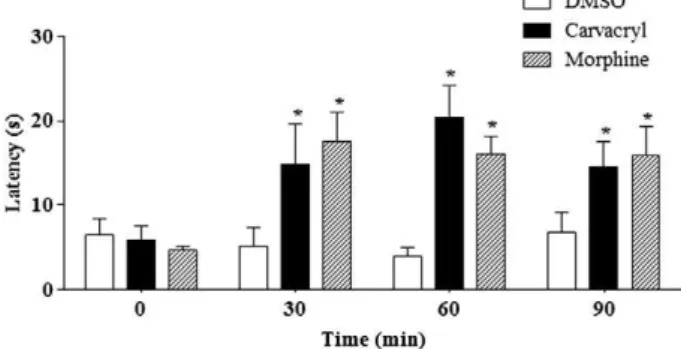
Hot plate test
Pretreatment with the carvacryl acetate (75 mg/kg, i.p.) significantly increased latency time on the plate in all the evaluated times (30, 60 and 90 min), as compared to time zero. Likely, treatment with morphine (5 mg/kg, s.c.) induced a significant increase in latency time in the hot plate test, throughout the experimental period (Fig. 5).
Formalin-induced licking
As shown inFig. 6a, the 2.8% formalin administration caused an ele-vated licking time in the paw in thefirst and in the second phase
(60.36 ± 12.91 and 37.28 ± 11.19 s, respectively;Fig. 6a). The pre-treatment with carvacryl acetate (75 mg/kg, i.p.) reverted significantly the licking time in both the neurogenic (63.99%,Pb0.05) and infl am-matory (93.30%,Pb0.01) phases. Similarly, the morphine administra-tion also reverted the pain induced by formalin, decreasing the licking times in 57.38% (Pb0.05) in the phase 1 and 98.23% (pb0.05) in the phase 2. TheFig. 6b shows that the pretreatment of animals with nalox-one (3 mg/kg, s.c.), a nonselective opioid antagonist, did not change the carvacryl acetate-mediated antinociceptive effect, however largely re-versed the anti-nociceptive effect caused by morphine when analyzed in the early phase of the formalin test (3.88 ± 1.49 s in morphine group versus 26.57 ± 4.75 s in naloxone group).
Capsaicin-induced nociception
Fig. 7illustrates that the capsaicin (3μg/paw) administration caused accentuated hypernociception in mice, with a licking time of the paw of 33.08 ± 1.27 s. In addition, the carvacryl acetate (75 mg/kg, i.p.) admin-istration 30 min before of capsaicin promoted accentuated decrease in licking response, with an inhibition percentage of 53.27% (Pb0.001). As expected, the morphine also reverted significantly the hypernociception (87.34%,Pb0.001).
Glutamate-induced nociception
In the glutamate-induced hyperalgesia (Fig. 8), the carvacryl acetate (75 mg/kg, i.p.) caused pronounced anti-nociception, with 55.92% inhi-bition (Pb0.05) in licking response (56.05 ± 11.21 s in glutamate group versus 24.71 ± 9.45 s in carvacryl acetate-treated group). The MK-801, a non-competitive antagonist of glutamate receptor, also inhibited in a significant way the hyperalgesia caused by glutamate (65.96% reduction,Pb0.05).
Evaluation of the motor performance
The carvacryl acetate (75 mg/kg) did not affect the number of falls or time of stay on the rotating bar, when compared with the vehicle in rotarod test. As opposed, diazepam administration, positive control for Fig. 3.Anti-inflammatory effect of carvacryl acetate on carrageenan-induced peritonitis in
mice. Mice were injected i.p. with dimethyl sulfoxide (DMSO), carvacryl acetate (carvacryl; 75 mg/kg), or indomethacin (10 mg/kg, reference control) and were injected with 250μl of carrageenan (500μg/cavity, i.p.) 30 min later. Neutrophil migration was evaluated after 4 h. The white bars represent the peritoneal neutrophils in animals injected with DMSO (untreated group). (a) Total counts and (b) differential counts. The values are means ± SEM offive animals for each group (#P
b0.05 compared to the DMSO group;⁎Pb0.05 compared to the carrageenan group).
Table 2
Effect of carvacryl acetate on the myeloperoxidase (MPO), interleukin (IL)-1β, tumor necrosis factor (TNF)-αand interleukin (IL)-10 levels in the peritoneal exudate after carrageenan injection.
Treatment Dose (mg/kg) MPO (U/ml) IL-1β(pg/ml) TNF-α(pg/ml) IL-10 (pg/ml)
DMSO 2.14 ± 0.24 53.78 ± 32.93 36.67 ± 12.57 16.32 ± 04.28
Control (Cg) 7.69 ± 0.80 1995.0 ± 13.19 202.20 ± 19.97 28.03 ± 08.18
Indomethacin 10 4.37 ± 1.45* (43.1) 1192.0 ± 175.20*** (40.2) 80.77 ± 27.32* (60.0) 65.21 ± 09.26* Carvacryl Acetate 75 4.10 ± 0.66* (46.6) 951.10 ± 143.30*** (52.3) 119.10 ± 51.86 (41.0) 71.46 ± 11.69*
Results are expressed as the mean ± SEM offive animals per group (*pb0.05 compared to the carrageenan group. ***pb0.001 compared to the carrageenan group).
Fig. 4.Effect of carvacryl acetate on the writhing response induced by acetic acid in mice. Mice received dimethyl sulfoxide (DMSO), carvacryl acetate (carvacryl; 75 mg/kg, i.p.), or morphine (5 mg/kg, s.c.; reference control), 30 min prior to 0.6% acetic acid (250μl/cavity; i.p.). Data are expressed as the means ± SEM offive animals for each group (⁎Pb0.05 indicates a significant difference from the acetic acid group).
motor impairment, changed significantly the motor performance of the tested animals, increasing the number of falls and decreasing time of stay on the rotating bar.
Discussion
In the present study, we evaluated the potential anti-inflammatory and anti-nociceptive effects of carvacryl acetate, a derivative of carvacrol, through the use of several pharmacological tools. Our results showed that the carvacryl acetate presents significant anti-inflammatory and anti-nociceptive effects.
Inflammation is a biological reaction to an injured tissue homeosta-sis (Medzhitov, 2008), which involves redness, edema, rise in tempera-ture, pain and loss of function (Bano et al., 2013). Among these
parameters triggered by inflammation, edema and pain formation are targets for studies in the search for new compounds with anti-inflammatory and analgesic activities.
Carrageenan-induced paw edema is widely used for determining the biphasic event of inflammation that involves the participation of diverse set of inflammatory mediators and intense neutrophil infiltration (Morris et al., 2003). Thefirst phase of this experimental model has been attributed to the action of mediators such histamine, serotonin and bradykinin on vascular permeability (Niazi et al., 2009). The second phase is characterized by an excess production of prostaglandins and an intense neutrophil infiltrate (Perez-Gurrero et al., 2001; Déciga-Campos Fig. 5.Effect of carvacryl acetate on reaction times to thermal stimuli (hot plate). Mice
re-ceived dimethyl sulfoxide (DMSO), carvacryl acetate (carvacryl; 75 mg/kg, i.p.), or mor-phine (5 mg/kg, s.c.). Data are expressed as the means ± SEM offive animals for each group (⁎Pb0 .05 when compared with the time zero).
Fig. 6.Effect of carvacryl acetate on the formalin test in mice. The time spent licking was determined during thefirst 0–5 min (phase 1; panel a) and during 20–25 min (phase 2; panel b) after injection with 2.8% formalin. Dimethyl sulfoxide (2% DMSO), carvacryl acetate (carvacryl; 75 mg/kg, i.p.), or morphine (5 mg/kg, s.c.; positive control) were administered 30 min before subcutaneously administration of formalin. Data are expressed as the mean ± SEM offive animals for each group (⁎Pb0.05 indicates significant difference from the formalin group).
Fig. 7.Evaluation of the opioid pathway on the analgesic effect of carvacryl acetate. Mice were pretreated with 2% DMSO or naloxone (3 mg/kg, s.c., opioid antagonist). After 30 min, the animals were treated with carvacryl acetate (carvacryl; 75 mg/kg, i.p.) or morphine (5 mg/kg, s.c.). After 60 min, the time spent licking was determined during the first 0–5 min (phase 1) after injection with 2.8% formalin. Data are expressed as the means ± SEM of six animals for each group (⁎Pb0.05 indicates significant difference from the formalin group;#P
et al., 2007). In the present work, we evaluated the inhibitory potential of the carvacryl acetate on the carrageenan edema, where we observed that the carvacryl acetate caused significant reduction of edema at all times evaluated, suggesting that its anti-edematogenic effect is proba-bly due to the inhibition of different aspects and chemical mediators of inflammation.
To confirm the involvement of these mediators in the anti-inflammatory effect provided by carvacryl acetate we realize the paw edema induced by compound 48/80, histamine, serotonin and prosta-glandin E2. Compound 48/80 causes massive mast cell degranulation that promotes the release of inflammatory mediators such as histamine, serotonin, platelet-activating factor, leukotrienes and a variety of cyto-kines that can elicit many events associated with inflammation (Datti et al., 2002; Coussens and Werb, 2002). Among these agents, histamine and serotonin are important inflammation mediators; they are potent vasodilator substances and also increase the vascular permeability (Linardi et al., 2000). The carvacryl acetate significantly reversed the edema induced by compound 48/80, histamine and serotonin. Thus, it is possible to confirm the involvement of vasoactive mediators derived from mast cell granules, such as histamine and serotonin, in the carvacryl acetate anti-inflammatory effect.
PGE2is synthesized in substantial amounts at the sites of infl amma-tion, and it acts as a potent vasodilator, inducing the production of vari-ous chemo-attractants, such as leucocytes and pro-inflammatory cytokines including IL-1βand TNF-α(Kaur et al., 2004). This mediator also participates in acute pain, sensitising the nerve endings of nocicep-tivefibres Aδand C through its receptor EP1 (Rady et al., 2001). Thus, high levels of PGE2have been found in inflammatory exudates, and the injection of PGE2directly into tissue has been shown to induce a number of classical sign of inflammation (Claudino et al., 2006). The carvacryl ac-etate also significantly reduced the edema induced by PGE2, demonstrat-ing that the carvacryl acetate-mediated anti-inflammatory effect also involves the decrease of this important inflammatory mediator.
Leukocyte migration to injured tissue is another important aspect of the inflammatory process (González et al., 2013). To evaluate the partic-ipation of leukocyte migration in the anti-inflammatory activity mediat-ed by carvacryl acetate, we performmediat-ed the carrageenan-inducmediat-ed peritonitis model. Intraperitoneal administration of carrageenan pro-duces a sustained increase in postcapillary venule permeability, thereby leading to increased cellular infiltration, particularly of neutrophils (Malech and Gallin, 1987). Thus, this model of acute inflammation al-lows the quantification of leukocytes that migrate into the peritoneal cavity under the action of chemotactic agents, mainly leukotrienes and cytokines, and is sensitive to the action of non-steroidal anti-inflammatory drugs (Brooks and Day, 1991). The results of present study showed that pre-treatment with carvacryl acetate significantly decrease the leukocytes count, as well as neutrophils that migrated
into the peritoneal cavity. These data suggest that the carvacryl acetate-mediated anti-inflammatory effect involves, in part, inhibition of neutrophil migration into the injury site.
To confirm these results, we evaluated the levels of myeloperoxidase in inflammatory exudate. Myeloperoxidase (MPO) is an enzyme secret-ed by neutrophils at inflammatory sites that induce damage to adjacent tissue, contributing to the pathogenesis of inflammation; it is also widely used as a marker of neutrophil infiltration (Vlasova et al., 2012). Our re-sults showed that pretreatment with carvacryl acetate significantly re-duced levels of MPO in the peritoneal exudate when compared with the carrageenan group. The results of this evaluation confirm the perito-nitis test, indicating that the anti-inflammatory activity of carvacryl ace-tate involves, among other parameters, decreased neutrophil migration. In addition to cell migration, carrageenan-induced peritonitis also involves plasma exudation and production of mediators such as nitric oxide, IL-1β, TNF-αand IL-6 (Salvemini et al., 1996; Loram et al., 2007). Considering this information, we evaluated the effect of carvacryl acetate on IL-1βand TNF-αlevels in the peritoneal exudate. The results found showed that the carvacryl acetate significantly decreased the concentrations of IL-1β, when compared with carrageenan group; how-ever, it did not reduced TNF-αlevel. These data suggest that the anti-inflammatory effect of the carvacryl acetate may be due to the inhibition of the production of IL-1βat inflammatory sites. IL-1βis a pleiotropic cytokine that plays a key role in the innate immune response and is as-sociated with the reduction of cell migration and exudation (Vigil et al., 2008). This pro-inflammatory cytokine promotes the expression of ad-hesion molecules, leukocyte migration, increased vascular permeability and transendothelial migration (Hallegua and Weisman, 2002).
To limit the deleterious consequences of prolonged inflammatory reaction, the release of pro-inflammatory cytokines is followed by the release of anti-inflammatory cytokines, such as IL-4, IL-10, and IL-13. Studies have shown that IL-10 has been found as a potent leukocyte de-activator, which blocked TNF-α, IL-1, IL-6, IL-8 (Cyktor and Turner, 2011). Other authors demonstrated that the anti-inflammatory effect of carvacrol is, at least in part, dependent of increase of IL-10 levels (Lima et al., 2013). Thus, we investigated the possibility that IL-10 pro-duction also contributes to the anti-inflammatory effect of carvacryl ac-etate. Our presentfindings are consistent with the results ofLima et al. (2013). In our study, we also demonstrated that carvacryl acetate en-hanced the IL-10 levels in carrageenan-induced peritonitis in mice. Thus, carvacryl acetate, like carvacrol, can be able to modify the number of neutrophills and actively interfere with anti-inflammatory and pro-inflammatory signalling pathways by inducing production of IL-10 and reducing IL-1βrelease.
Mediators produced at the sites of inflammation have been known to produce pain through the activation or sensitization of nociceptors adjacent to the injured tissue (Carvalho et al., 2013). Experimental models of inflammatory pain in rodents have been successfully employed to reproduce this kind of pain and are used to search new anti-inflammatory and analgesic drugs (Andrade et al., 2012).
Acetic acid-induced writhing is a visceral pain model which has been largely used for the evaluation of peripheral anti-nociceptive activity because of its high sensitivity (Santos et al., 2011). Acetic acid induces local production of inflammatory mediators, including prostaglandins, serotonin, histamine, bradykinin and substance P, which will sensitize the nociceptors leading to hyperalgesia (Chu et al., 2008; Serhan and Haeggstrom, 2010). Pretreatment with the carvacryl acetate signifi cant-ly decreased the amount of writhing induced by acetic acid in mice. The anti-nociceptive effect demonstrated by carvacryl acetate in this exper-imental model suggests that it acts through the inhibition of the synthe-sis or action of inflammatory mediators that contribute to the development of inflammatory pain. Interestingly, thisfinding confirms our results obtained in experimental models of inflammation, where it was observed that the carvacryl acetate significantly reversed the edema of all inflammatory mediators tested, namely histamine, seroto-nin and prostaglandin E2.
Fig. 8.Effect of carvacryl acetate on the capsaicin-induced nociception in mice. The time spent licking was determined during thefirst 0–5 min after injection with capsaicin solution (3μg/paw). Dimethyl sulfoxide (DMSO, control), carvacryl acetate (carvacryl; 75 mg/kg, i.p.), or morphine (5 mg/kg, s.c.; positive control) were administered 30 min before intrader-mal administration of capsaicin. Data are expressed as the mean ± SEM offive animals for each group (⁎Pb0.05 indicates significant difference from the capsaicin group).
The hot plate test is a specific central nociceptive test in which agents exert their analgesic effects via supraspinal and spinal receptors (Mbiantcha et al., 2011). The results obtained showed that carvacryl ac-etate presented significant anti-nociceptive effect in the hot-plate test. These data suggest the participation of central mechanisms in the anti-nociceptive effects of the carvacryl acetate since both behavioral components that were evaluated in this experimental model, namely paw licking and jumping, are considered to be supraspinally integrated responses (Le Bars et al., 2001).
Another model of nociception that has been extensively used to verify the anti-nociceptive effect noted in new compounds is the formalin-induced paw licking test (Sani et al., 2012). This model evaluates two dis-tinct phase of nociception. Thefirst phase, classified as a neurogenic pain, is an acute response observed immediately after the administration of for-malin, persisting for 5 min and that occurs through direct chemical stim-ulation promoted by formalin on nociceptors, in type C and part of the Aδ
afferentfibers; it is also associated with the release of excitatory amino acids, as glutamate, aspartate, taurine and glycine that are known to be in-volved in the transmission of peripheral nociception (Verma et al., 2005; Malmberg and Yaksh, 1995). The second phase appears between 15 and 60 min after the formalin administration, classified as an inflammatory pain, and is a tonic response associated with the release of chemical mediators such as histamine, serotonin, bradykinin, prostaglandins and excitatory amino acids (Sani et al., 2012).
The achieved results in current study evidenced that the carvacryl acetate inhibited both phases of the formalin-induced nociception. Thesefindings strongly confirms its ability to act at central nociceptive level, by inhibiting the neurogenic phase, corroborating with the results found in the hot plate test, besides acting also at the peripheral level, by inhibiting the inflammatory phase, confirming the results found in the acetic acid-induced writhing test.
Notably opioids and non-steroidal anti-inflammatory drugs (NSAIDs) are the classic anesthetics currently used. However, there are several other mechanisms by which the nociception can be re-versed; thus, emerging therapies are designed to reduce the nociceptive process by targeting inflammatory mediators (kinins, prostanoids, prostaglandins, cytokines, chemokines) as well as ligand-gated ion channels (Transient Receptor Potential Vanilloid—TRPV, Neuronal Nico-tinic Receptors—NNRs, N-methyl-D-aspartate—NMDA, metabotropic glutamate receptor—mGluR) and voltage-regulated sodium and calci-um channels (Dray, 2008).
Based on these considerations, we assess the possible mechanisms of anti-nociception of the carvacryl acetate. For this purpose, we evalu-ated the participation of the opioid pathway in this effect. However, the administration of naloxone, a nonselective opioid antagonist, did not reverse the anti-nociceptive effect of carvacryl acetate, showing that there is no participation of the opioid pathway.
Because of this, there was the necessity to evaluate other possible mechanisms by which the carvacryl acetate exerts its analgesic effect. Thus, were performed tests of nociception capsaicin and glutamate for observing the participation of these pathways.
The capsaicin-induced nociception is brought about by the activa-tion of the capsaicin receptor, also known as the transient receptor po-tential vanilloid, denominated TRPV, a ligand-gated non-selective cation channel in primary sensory neurons (Capasso and Calignano, 1988). Some analgesia strategies aim at developing either TRPV agonists or antagonist drugs to attenuate excitability in sensoryfibers. TRPV ag-onists, at high doses, promote receptor desensitization or a reversible sensory nerve terminal degeneration because of the prolonged cation influx into the nerve, osmotic damage and metabolic collapse (Szallasi and Blumberg, 1999), while antagonists aim at attenuating peripheral nervefiber sensitivity by blocking TRPV signal transduction (Gavva et al., 2005; Wang et al., 2007). The data obtained in this work show that the carvacryl acetate produced a neurogenic inhibition against capsaicin-induced nociception indicating its ability to inhibit nocicep-tive transmission initiated by TRPV activation.
Another attempt in the modulation of carvacryl acetate-mediated antinociception was to evaluate the involvement of the glutamatergic system. Glutamate is a major excitatory neurotransmitter in the central nervous system (Beirith et al., 2002). The glutamatergic receptors, both ionotropic and metabotropic glutamate receptors (iGluR and mGluRs), significantly contribute to nociceptive neurotransmission under the de-velopment and maintenance of pain responsiveness (Mao et al., 1992). The nociception induced by glutamate involves peripheral, spinal and supraspinal sites of action (Santos et al., 2005). Thus, activation of gluta-mate receptors, especially NMDA receptors, is involved in the develop-ment of spinal hyperexicitability and persistent pain transmission (Petrenko et al., 2003). Our results provide evidence that carvacryl ace-tate exerts a pronounced anti-nociception in glutamate-induced pain, indicating that there is the involvement of the glutamatergic pathway.
A relevant observation about the behavioral effects of carvacryl ace-tate evaluated in this work is the fact that it did not impair motor perfor-mance of mice evaluated in the rotarod test, which was used to exclude the possibility that carvacryl acetate caused motor relaxation and seda-tion (Sulaiman et al., 2010). Thus, the results of the nociception tests of carvacryl acetate would be not influenced by induced motor impairment.
Conclusion
In summary, our results show that the carvacryl acetate presents anti-inflammatory and anti-nociceptive effects. The present data provide evidence that in the carvacryl acetate-mediated anti-inflammatory effect, the mechanisms involved are inhibition of infl am-matory the mediators histamine, serotonin and prostaglandins, as well as a decrease of mast cell degranulation, pro-inflammatory cytokines and neutrophil migration. Our results also show that the carvacryl acetate-mediated anti-nociceptive effect occurs centrally and peripher-ally, through the involvement of capsaicin and glutamate pathways and decreased inflammatory mediators.
Conflict of interest statement
The authors report no conflict of interest.
Acknowledgments
The authors gratefully acknowledge to the National Counsel of Tech-nological and Scientific Development, CNPq (Brazil) and to the Research Foundation for the State of Piauí-Brazil (FAPEPI) for financially supporting this work.
References
Andrade EL, Meotti FC, Calixto JB.TRPA1 antagonists as potential analgesic drugs. Pharmacol Ther 2012;133:189–204.
Bano S, Javed K, Ahmad S, Rathish IG, Singh S, Chaitanya M, et al.Synthesis of some novel chalcones,flavanones andflavones and evaluation of their anti-inflammatory activi-ty. Eur J Med Chem 2013;65:51–9.
Beena N, Kumar D, Rawat DS.Synthesis and antioxidant activity of thymol and carvacrol based Schiff bases. Bioorg Med Chem Lett 2013;23:641–5.
Beirith A, Santos ARS, Calixto JB.Mechanisms underlying the nociception and paw edema caused by injection of glutamate into the mouse paw. Brain Res 2002;924:219–28. Bradley PP, Priebat DA, Christensen RD, Rothstein G.Measurement of cutaneous infl
am-mation: estimation of neutrophil content with an enzyme marker. J Invest Dermatol 1982;78:206–9.
Brooks PM, Day RO.Non-steroidal anti-inflammatory drugs differences and similarities. N Engl J Med 1991;324:1716–25.
Calixto JB.Twenty-five years of research on medicinal plants in Latin America: a personal review. J Ethnopharmacol 2005;100:131–4.
Capasso A, Calignano A.Synergism between the sedative action of kava extract andD, L-kavain. Acta Theriol 1988;14:249–56.
Carvalho V, Fernandes L, Conde T, Zamith H, Silva R, Surrage A, et al.Antinociceptive ac-tivity ofStephanolepis hispidusskin aqueous extract depends partly on opioid system activation. Mar Drugs 2013;11:1221–34.
Chu C, Huang Y, Chen YF, Wu JH, Rahman K, Zheng HC, et al.Antinociceptive activity of aqueous fraction from the MeOH extracts ofPaederia scandensin mice. J Ethnopharmacol 2008;118:177–80.
Claudino RF, Kassuya CAL, Ferreira J, Calixto JB.Pharmacological and molecular character-ization of the mechanisms involved in prostaglandin E2-induced mouse paw edema. J Pharmacol Exp Ther 2006;318:611–8.
Coussens LM, Werb Z.Inflammation and cancer. Nature 2002;420:860–7.
Cyktor JC, Turner J.Interleuk10 and immunity against prokaryotic and eukaryotic in-tracellular pathogens. Infect Immun 2011;79:2964–73.
Datti F, Datti M, Antunes E, Teixeira NA.Influence of chronic unpredictable stress on the allergic responses in rats. Physiol Behav 2002;77:79–83.
Déciga-Campos M, Palacios-Espinosa JF, Reyes-Ramírez A, Mata R.Antinociceptive and anti-inflammatory effects of compounds isolated fromScaphyglottis lividaand Maxillaria densa. J Ethnopharmacol 2007;114:161–8.
Dray A.New horizons in pharmacologic treatment for rheumatic disease pain. Rheum Dis Clin North Am 2008;34:481–505.
Dunham NW, Miya TS.A note on a simple apparatus for detecting neurological deficit in rats and mice. J Am Pharm Assoc 1957;46:208–10.
Eddy NB, Leimbach D.Synthetic analgesics. II. Dithienylbutenyl- and dithienylbutylamines. J Pharmacol Exp Ther 1953;107:385–93.
Fasmer OB, Berge OG, Hole K.Changes in nociception after lesions of descending seroto-nergic pathways induced with 5,6-dihydroxytryptamine. Different effects in the for-malin and tail-flick tests. Neuropharmacology 1986;24:729–34.
Gavva NR, Tamir R, Qu Y, Klionsky L, Zhang TJ, Immke D, et al. AMG 9810 [(E)-3-(4-t-butylphenyl)-N-(2,3-dihydro-benzo[b][1,4] dioxin-6-yl)acrylamide], a novel vanilloid receptor 1 (TRPV1) antagonist with antihyperalgesic properties. J Pharmacol Exp Ther 2005;313:473–84.
González CP, Vega RS, González-Chávez M, Sánchez MAZ, Gutiérrez SP.Antiinflammatory activity and composition ofSenecio salignus Kunth. BioMed Res Int 2013;2013:1–4. Hallegua DS, Weisman MH.Potential therapeutic uses of interleukin 1 receptor
antago-nists in human diseases. Ann Rheum Dis 2002;61:960–7.
Hunskaar S, Fasmer OB, Hole K.Formalin test in mice, a useful technique for evaluating mild analgesics. J Neurosci Methods 1985;14:69–76.
Ipek E, Zeytinoglu H, Okay S, Tuylu BA, Kurkcuoglu M, Baser KHC.Genotoxicity and antigenotoxicity of Origanum oil and carvacrol evaluated by Ames Salmonella/ microsomal test. Food Chem 2005;93:551–6.
Kaur G, Hamid H, Ali A, Alam MS, Athar M.Antiinflammatory evaluation of alcoholic ex-tract of galls ofQuercus infectoria. J Ethnopharmacol 2004;90:285–92.
Klein G, Rüben C, Upmann M.Antimicrobial activity of essential oil components against potential food spoilage microorganisms. Curr Microbiol 2013;67:200–8.
Koster R, Anderson M, Debeer EI.Acetic acid for analgesic screening. Fed Proc 1959;18: 412–8.
Landa P, Kokoska L, Pribylova M, Vanek T, Marsik P.In vitro anti-inflammatory activity of carvacrol: inhibitory effect on cox-2 catalyzed prostaglandin E2biosynthesis. Arch Pharm Res 2009;32:75–8.
Le Bars D, Gozariu M, Cadden SW.Animal models of nociception. Pharmacol Rev 2001;53: 597–652.
Lima MS, Quintans-Júnior LJ, Santana WA, Kaneto CM, Soares MBP, Villarreal CF. Anti-inflammatory effects of carvacrol: evidence for a key role of interleukin-10. Eur J Pharmacol 2013;699:112–7.
Linardi A, Costa SKP, De Silva GR, Antunes E.Involvement of kinins, mast cells and sensory neurons in the plasma exudation and paw edema induced by staphylococcal entero-toxin B in the mouse. Eur J Pharmacol 2000;399:235–42.
Loram LC, Fuller A, Fick LG, Cartmell T, Poole S, Mitchell D.Cytokine profiles during carrageenan-induced inflammatory hyperalgesia in rat muscle and hind paw. J Pain 2007;8:127–36.
Malech HL, Gallin JI.Current concepts: immunology-neutrophils in human diseases. N Engl J Med 1987;317:687–94.
Malmberg AB, Yaksh TL.Cyclooxygenase inhibition and the spinal release of prostaglan-din E2 and amino acid evoked by paw formalin injection: a microdialysis study in un-anesthetized rats. J Neurosci 1995;14:2768–76.
Mao J, Price DD, Hayes RL, Lu J, Mayer DJ.Differential roles of NMDA and non-NMDA re-ceptor activation in induction and maintenance of thermal hyperalgesia in rats with painful peripheral mononeuropathy. Brain Res 1992;598:271–8.
Mbiantcha M, Kamanyi A, Teponno RB, Tapondjou AL, Watcho P, Nguelefack TB.Analgesic and anti-inflammatory properties of extracts from the bulbils ofDioscorea bulbiferaL. var sativa (Dioscoreaceae) in mice and rats. Evid Based Complement Alternat Med 2011;10:1–9.
Medzhitov R.Origin and physiological roles of inflammation. Nature 2008;454:428–35. Melo FHC, Rios ERV, Rocha NFM, Citó MCO, Fernandes ML, Sousa DP, et al.Antinociceptive
activity of carvacrol (5-isopropyl-2-methylphenol) in mice. J Pharm Pharmacol 2012;64:1722–9.
Moraes J, Carvalho AA, Nakano E, de Almeida AA, Marques TH, Andrade LN, et al. Anthel-mintic activity of carvacryl acetate againstSchistosoma mansoni. Parasitol Res 2013;112:603–10.
Morris CJ, Ferreira SH, Vane JR.Carrageenan-induced paw edema in the rat and mouse. Methods Mol Biol 2003;225:115–21.
Niazi J, Singh P, Bansal Y, Goel RK.Anti-inflammatory, analgesic and antipyretic activity of aqueous extract of fresh leaves of Cocciniaindica. Inflammopharmacol 2009;17: 239–44.
Perez-Gurrero C, Herrera MD, Ortiz R, Alvarez de Sotomayor M, Fernandez MA.A pharma-cological study ofCecropia obtusifoliaBetrol aqueous extract. J Ethnopharmacol 2001;76:279–84.
Petrenko AB, Yamakura T, Baba H, Shimoji K.The role of N-methyl-D-aspartate (NMDA) receptors in pain: a review. Anesth Analg 2003;97:1108–16.
Rady JJ, Campbell WB, Fujimoto JM.Antianalgesic action of nociception originating in the brain is mediated by spinal prostaglandin E2in mice. J Pharmacol Exp Ther 2001;296: 7–14.
Rodriguez-Vita J, Lawrence T.The resolution of inflammation and cancer. Cytokine Growth Factor Rev 2010;21:61–5.
Sakurada T, Matsumura T, Moriyama T, Sakurada C, Ueno S, Sakurada S.Differential effects of intraplantar capsazepine and ruthenium red on capsaicin-induced desensi-tization in mice. Pharmacol Biochem Behav 2003;75:115–21.
Salvemini D, Wang ZQ, Wyatt PS, Bourdon DM, Marino MH, Manning PT, et al.Nitric oxide: a key mediator in the early and late phase of carrageenan-induced rat paw inflammation. Br J Pharmacol 1996;118:829–38.
Sani MH, Zakaria ZA, Balan T, The LK, Salleh MZ.Antinociceptive activity of methanol ex-tract ofMuntingia calaburaleaves and the mechanisms of action involved. Evid Based Complement Alternat Med 2012;2012:1–10.
Santos AR, Gadotti VM, Oliveira GL, Tibola D, Paszcuk AF, Neto A, et al.Mechanisms in-volved in the antinociception caused by agmatine in mice. Neuropharmacology 2005;48:1021–34.
Santos EN, Lima JCS, Noldin VF, Cechinel-Filho V, Rao VSN, Lima EF, et al. Antiinflammatory, antinociceptive, and antipyretic effects of methanol extract of Cariniana rubrastem bark in animal models. An Acad Bras Cienc 2011;2:557–66. Serhan CN, Haeggstrom JZ.Lipid mediators in acute inflammation and resolution:
eicosa-noids, PAF, resolvins and proteins. Cambridge: Cambridge University Press; 2010153–74.
Silva VG, Silva RO, Damasceno SRB, Carvalho NS, Prudêncio RS, Aragão KS, et al. Anti-inflammatory and antinociceptive activity of epiisopiloturine, an imidazole alka-loid isolated fromPilocarpus microphyllus. J Nat Prod 2013;76:1071–7.
Sulaiman MR, Padzil A, Shaari K, Khalid S, Shaikmossadeq W, Shahmohamad A, et al. Antinociceptive activity ofMelicopep telefoliaethanolic extract in experimental ani-mals. J Biomed Biotechnol 2010;2010:1–6.
Szallasi A, Blumberg PM.Vanilloid (Capsaicin) receptors and mechanisms. Pharmacol Rev 1999;51:159–212.
Verma PR, Joharapurkar AA, Chatpalliwar VA, Asnani AJ.Antinociceptive activity of alco-holic extract ofHemidesmus indicusR. Br. in mice. J Ethnopharmacol 2005;102: 298–301.
Vigil SVG, Liz R, Medeiros YS, Frode TS.Efficacy of tacrolimus in inhibiting inflammation caused by carrageenan in a murine model of air pouch. Transpl Immunol 2008;19:25–9. Vincenzi MD, Stammati A, Vincenzi AD, Silano M.Constituents of aromatic plants:
carva-crol. Fitoterapia 2004;75:801–4.
Vlasova II, Sokolov AV, Arnhold J.The free amino acid tyrosine enhances the chlorinating activity of human myeloperoxidase. J Inorg Biochem 2012;106:76–83.
Vogel AI, Tatchell AR, Furnis BS, Hannaford AJ, Smith PWG.Vogel's text book of practical organic chemistry. 5th ed. Englewood Cliffs: Prentice-Hall; 1996.
Wang HL, Katon J, Balan C, Bannon AW, Bernard C, Doherty EM, et al.Vanilloid receptor-1 antagonists: 3. The identification of a second-generation clinical candidate with im-proved physicochemical and pharmacokinetic properties. J Med Chem 2007;50: 3528–39.
Winter CA, Risley EA, Nuss GW.Carrageenin-induced edema in hind paw of the rat as an assay for antiiflammatory drugs. Proc Soc Exp Biol Med 1962;111:544–7. Yanishlieva N, Marinova EM, Gordon MH, Raneva VG.Antioxidant activity and
mecha-nism of action of thymol and carvacrol in two lipid systems. Food Chem 1999;64: 59–66.