Activation of Mu or Delta Opioid Receptors in
the Lumbosacral Spinal Cord Is Essential for
Ejaculatory Reflexes in Male Rats
Natalie Kozyrev1,2, Lique M. Coolen1,2,3,4*
1Department of Anatomy & Cell Biology, the University of Western Ontario, London, Ontario, Canada,
2Department of Physiology, University of Michigan, Ann Arbor, Michigan, United States of America,
3Department of Neurobiology & Anatomical Sciences, University of Mississippi Medical Center, Jackson, Mississippi, United States of America,4Department of Physiology & Biophysics, University of Mississippi Medical Center, Jackson, Mississippi, United States of America
Abstract
Ejaculation is controlled by a spinal ejaculation generator located in the lumbosacral spinal cord, consisting in male rats of lumbar spinothalamic (LSt) cells and their inter-spinal projec-tions to autonomic and motor centers. LSt cells co-express several neuropeptides, including gastrin releasing peptide (GRP) and enkephalin. We previously demonstrated in rats that GRP regulates ejaculation by acting within the lumbosacral spinal cord. In the present study, the hypothesis was tested that enkephalin controls ejaculation by acting on mu (MOR) or delta opioid receptors (DOR) in LSt target areas. Adult male rats were anesthe-tized and spinalized and received intrathecal infusions of vehicle, MOR antagonist CTOP (0.4 or 4 nmol), DOR antagonist (TIPP (0.4, 4 or 40 nmol), MOR agonist DAMGO (0.1 or 10 nmol), or DOR agonist deltorphin II (1.3 or 13 nmol). Ejaculatory reflexes were triggered by stimulation of the dorsal penile nerve (DPN) and seminal vesicle pressure and rhythmic con-tractions of the bulbocavernosus muscle were analyzed. Intrathecal infusion of MOR or DOR antagonists effectively blocked ejaculatory reflexes induced by DPN stimulation. Intra-thecal infusion of DAMGO, but not deltorphin II triggered ejaculation in absence of DPN stimulation. Both MOR and DOR agonists facilitated ejaculatory reflexes induced by sub-threshold DPN stimulation in all animals. Overall, these results support the hypothesis that enkephalin plays a critical role in the control of ejaculation in male rats. Activation of either MOR or DOR in LSt target areas is required for ejaculation, while MOR activation is suffi-cient to trigger ejaculation in the absence of sensory stimulation.
Introduction
Ejaculation is a complex physiological phenomenon that is highly rewarding and culminates in the ejection of seminal contents from the urethral meatus [1–3]. Ejaculation involves two phases,
emission and expulsion. During the emission phase, seminal fluids are secreted from the
OPEN ACCESS
Citation:Kozyrev N, Coolen LM (2015) Activation of Mu or Delta Opioid Receptors in the Lumbosacral Spinal Cord Is Essential for Ejaculatory Reflexes in Male Rats. PLoS ONE 10(3): e0121130. doi:10.1371/ journal.pone.0121130
Academic Editor:David Brown, University of New South Wales, AUSTRALIA
Received:July 14, 2014
Accepted:February 10, 2015
Published:March 31, 2015
Copyright:© 2015 Kozyrev, Coolen. This is an open access article distributed under the terms of the Creative Commons Attribution License, which permits unrestricted use, distribution, and reproduction in any medium, provided the original author and source are credited.
Data Availability Statement:All relevant data are within the paper.
Funding:The authors received no specific funding for this work.
accessory sex glands, including the prostate, seminal vesicles and vas deferens and the bladder neck and external urethral sphincter shut to avert retrograde ejaculation. During the expulsion phase, rhythmic contractions of the striated perineal muscles, and the bulbocavernosus muscle in particular, forcefully expel semen from the urethral meatus [4–7]. Ejaculation is a reflex
controlled by a spinal ejaculation generator in the lumbosacral spinal cord [2,8]. The spinal ejac-ulation generator is thought to control ejacejac-ulation by closely coordinating sympathetic, parasym-pathetic, and motor components to initiate emission and expulsion [3,9–13]. In addition, it is
postulated that the spinal ejaculation generator triggers ejaculation by integrating sensory inputs conveyed by the dorsal penile nerve (DPN), the sensory branch of the pudendal nerve, during copulation with autonomic and motor outflow [14–16]. The spinal ejaculation generator consists
of a set of interneurons in lamina 10 and the medial portion of lamina 7 of lumbar segments 3 and 4 (L3-4) that are integral in the control of ejaculation. These lumbar interneurons are re-ferred to as lumbar spinothalamic (LSt) cells due to their anatomical position in the lumbar spi-nal cord and projections to the subparafascicularparvocellular nucleus of the thalamus [17–20].
One proposed role of LSt cells is to transform sensory signals associated with the summation of sexual activity into motor or secretory outputs [17]. LSt cells display neural activation specifically with ejaculation but not with other components of sexual behavior following copulation [20] or following electrical stimulation of the dorsal penile nerve (DPN) in male rats [14]. Markers of neural activation in LSt cells with ejaculation include cFos [20] and phosphorylated extracellular signal-regulated kinases 1 and 2 (pERK) [14] and ejaculation is triggered through activation of the MAP kinase pathway [14] following mating in intact animals or DPN stimulation in anesthe-tized and spinalized male rats. Furthermore, LSt cells have axonal projections to LSt target re-gions including preganglionic sympathetic, parasympathetic and motor neurons in the thoracolumbar and lumbosacral spinal cord [12,17,21–24].
LSt cells express several neuropeptides including galanin, cholecystokinin, gastrin releasing peptide (GRP), and enkephalin [17–20]. Thus, we hypothesize that these neuropeptides act in
the LSt target areas to trigger or modulate ejaculatory reflexes. Indeed, studies from our labora-tory revealed a critical role for GRP in the control of ejaculation in male rats [25]. Intrathecal in-fusions of GRP antagonist RC-3095 completely abolished ejaculatory reflexes while intrathecal infusion of GRP agonist GRP2029triggered ejaculatory reflexes in 43–66% of animals and
facili-tated ejaculatory reflexes in response to subthreshold frequencies (5–10 Hz) of DPN stimulation
intraspinal projections to autonomic and motor areas are critically involved in the ejaculatory reflexes induced by DPN stimulation in male rats, further validating the use of the DPN stimula-tion paradigm to study ejaculatory reflexes in rats [16]. First, we tested whether enkephalin is re-quired for ejaculatory reflexes in male rats using intrathecal delivery of MOR and DOR
antagonists during simultaneous recordings of seminal vesicle (SV) pressure, a marker of emis-sion, and rhythmic contractions of the bulbocavernosus muscle (BCM), a marker of expulemis-sion, in anesthetized and acutely spinalized male rats. Next, it was tested whether enkephalin is suffi-cient to trigger ejaculation in male rats in the absence of DPN stimulation or following sub-threshold levels of DPN stimulation (5–10 Hz) that do not trigger ejaculatory reflexes in male
rats in control conditions [27] and intrathecal infusions of specific MOR and DOR agonists in the lumbosacral spinal cord during recordings of SV pressure and BCM contractions.
Materials and Methods
Animals
Adult male Sprague Dawley rats (225–250 grams) were acquired from Charles River
(Wilming-ton, MA, USA) and pair-housed in standard housing cages on a 12-hour light/dark cycle with lights off at 9pm.Food and water were available ad libitum. All procedures were approved by the University Committee on Use and Care of Animals at the University of Michigan and the University of Western Ontario and conformed to the guidelines outlined by the National Insti-tutes of Health.
Pharmacological Experiments
Surgical procedures. Procedures were similar to our previous studies [14–16,25]. Male
rats were anesthetized with urethane (1.5 g/kg, i.p.) and a laminectomy was performed between the sixth and the eighth thoracic spinal segments. A complete transection of the spinal cord was performed at the seventh thoracic spinal segment. Subsequently, animals were prepared for electromyographic (EMG) recordings of bulbocavernosus muscle (BCM) bursting and sem-inal vesicle pressure (SVP); indicators of expulsion and emission components of ejaculation re-spectively [8,30,33]. The BCM and the dorsal penile nerve (DPN) were surgically exposed and the surrounding connective tissue was removed. Silver recording electrodes, connected to the PowerLab/7SP Data Acquisition System (AD Instruments, Inc., Colorado Springs, CO, USA) were placed bilaterally into the BCM and a ground electrode was inserted into the muscle of the right thigh. In a subset of animals, the right seminal vesicle was exposed by coeliotomy and a pressure catheter (AD Instruments Inc., model number: SPR-671(1.4 F, Single, Straight, 15 cm, Ny) connected to a catheter interface cable (AD Instruments Inc., model number: AEC-IOD) and attached to a Bridge AMP (AD Instruments Inc.), was gently guided into the lumen of the SV and fixed in place. Finally, for stimulation of the DPN, a bipolar stimulating electrode connected to the SD9 Square Pulse Stimulator (Grass Technologies, West Warwick, RI, USA) was arranged directly above the DPN. 2–4 Hours prior to any of the experiments, the
DPN was stimulated (4 V at 60 Hz square wave pulses of 1 ms duration, for total of 10 seconds) in order to verify the completeness of the spinal cord transection. These established stimulation parameters consistently trigger rhythmic bursting of the BCM, indicative of the expulsion phase of ejaculation, in all animals [14–16,25].
mid-lumbar levels, such that saline or drug infused through the catheter would bathe the entire lumbosacral spinal cord. First, 10μL of saline was delivered (at 10μl/minute rate) and BCM
EMG activity was recorded for 25 minutes. Next, the DPN was stimulated at 30 Hz and 60 Hz (4 V, square wave pulses of 1 ms duration, for a total of 10 seconds) in a counterbalanced man-ner, with 5-minutes between the stimulations (testing trial 1; control trial). BCM EMG and SVP were recorded. An hour later, the procedure was performed again in the same animals, which were now administered a 10μL infusion of opioid receptor antagonists, after which the
DPN was stimulated at 30 Hz and 60 Hz in a counterbalanced manner, and BCM EMG activity and SVP were recorded (testing trial 2; drug trial). We previously established that repeated stimulations and infusions of saline did not affect BCM EMG bursting in response to or with-out DPN stimulation [25], therefore all animals received drug treatment in testing trial 2 (drug trial). In experiment 1, one of two doses of the mu opioid receptor antagonist H-D-Phe-Cys-Tyr-D-Trp-Orn-Thr-Pen-Thr-NH2(CTOP; Bachem Americas, Inc. Torrance, CA, USA)
0.4nmol (N = 7) or 4nmol (N = 8) were infused and BCM EMG (but not SVP) was recorded. In experiment 2, the most effective dose of CTOP was administered, i.e. 4 nmol, and BCM EMG and SVP were recorded simultaneously (N = 6). In experiment 3, one of three doses of the delta opioid receptor antagonist H-Tyr-Tic-Phe-Phe-OH (TIPP), [Bachem Americas, Inc. Torrance, CA, USA], 0.4 nmol (N = 6), 4 nmol (N = 7), or 40 nmol (N = 6) were infused and BCM EMG and SVP were recorded.
Intrathecal infusion of opioid receptor agonists. Next, it was tested whether activation of opioid receptors is sufficient to trigger ejaculation in male rats in the absence of DPN stimula-tion or following subthreshold levels of DPN stimulastimula-tion (5–10 Hz) that do not trigger
ejacula-tory reflexes in male rats in control conditions [25]. In addition, the effects of opioid receptor agonists were tested on the DPN stimulation parameters that do reliably trigger ejaculatory re-flexes (30 and 60 Hz).Four separate groups of male rats were anesthetized, spinalized, and pre-pared as described above. In testing trial 1 (control trial), all male rats received 10μL intrathecal
infusions of 0.9% saline (N = 30) and BCM EMG and SVP were recorded for 10 minutes after infusion. Next, the DPN was stimulated at 5 HZ, 10 HZ, 30 HZ and 60 HZ in a randomized and counterbalanced order, and BCM EMG and SVP were recorded for the duration of 90 seconds following stimulation (testing trial 1; control trial). We previously determined that repeated stimulation using these parameters did not affect BCM EMG [25]. One hour following the final DPN stimulation, the procedure was repeated in the same groups of animals, which now re-ceived 10μL intrathecal infusions of one of two doses of mu opioid receptor agonist (D-Ala2,
N-Me-Phe4, glycinol5)—Enkephalin [DAMGO 0.1 nmol, N = 7; 10 nmol, N = 8; (Bachem
Americas, Inc. Torrance, CA, USA)] or one of two doses of the delta opioid receptor agonist [DAla2] Deltorphin II, 1.3 nmol, N = 8; 13 nmol, N = 8; [American Peptide Co, Sunnyvale, CA, USA] and BCM EMG and SVP were recorded for 10 minutes after infusion (testing trial 2; drug trial). Finally, the DPN was stimulated at 5, 10, 30, and 60 HZ in a counterbalanced manner as described above and previously (testing trial 2; drug trial [25]).
Analysis: BCM EMG and SVP increases. BCM EMG and SVP recordings were analyzed using methods we previously described [25]. Briefly, data were analyzed for the 25 or 10 min-utes following the infusion of the antagonist or agonist, respectively. In addition, EMG and SVP were analyzed for 90 seconds following each DPN stimulation, which is the time span of a characteristic DPN-stimulation induced ejaculatory reflex in control male rats [14–16,25]. The
For the antagonist experiments, the effects of antagonist treatments on the numbers of events, bursts and SVP increases were statistically compared using Two-Way Repeated Anova (factors: Testing trial and Drug dose) and Holm-Sidak post hoc tests, separately for each 30 and 60 Hz stimulation, both within animals (within each drug dose group, but between testing trial 1 (control trial) and testing trial 2 (drug trial)) and between groups (between different drug dose groups, but within testing trial 1 or within testing trial 2). For the agonist experi-ments, numbers of events, bursts and SVP increases were compared separately for each stimu-lation setting (following infusion without stimustimu-lation, 5, 10, 30, 60 Hz), using a Two-Way Repeated Anova (factors: Testing trial and Drug Dose) and Holm-Sidak post hoc tests, both within animals (between testing trial 1 and testing trial 2) and between treatment groups (be-tween different dose groups within testing trial 1 or within testing trial 2). A 95% confidence level was used for all tests.
Immunohistochemistry Experiment
To verify that mu opioid receptor antagonist CTOP and delta opioid receptor antagonist TIPP did not block ejaculation by preventing the activation of LSt cells, expression of phosphoryla-tion of ERK (pERK) in LSt cells was examined following stimulaphosphoryla-tion of the DPN [14,25]. Fur-thermore, previous experiments have shown that pERK is not expressed in LSt cells under baseline conditions and that spinalization and the surgical exposure of the DPN and BCM in the absence of electrical stimulation of the DPN do not induce pERK expression in LSt cells [14]. Therefore, pERK expression in LSt cells is specifically induced by ejaculatory behavior or DPN stimulation causing ejaculatory reflexes and negative control groups were not included in this experiment. Male rats (N = 9) were anesthetized and spinalized as described above. Two hours after spinalization, the DPN and BCM were exposed and vehicle (saline, N = 4) or drug (CTOP 4nmol; N = 3, TIPP 40 nmol; N = 2, Bachem Americas, Inc. Torrance, CA, USA) were infused in a volume of 10μL intrathecally as described above. Following a 25-minute infusion,
the DPN was stimulated at 30 Hz and BCM EMG activity and SVP were recorded for 90 sec-onds. Five minutes following DPN stimulation, animals were perfused transcardially with 5 mL 0.9% saline solution and 500 mL of 4% paraformaldehyde in 0.1 M PB. Spinal cords were removed and postfixed for 1hour in the same fixative and then transferred into a cryoprotective solution (20% sucrose in 0.1 PB with 0.01% sodium azide) until further processing for immu-nohistochemical visualization of pERK and galanin (marker for LSt cells, as it is expressed ex-clusively in LSt cell bodies [17,20]. The stimulation parameters and time of perfusion were previously shown to be optimal for detection of pERK expression in LSt cells [14,25].
Immunohistochemistry: Galanin/pERK dual fluorescence. Spinal cords were cut using a freezing microtome (Thermo Fisher Scientific, Walldorf, Germany) into 12 parallel series of 35μm coronal sections in cryoprotectant solution (30% sucrose, 30% ethylene glycol in 0.1 M
PerkinElmer LifeSciences, Boston, MA, USA), and Alexa 488-conjugated streptavidin (1:100 in PBS; 30 minutes (Jackson ImmunoResearch Laboratories, WestGrove, PA, USA). Next, sec-tions were incubated with rabbit anti-galanin (1:3,000; overnight, T-4334; Bachem,Torrance, CA, USA) and Alexa 555-conjugated goat anti-rabbit (1:100 in PBS; 30 minutes; Jackson Immuno-Research Laboratories) and mounted on plus charged slides, cover-slipped with gel-vatol and stored in the dark at 4°C. For all immunohistochemistry procedures, the omission of primary antibodies resulted in a complete loss of signal at specific wavelengths and all primary antibodies have been previously validated [14,25].
Analysis: pERK Expression in LSt cells. pERK expression in LSt cells was analyzed on a DM5000B Leica fluorescent microscope (Leica Microsystems, Wetzlar, Germany). Specifically, all lumbar spinal cord cells expressing galanin-ir were analyzed for expression of pERK, as cyto-plasmic galanin-ir is exclusively expressed in LSt cells [20]. Data are expressed as the mean per-centages of LSt cells that display pERK for each animal. The group means were calculated and compared between animals that received an infusion of CTOP (4 nmol) or TIPP (40 nmol) and saline-treated controls, using One Way ANOVA with 95% confidence levels.
Results
Mu Opioid Receptor Antagonist CTOP Suppressed Ejaculatory Reflexes
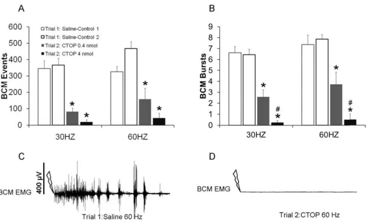
Experiment 1. Mu opioid receptor antagonist CTOP significantly decreased DPN stimulation-induced ejaculatory reflexes at both 30 and 60 Hz stimulation frequencies as re-flected in significantly reduced numbers of BCM events and bursts.
Two-Way ANOVA revealed main effects of testing trial (control trial 1 versus drug trial 2) on the numbers of BCM events for both the 30 Hz (F(1, 29) = 88.436;P<0.001;Fig. 1A) and
60 Hz stimulation frequencies (F(1, 29) = 62.013;P<0.001;Fig. 1A). Post hoc analyses revealed
that animals treated with either dose of CTOP during the testing trial 2, i.e. drug trial, had signif-icantly decreased BCM events in response to 30 or 60 Hz DPN stimulation compared to their DPN stimulation induced BCM events following saline treatment in the first control testing trial (trial 1; 60 Hz:P<0.001 (0.4 nmol);P= 0.009 (4 nmol); 30 Hz:P=<0.001(0.4 nmol);P<0.001
(4 nmol);Fig. 1A). There were no significant effect of drug dosage and no differences between the two groups treated with the lower and higher doses of CTOP for each
stimulation frequency.
Similarly, there were main effects of testing trial on the numbers of BCM bursts for both 30 Hz (F(1, 29) = 108.732;P<0.001;Fig. 1B) and 60 Hz (F(1, 29) = 123.431;P<0.001;Fig. 1B)
stimulation frequencies. In addition, there were significant interactions between testing trial and drug dosage for both stimulation frequencies (30Hz: F(1.29) = 11.83;P =0.023; 60 Hz: F(1,29) = 23.87;P =0.016). CTOP during trial 2 (drug trial) significantly reduced BCM bursts compared to trial 1 (control trial) with saline treatment (30 Hz:P<0.001 [0.4nmol], 30 Hz:
P<0.001 [4 nmol], 60 Hz:P<0.001 [0.4 nmol], 60 Hz:P<0.001 [4nmol];Fig. 1B, C, D).
There was an effect of dose on the numbers of BCM bursts in testing trial 2: drug trial. Specifi-cally, there were significantly fewer BCM bursts following the higher dose compared to the lower dose of CTOP (30 Hz:P= 0.003;Fig. 1B, 60 Hz:P= 0.006;Fig. 1B). Finally, during the control testing trial (trial 1), there were no significant differences between groups in the num-bers of BCM events or bursts.
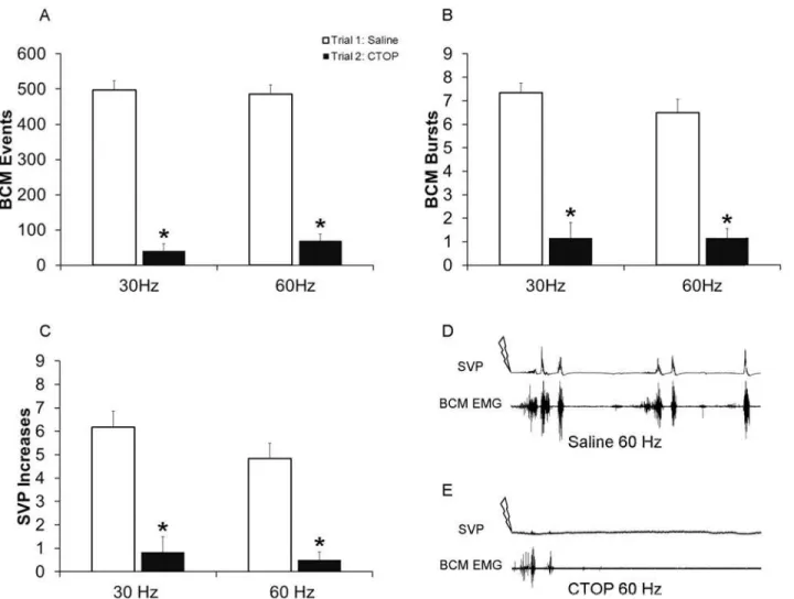
Fig. 2C, D, E), as well as BCM events (F(1, 23) = 513.688;P<0.001;Fig. 2A, D, E), and BCM
bursts (F(1, 23) = 118.433;P<0.001;Fig. 2B, D, E). Post hoc analyses revealed that CTOP
(4 nmol) in trial 2 (drug trial), significantly reduced numbers of SVP increases, BCM events and bursts compared to control trial 1, for both 30 Hz (events:P<0.001; bursts:P<0.001;
SVP:P<0.001;Fig. 2A-E) and 60 Hz (events:P<0.001; bursts:P<0.001; SVP:P<0.001; Fig. 2A-E). Overall, these results confirm the hypothesis that intrathecal CTOP suppresses both the emission and expulsion components of ejaculation and that activation of mu opioid receptors in the lumbosacral spinal cord is required for ejaculatory reflexes in male rats.
Delta Opioid Receptor Antagonist TIPP Suppressed Ejaculatory
Reflexes
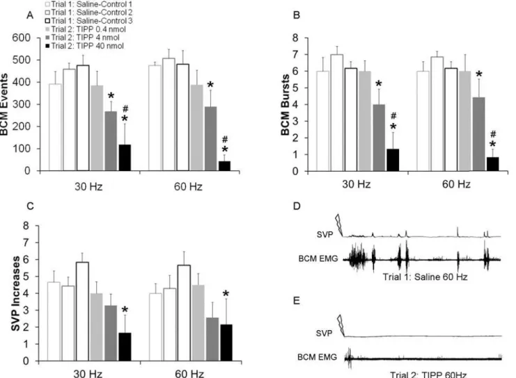
Similar to the effects of the mu opioid antagonist, inthrathecal infusion of delta opioid receptor antagonist TIPP significantly decreased DPN stimulation-induced ejaculatory reflexes at both 30 and 60 Hz stimulation frequencies as reflected in the reduced numbers of BCM events, BCM bursts, and SVP increases.
There were main effects of testing trial on the numbers of BCM events for both 30 Hz (F(1,37) = 20.131;P<0.001;Fig. 3A) and 60 Hz stimulation frequencies (F(1, 37) = 26.666;
P<0.001;Fig. 3A), main effect of drug dosage for 60 Hz (F2,37) = 6.18; P = 0.01) and
signifi-cant interactions between testing trial and drug dosage for both 30 and 60 Hz (30 Hz: (F(2,37) = 5.861;P= 0.012; 60 Hz ((F(2,37) = 4.349;P= 0.031). Animals treated with the middle (4 nmol)
Fig 1. Quantitative analyses of BCM events (A) and bursts (B) in response to 30 and 60 Hz DPN stimulation following infusions of saline in trial 1 (Saline-Controls 1 and 2; white bars) or one of two doses of CTOP (0.4 or 4 nmol) in trial 2 (drug trial; filled bars).Trials 1 and 2 were conducted in the same animals, hence Saline-Control 1 are data for animals subsequently receiving CTOP 0.4 nmol in trial 2, and Saline-Control 2 are data for animals receiving CTOP 4 nmol. Data are presented as Mean±SEM.*denotes significant differences from trial 1 within the same treatment group, while # indicates significant differences between treatment groups within the same testing trial. Representative BCM EMG traces (90 seconds duration) following 60 Hz DPN stimulation (arrow) after an intrathecal infusion of saline (C: control trial) and CTOP (D: same animal as in C).
and higher (40 nmol) but not the lower (0.4 nmol) dose of TIPP during trial 2 (drug trial) had significantly decreased BCM events in response to 30 Hz (P= 0.012: 4 nmol;P=<0.001: 40
nmol;Fig. 3A) and 60 Hz (60 Hz:P= 0.014, 4 nmol;P<0.001, 40 nmol;Fig. 3A) stimulation
frequencies compared to saline treatment in trial 1 (control trial). In addition, there was an ef-fect of dosage, as animals treated with the highest dose of TIPP (40 nmol) in the second trial (drug trial) displayed significantly fewer BCM events compared to those treated with the lower dose of TIPP (0.4 nmol) for both the 30 Hz (P= 0.004, 40 nmol;Fig. 3A) and 60 Hz
(P<0.001, 40 nmol;Fig. 3A) stimulation frequencies and compared to animals treated with
the middle dose of TIPP (4 nmol) in response to the 60 Hz (P= 0.003, 4 nmol;Fig. 3A) stimula-tion (with a trend for the 30 Hz (P= 0.075, 4 nmol;Fig. 3A)).
Similarly, numbers of BCM bursts were reduced for both 30 Hz (F(1, 37) = 23.209;
P<0.001;Fig. 3B) and 60 Hz (F(1, 37) = 21.298;P<0.001;Fig. 3B) stimulation frequencies.
Males treated with the middle (4 nmol) and the higher (40 nmol) but not the lower (0.4 nmol) dose of TIPP during trial 2 (drug trial) had significantly fewer BCM bursts compared to trial
Fig 2. Quantitative analyses of BCM events (A) bursts (B) and SVP increases (C) in response to 30 and 60 Hz DPN stimulation following infusions of saline in trial 1 (control trial; white bars) or CTOP (4 nmol) in trial 2 (drug trial; filled bars).Data are presented as Mean±SEM.*denotes significant
differences from trial 1 (control trial). BCM EMG and concurrent SVP traces of 90 seconds duration following 60 Hz DPN stimulation (arrow) after an intrathecal infusion of saline (Dcontrol trial) and CTOP (Esame animal as in D).
1 (control trial) with saline treatment (30 Hz:P= 0.004 [4 nmol];Fig. 3B, 30 Hz:P<0.001
[40 nmol];Fig. 3B; 60 Hz:P= 0.018 [4 nmol];Fig. 3B, 60 Hz:P<0.001 [40 nmol];Fig. 3B, D, E).
Finally, TIPP reduced SVP increases and there were main effects of testing trial for both 30 Hz (F(1, 37) = 19.041;P<0.001;Fig. 3C) and 60 Hz (F(1, 37) = 9.059;P= 0.008;Fig. 3C)
stim-ulation frequencies. Males treated with the highest (40 nmol) but not the middle (4 nmol) or lower (0.4 nmol) doses of TIPP during trial 2 (drug trial) had significantly fewer SVP increases compared to trial 1 (control trial) with saline treatment (30 Hz:P<0.001 [40 nmol], 60 Hz:
P= 0.002 [40nmol];Fig. 3C, D, E).
There were no significant differences for any of the ejaculation parameters between the groups of animals during trial 1 (control trial) when the groups received saline, indicating that group differences in trial 2 were caused by drug treatment.
Fig 3. Quantitative analyses of BCM events (A) bursts (B) and SVP increases (C) in response to 30 and 60 Hz DPN stimulation following infusions of saline in trial 1 (Saline-Controls 1–3; white bars) or one of three doses of TIPP (0.4, 4 or 40 nmol) in trial 2 (drug trial; filled bars).Trials 1 and 2
were conducted in the same animals, hence Saline-Control 1 are data for animals subsequently receiving TIPP 0.4 nmol in trial 2; Saline-Control 2 are data for animals receiving TIPP 4 nmol in trial 2; and Saline-Control 3 are data used for animals receiving TIPP 40 nmol in trial 2. Data are presented as Mean±SEM.*denotes significant differences from trial 1 within the same treatment group, while # indicates significant differences between treatment groups within the same testing trial. BCM EMG traces of 90 seconds duration following 60 Hz DPN stimulation (arrow) after an intrathecal infusion of saline (D: control trial) and TIPP (E: same animal as in C).
CTOP and TIPP Did Not Affect DPN Stimulation-Induced pERK in LSt
Cells
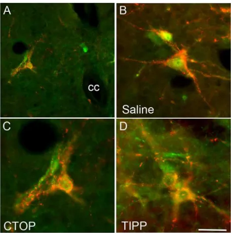
In order to test that mu opioid receptor antagonist CTOP and delta opioid receptor antagonist TIPP suppressed BCM bursting and SV pressure via actions in LSt target areas rather than by preventing activation of LSt cells, DPN stimulation-induced activation of LSt cells was exam-ined. Previous experiments have shown that pERK expression in LSt cells is specifically in-duced by DPN stimulation or by ejaculatory behavior, and is not present in baseline conditions [14]. DPN stimulation induced pERK in control saline-treated males, in 95.4 ± 2.7% of LSt cells (Fig. 4), as we reported previously [14,25]. Neither CTOP or TIPP infusions prior to DPN stimulation affected LSt activation and percentages of LSt cells expressing DPN stimulation-induced pERK in CTOP- (96.5 ± 3.5% of LSt cells;Fig. 4) or TIPP-treated (100 ± 0% of LSt cells;Fig. 4) males did not significantly differ from saline-treated controls.
Mu Opioid Receptor Agonist DAMGO Triggered and Facilitated
Ejaculatory Reflex
The effects of Mu opioid receptor agonist DAMGO on ejaculatory reflexes was examined in conditions in which such reflexes are not triggered in control conditions; i.e. sub threshold
Fig 4. Opioid receptor antagonist infusions did not prevent the activation of pERK in LSt cells by DPN stimulation in male rats.Images demonstrate pERK expression (green) induced by DPN stimulation in LSt cells (visualized by galanin immunoreactivity; red) and the general location of the LSt cells, lateral to the central canal (cc) in laminae X and VII (A; in CTOP-treated male). Images show representative pERK-positive LSt cells following infusions of saline (B), the mu opioid receptor antagonist CTOP (C), or the delta opioid receptor antagonist TIPP (D). The pattern of pERK labeling is characteristic of all CTOP, TIPP, or saline-treated animals. Scale bar indicates 50 (A) or 15 (B-D)μm.
DPN stimulation (5–10 Hz), or in the absence of DPN stimulation (Infusion only). In addition,
effects of DAMGO were tested on ejaculatory reflexes triggered by DPN stimulation parame-ters that reliably trigger such reflexes (30–60 Hz). The mu opioid receptor agonist DAMGO
triggered BCM events, bursts and SVP increases in the absence of DPN stimulation (Infusion;
Fig. 5). In addition, DAMGO dose-dependently facilitated BCM events, bursts and SVP in-creases induced by subthreshold levels of DPN stimulation (5–10 Hz;Fig. 5). For the numbers
of BCM events, there were main effects of testing trial for all stimulation frequencies (Infusion (0 Hz: (F(1, 29) = 6.400;P= 0.025); 5 Hz: (F(1, 29) = 11.407;P= 0.005); 10 Hz:(F(1, 29) = 41.312;P<0.001); 30 Hz (F(1, 29) = 42.443;P<0.001); and 60 Hz (F(1, 29) = 73.256;
P<0.001);Fig. 5A). Moreover, there were effects of drug dosage (5 Hz: (F(1, 29) = 8.637;
P= 0.012), 10 Hz:(F(1, 29) = 29.005;P<0.001) and 30 Hz (F(1, 29) = 4.763;P= 0.048).
DAMGO induced BCM events in the absence of DPN stimulation, but only following the higher dose (10 nmol) and in 100% of males (Infusion:P= 0.009 compared to saline in trial 1; p = 0.024 compared to lower dosage in trial 2;Fig. 5A). DAMGO treatment also induced BCM events following subthreshold DPN stimulation; but this effect was only observed with the lower dosage (0.1 nmol) and not the higher dose (10 nmol) (5–10 Hz:P<0.001; compared to
saline treatment in trial 1; 5–10 Hz:P<0.001 compared to higher dosage in trial 2;Fig. 5A). In
contrast, DAMGO reduced numbers of BCM events following 30 and 60 Hz DPN stimulation frequencies, which reliably triggered BCM activity following saline in trial 1 (Fig. 5A; 30Hz:
P<0.001, 10 nmol compared to saline in trial 1 and compared to lower dosage in trial 2; 60
Hz:P<0.001, 10 nmol;P<0.001, 0.1 nmol; compared to saline in trial 1).
The effects of DAMGO on BCM bursts were nearly identical to those described above for BCM events with main effects of testing trial and drug dosage for all stimulation frequencies ex-cept 30 Hz (Fig. 5B; Testing trial: Infusion, 0 Hz: (F(1, 29) = 10.726;P= 0.006; 5 Hz: (F(1, 29) = 16.136;P= 0.001; 10 Hz:(F(1, 29) = 80.755;P<0.001; and 60 Hz (F(1, 29) = 78.692;P<0.001);
Drug dosage: and main effects of drug dosages: Infusion: F(1, 29) = 4.960;P= 0.044; 5 Hz: (F(1, 29) = 11.152;P= 0.005; 10 Hz:(F(1, 29) = 69.792;P<0.001; and 60 Hz (F(1, 29) = 8.074;
P= 0.014). Post hoc tests showed that DAMGO induced BCM bursts in the absence of DPN stimulation in males treated with the higher dose (10 nmol) but not the lower dose (0.1 nmol; Infusion:P= 0.001; compared to saline in trial 1;P =0.001 compared to lower dosage in trial 2,
Fig. 5B). DAMGO also facilitated BCM bursts following subthreshold DPN stimulations, but only with the lower (0.1 nmol) and not the higher (10 nmol) dose (5 Hz:P<0.001; 10 HzP<
0.001; compared to saline in trial 1 or higher dose in trial 2,Fig. 5B). DAMGO reduced numbers of BCM bursts following 60 Hz DPN stimulation (lower dose (0.1 nmol):P<0.001 compared
to saline in trial 1; higher dose (10 nmol):P<0.001 compared to saline in trial 1 andP =0.014
compared to lower dose in trial 2,Fig. 5B).
Finally, effects of DAMGO on SVP increases mirrored those on BCM activity (Fig. 5C). There were main effects of testing trial (Infusion (0 Hz: (F(1, 27) = 8.168;P= 0.014; 5 Hz: (F(1, 27) = 28.106;P<0.001; 10 Hz:(F(1, 27) = 63.157;P<0.001; 30 Hz (F(1, 27) = 6.866;
P= 0.022; and 60 Hz (F(1, 27) = 16.471;P<0.002) and of drug dosage (Infusion: (F(1, 27) =
8.168;P= 0.014; 5 Hz: (F(1, 27) = 5.807;P= 0.033; 10 Hz:(F(1, 27) = 31.383;P<0.001; 30 Hz:
(F(1, 27) = 18.103;P= 0.001; and 60 Hz:(F(1, 27) = 9.874;P= 0.008). DAMGO infusions alone, in the absence of DPN stimulation, caused increases in SVP in males treated with the higher dose (10 nmol) but not the lower dose (0.1 nmol) (Infusion:P<0.001; compared to
sa-line in trial 1 or to the lower dose in trial 2,Fig. 5C). Furthermore, DAMGO increased SVP fol-lowing subthreshold DPN stimulation (5 Hz:P<0.001, 0.1 nmol;P= 0.047, 10 nmol; 10 Hz:
P<0.001; 0.1 nmol) compared to saline treatment in trial 1. The lower dosage of DAMGO was
significantly more effective in causing SVP increases than the higher dosage (5 Hz:P<0.001;
induced by threshold DPN stimulation (30 Hz (P<0.001) and 60 Hz (P<0.001) compared to
saline in trial 1 or to the lower dose in trial 2,Fig. 5C).
During trail 1 (saline control), there were no significant differences in BCM events, bursts, or SVP increases between groups, indicating that the differences observed in trial 2 were due to the effects of DAMGO.
Fig 5. Quantitative analyses of BCM events (A) bursts (B) and SVP increases (C) in response to infusion, 5 Hz, 10 Hz, 30 Hz and 60 Hz DPN stimulation following intrathecal infusions of saline in trial 1 (Saline-Controls 1 and 2; white bars) or one of two doses of DAMGO (0.1 or 10 nmol) in trial 2 (drug trial; filled bars).Trials 1 and 2 were conducted in the same animals, hence Saline-Control 1 are data for animals subsequently receiving DAMGO 0.1 nmol in trial 2 and Saline-Control 2 are data for animals receiving DAMGO 10 nmol in trial 2. Note absence of white bars for infusions, 5, and 10 Hz, as these stimulation frequencies did not trigger BCM events, bursts, nor SVP increases in control conditions. Data are presented as Mean±SEM.
*denotes significant differences from trial 1 within the same treatment group, while # indicates significant differences between treatment groups within the same testing trial. Representative BCM EMG traces (180 seconds duration) following an intrathecal infusion of saline (D: control trial) and DAMGO (E: 10 nmol: same animal as in D) in the absence of DPN stimulation. EMG and concurrent SVP traces (90 seconds duration) following 10 Hz DPN stimulation (arrow) after an intrathecal infusion of saline (F: control trial) and DAMGO (G: 0.1 nmol: same animal as in F).
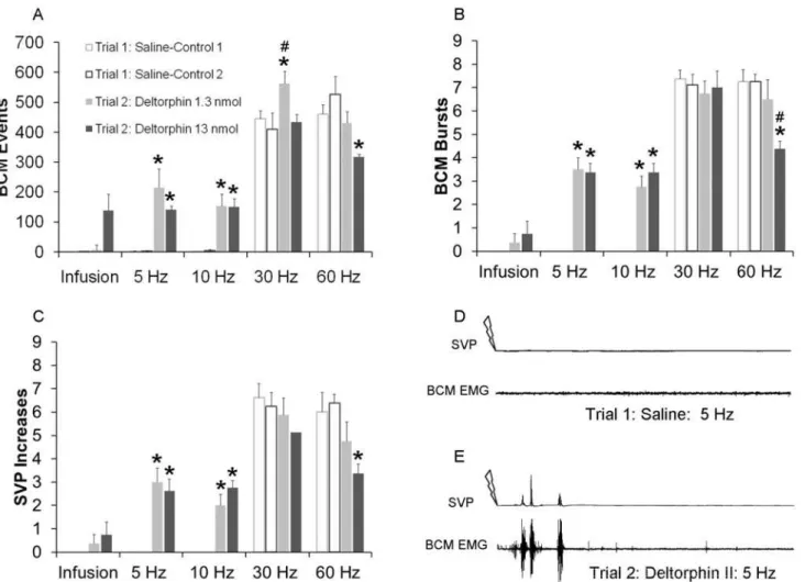
Delta Opioid Receptor Agonist Deltorphin II Facilitated Ejaculatory
Reflexes
In contrast to the effects of DAMGO, deltorphin II did not induce BCM activity or SVP in-creases in the absence of DPN stimulation (Infusion;Fig. 6). However, deltorphin II enhanced the emission and expulsion components of the ejaculatory reflex following subthreshold DPN stimulation (5–10 Hz;Fig. 6). There were main effects of testing trial on BCM events (5 Hz:
(F(1, 31) = 33.286;P<0.001; 10 Hz:(F(1, 31) = 56.543;P<0.001; 30 Hz (F(1, 31) = 6.059;
P= 0.027; 60 Hz (F(1, 31) = 5.710;P<0.031;Fig. 6A), BCM bursts (5 Hz: (F(1, 31) = 121.00;
P<0.001; 10 Hz:(F(1, 31) = 108.432;P<0.001; 60 Hz (F(1, 31) = 14.608;P<0.002;Fig. 6B),
and SVP increases (5 Hz: (F(1, 31) = 52.417;P<0.001; 10 Hz:(F(1, 31) = 54.000;P<0.001;
and 60 Hz (F(1, 31) = 8.590;P= 0.011;Fig. 6C), and a significant interaction between testing trial and drug dosage on BCM bursts for 60 Hz (F(1, 31) = 5.020;P= 0.042) stimulation. Del-torphin II (1.3 nmol or 13 nmol) significantly increased numbers of BCM events, burst and
Fig 6. Quantitative analyses of BCM events (A) bursts (B) and SVP increases (C) following infusion, 5 Hz, 10 Hz, 30 Hz and 60 Hz DPN stimulation after intrathecal infusions of saline in trial 1 (Saline-Controls 1 and 2; white bar) or deltorphin II (1.3 or 13 nmol) in trial 2 (drug trial; filled bars).
Trials 1 and 2 were conducted in the same animals, hence Saline-Control 1 are data for animals subsequently receiving deltorphin II 1.3 nmol in trial 2 and Saline-Control 2 are data for animals receiving deltorphin II 13nmol in trial 2. Data are presented as Mean±SEM. Note absence of white bars for infusions, 5, and 10 Hz, as these stimulation frequencies did not trigger BCM events, bursts, nor SVP increases in control conditions.*denotes significant differences from trial 1 (control trial), while # indicates significant differences from lower dosage within the same testing trial. BCM EMG and SVP traces (90 seconds duration) following 5 Hz DPN stimulation (arrow) after an intrathecal infusion of saline (D: control trial) and deltorphin II (E: same animal as in D).
SVP increases following 5 Hz (events: 1.3 nmol:P<0.001; 13 nmol:P= 0.006; bursts: 1.3 nmol:
P<0.001; 13 nmol:P<0.001; SVP:1.3 nmol:P<0.001; 13 nmol:P<0.001;Fig. 6A-E) and
10 Hz (events: 1.3 nmol:P<0.001; 13 nmol:P<0.001; bursts: 1.3 nmol:P<0.001; 13 nmol:
P<0.001; SVP:1.3 nmol:P<0.001; 13 nmol:P<0.001;Fig. 6C) stimulation frequencies.
Del-torphin II did also slightly increase BCM events following 30 Hz, but only after the lower dos-age (P =0.012), and without effects on BCM bursts or SVP increases. Finally, the higher dose of deltorphin II (13 nmol) reduced BCM activity and SVP increases following 60 Hz DPN stim-ulation (events:P= 0.011; bursts:P<0.001; SVP increases:P= 0.011) compared to saline
treat-ment in trial 1. As noted for each of the experitreat-ments presented above, there were again no differences in BCM or SVP parameters between groups during the saline control trial 1.
Discussion
The results of these experiments support the hypothesis that enkephalin plays a critical role in the control of ejaculation by acting on mu and delta opioid receptors in LSt target areas in the lumbosacral spinal cord. Antagonist studies showed that activation of either mu or delta opioid receptors is critical for the emission and expulsion phases of ejaculation in response to sensory stimulation in male rats. In addition, activation of either mu or delta receptor by agonist ad-ministration at lower dosages facilitated both the emission and expulsion phases of ejaculation following sub- threshold sensory stimulation. Finally, stimulation of mu, but not delta, opioid receptors was sufficient to trigger ejaculatory reflexes in the absence of sensory stimulation. To-gether, these findings demonstrate that endogenous activation of mu or delta opioid receptors plays a facilitative role in the control of ejaculation.
A first main finding of the current study was that intrathecal infusions of either mu or delta opioid antagonists CTOP or TIPP, severely disrupted ejaculatory reflex induced by DPN stim-ulation, using nerve stimulation parameters that reliable triggered ejaculation under control conditions. Conversely, facilitation of ejaculatory reflexes by infusions of mu or delta receptor agonists DAMGO or deltorphin II, was observed after DPN stimulation at sub-threshold pa-rameters that do not trigger ejaculation in control animals [14–16,25]. These data support our
hypothesis that endogenous opioids are acting in LSt target areas to regulate ejaculatory re-flexes. This was further confirmed by the finding that antagonist infusions did not disrupt acti-vation of LSt cells, indicating that opioid antagonists acted on neurons downstream from LSt cells, rather than on inputs to LSt cells. The exact locations of the opioid receptors within the spinal ejaculation generator are currently unknown. Hence, a detailed examination of the local-ization of mu and delta receptors specifically within the LSt target areas is a critical next step. The finding that both mu and delta receptors are involved in mediating ejaculation, indicates that enkephalin may act on both receptors and suggest the possibility of interactions between the opioid receptors during control of ejaculation. Indeed, studies have shown that opioid re-ceptors may form heterodimers [34–36]. Mu and delta opioid receptors demonstrate a high
de-gree of sequence homology and a common opioid receptor binding site within the helical transmembrane core has been proposed to account for the ligand-directed signaling or biased agonism which occurs at opioid receptors [36]. Moreover, there is evidence for cooperation be-tween delta and mu opioid receptors [37], such that activation of one receptor causes allosteric enhancement of ligand binding and activity of the other receptor [38].
or facilitate ejaculatory reflexes is by means of inhibition of inhibitory neurons in LSt target re-gions. For example, opioids can exert analgesia through inhibition of inhibitory GABA (γ—
aminobuteric acid) neurons [40]. Opioids inhibit GABA-mediated synaptic transmission by reducing the likelihood of presynaptic neurotransmitter release [41,42] and this effect is medi-ated by voltage-dependent potassium conductance [42]. Mu but not delta opioid receptors are specifically coupled to this potassium conductance [43] and the subsequent inhibition of GABAergic synaptic transmission; therefore we speculate that this may explain that intrathecal infusions of DAMGO, a selective MOR agonist, but not deltorphin II, a selective DOR agonist, triggered ejaculatory reflexes in the absence of DPN stimulation. However, localization of pre-synaptic MOR on LSt axons has not been confirmed and further studies identifying the exact pre- or postsynaptic localization of these receptors are critical to elucidate the mechanisms by which opioids influence ejaculatory function.
In apparent contrast to the inhibitory effects of opioid receptor antagonists and the facilita-tive effects of the lower dosages of the opioid receptor agonists, infusions of higher dosages re-ceptor agonists for either the mu or delta opioid rere-ceptor disrupted the ability of DPN stimulation to trigger ejaculation. This finding is in conformity with previous studies which showed that systemic administration of morphine exerts inhibitory effects on ejaculatory be-havior [44] and reflexes in male rats [26]. The effects of systemic morphine have been proposed to be partially mediated via actions on peripheral tissues [27]. The current findings demon-strate that the inhibitory effects of intrathecal administration of high doses of opioid receptor agonists are mediated via central actions within the lumbosacral spinal cord. Noteworthy, our previous studies showed similar inhibitory effects of higher doses of GRP agonists on ejaculato-ry reflexes, while lower doses of GRP agonists triggered or facilitated ejaculation. Therefore, it is possible that high doses of GRP or opioid receptor ligands cause mechanisms of desensitiza-tion of the G-protein coupled receptors at the LST target sites [45]; thereby preventing the abil-ity of DPN stimulation to trigger further action of endogenous ligands on receptor activation. It is also possible that infusion of the higher dose of mu and delta receptor agonists prevented DPN stimulation-induced ejaculatory reflexes by acting on the processing of the sensory inputs relayed via the DPN. The DPN is comprised of Aδand C-fibers [46] but the specific contribu-tions of Aδand C-fibers to ejaculation have not been investigated. Intrathecal infusions of opi-oid agonists inhibit Aδand C-fibers [47] raising the possibility that the disruption of
ejaculatory reflexes to threshold sensory stimulation after the higher dose but not the lower dose of opioid agonists is mediated by inhibition of Aδand C-fibers in the dorsal horn of the lumbosacral spinal cord. In addition, the cell bodies of Aδand C-fibers in the dorsal root gan-glia express mu and delta opioid receptors [48], indicating that presynaptic inhibition of DPN synaptic transmission may have contributed to disruption of ejaculation following intrathecal infusions of higher dosages of opioid receptor agonists. Finally, the primary excitatory neuro-transmitter released from Aδand C-fibers is glutamate [39] which activates NMDA receptors in LSt cells to trigger ejaculatory reflexes in male rats [15]. Opioids in turn, can inhibit the re-lease of glutamate [39] thereby potentially blocking the activation of LSt cells in the lumbosa-cral spinal cord. This possibility was not tested in the current study and requires
further investigation.
including galanin and cholecystokinin, in the control of ejaculation remain an open question. Finally, these data indicate that mu and delta opioid antagonists may be useful for the treat-ment of ejaculatory dysfunction, specifically to delay the onset of ejaculation in men afflicted with premature ejaculation.
Author Contributions
Conceived and designed the experiments: NK LMC. Performed the experiments: NK LMC. Analyzed the data: NK LMC. Contributed reagents/materials/analysis tools: NK LMC. Wrote the paper: NK LMC.
References
1. Marberger H. Mechanisms of Ejaculation. Physiol Genet Reprod: 1974;99–110. 2. McKenna KE. Ejaculation. Encycl Reprod: 1999;1002–1008.
3. Veening JG, Coolen LM. Neural mechanisms of sexual behavior in the male rat: Emphasis on ejacula-tion-related circuits. Pharmacol Biochem Behav 2014; 121: 170–183. doi:10.1016/j.pbb.2013.12.017
PMID:24368305
4. Holmes GM, Sachs BD. The ejaculatory reflex in copulating rats: normal bulbospongiosus activity with-out apparent urethral stimulation. Neurosci Lett 1991; 125: 195–197. PMID:1881597
5. Holmes GM, Chapple WD, Leipheimer RE, Sachs BD. Electromyographic analysis of male rat perineal muscles during copulation and reflexive erections. Physiol Behav 1991; 49: 1235–1246. PMID:
1896506
6. Bohlen JG, Held JP, Sanderson MO. The male orgasm: pelvic contractions measured by anal probe. Arch Sex Behav 1980; 9: 503–521. PMID:7458658
7. Gerstenberg TC, Levin RJ, Wagner G. Erection and ejaculation in man. Assessment of the electromyo-graphic activity of the bulbocavernosus and ischiocavernosus muscles. Br J Urol 1990; 65: 395–402.
PMID:2340374
8. Pescatori ES, Calabro A, Artibani W, Pagano F, Triban C, Italiano G. Electrical stimulation of the dorsal nerve of the penis evokes reflex tonic erections of the penile body and reflex ejaculatory responses in the spinal rat. J Urol 1993; 149: 627–632. PMID:8437281
9. Coolen LM. Neural control of ejaculation. J Comp Neurol 2005; 493: 39–45. PMID:16255008 10. Coolen LM, Allard J, Truitt WA, McKenna KE. Central regulation of ejaculation. Physiol Behav 2004;
83: 203–215. PMID:15488540
11. Giuliano F. Neurophysiology of Erection and Ejaculation. J Sex Med 2011; 8: 310–315. doi:10.1111/j.
1743-6109.2011.02450.xPMID:21967393
12. Allard J, Truitt WA, McKenna KE, Coolen LM. Spinal cord control of ejaculation. World J Urol 2005; 23: 119–126. PMID:15947961
13. Giuliano F, Clement P. Neuroanatomy and physiology of ejaculation. Annu Rev Sex Res 2005; 16: 190–216. PMID:16913292
14. Staudt MD, de Oliveira C V, Lehman MN, McKenna KE, Coolen LM. Activation of MAP kinase in lumbar spinothalamic cells is required for ejaculation. J Sex Med 2010; 7: 2445–2457. doi:
10.1111/j.1743-6109.2010.01741.xPMID:20233289
15. Staudt MD, de Oliveira C V, Lehman MN, McKenna KE, Coolen LM. Activation of NMDA Receptors in Lumbar Spinothalamic Cells is Required for Ejaculation. J Sex Med. 2011; 8: 1015–1026. doi:10.1111/
j.1743-6109.2010.02168.xPMID:21235717
16. Staudt MD, Truitt WA, McKenna KE, de Oliveira CVR., Lehman MN, Coolen LM. A pivotal role of lumbar spinothalamic cells in the regulation of ejaculation via intraspinal connections. J Sex Med 2012; 9: 2256–2265. doi:10.1111/j.1743-6109.2011.02574.xPMID:22189051
17. Truitt WA, Coolen LM. Identification of a potential ejaculation generator in the spinal cord. Science (80-: ) 2002; 297: 1566–1569.
18. Ju G, Melander T, Ceccatelli S, Hökfelt T, Frey P. Immunohistochemical evidence for a spinothalamic pathway co-containing cholecystokinin- and galanin-like immunoreactivities in the rat. Neuroscience 1987; 20: 439–456. PMID:2438590
19. Coolen LM, Veening JG, Wells AB, Shipley MT. Afferent connections of the parvocellular subparafascicu-lar thalamic nucleus in the rat: evidence for functional subdivisions. J Comp Neurol 2003; 463: 132–156.
20. Truitt WA, Shipley MT, Veening JG, Coolen LM. Activation of a subset of lumbar spinothalamic neurons after copulatory behavior in male but not female rats. J Neurosci 2003; 23: 325–331. PMID:12514231 21. Newton BW. Galanin immunoreactivity in rat spinal lamina IX: emphasis on sexually dimorphic regions.
Peptides 1993; 14: 955–969. PMID:7506832
22. Xu C, Giuliano F, Yaici ED, Conrath M, Trassard O, Benoit G, et al. Identification of lumbar spinal neu-rons controlling simultaneously the prostate and the bulbospongiosus muscles in the rat. Neuroscience 2006; 138: 561–573. PMID:16364554
23. McKenna KE. Neural circuitry involved in sexual function. J Spinal Cord Med 2001; 24: 148–154.
PMID:11585233
24. Sakamoto H, Matsuda K, Zuloaga DG, Hongu H, Wada E, Wada K, et al. Sexually dimorphic gastrin re-leasing peptide system in the spinal cord controls male reproductive functions. Nat Neurosci 2008; 11: 634–636. doi:10.1038/nn.2126PMID:18488022
25. Kozyrev N, Lehman MN, Coolen LM. Activation of Gastrin-releasing Peptide Receptors in the Lumbo-sacral Spinal Cord is Required for Ejaculation in Male Rats. J Sex Med 2012; 9: 1303–1318. doi:10.
1111/j.1743-6109.2012.02688.xPMID:22429708
26. Carro-Juarez M, Rodriguez-Manzo G. Participation of endogenous opioids in the inhibition of the spinal generator for ejaculation in rats. J Sex Med 2009; 6: 3045–3055. PMID:20104655
27. Agmo A, Rojas J, Vázquez P. Inhibitory effect of opiates on male rat sexual behavior may be mediated by opiate receptors outside the central nervous system. Psychopharmacology (Berl) 1992; 107: 89–96.
PMID:1317041
28. Nicholas AP, Zhang X, Hökfelt T. An immunohistochemical investigation of the opioid cell column in lamina X of the male rat lumbosacral spinal cord. Neurosci Lett 1999; 270: 9–12. PMID:10454133 29. Cesselin F. Opioid and anti-opioid peptides. Fundam Clin Pharmacol 1995; 9: 409–433. PMID:
8617406
30. Giuliano F, Pfaus J, Srilatha B, Hedlund P, Hisasue S, Marson L, et al. Experimental models for the study of female and male sexual function. J Sex Med 2010; 7: 2970–2995. doi:10.1111/j.1743-6109.
2010.01960.xPMID:21050395
31. Herbert J. The role of the dorsal nerves of the penis in the sexual behaviour of the male rhesus monkey. Physiol Behav 1973; 10: 293–300. PMID:4350731
32. Wieder JA, Brackett NL, Lynne CM, Green JT, Aballa TC. Anesthetic block of the dorsal penile nerve in-hibits vibratory-induced ejaculation in men with spinal cord injuries. Urology 2000; 55: 915–917. PMID:
10840108
33. Clement P, Peeters M, Bernabe J, Denys P, Alexandre L, Giuliano F. Brain oxytocin receptors mediate ejaculation elicited by 7-hydroxy-2-(di-N-propylamino) tetralin (7-OH-DPAT) in anaesthetized rats. Br J Pharmacol 2008; 154: 1150–1159. doi:10.1038/bjp.2008.176PMID:18469843
34. Gomes I, Jordan BA, Gupta A, Trapaidze N, Nagy V, Devi LA. Heterodimerization of mu and delta opi-oid receptors: A role in opiate synergy. J Neurosci 2000; 20: RC110. PMID:11069979
35. Pfeiffer M, Kirscht S, Stumm R, Koch T, Wu D, Laugsch M, et al. Heterodimerization of Substance P andμ-Opioid Receptors Regulates Receptor Trafficking and Resensitization. J Biol Chem 2003; 278: 51630–51637. PMID:14532289
36. Pradhan AA, Smith ML, Kieffer BL, Evans CJ. Ligand-directed signalling within the opioid receptor fami-ly. Br J Pharmacol 2012; 167: 960–969. doi:10.1111/j.1476-5381.2012.02075.xPMID:22708627 37. Szentirmay AK, Király KP, Lenkey N, Lackó E, Al-Khrasani M, Friedmann T, et al. Spinal interaction
be-tween the highly selectiveμagonist DAMGO and severalδopioid receptor ligands in naive and mor-phine-tolerant mice. Brain Res Bull 2013; 90: 66–71. doi:10.1016/j.brainresbull.2012.09.006PMID:
22995282
38. Gomes I, Ijzerman AP, Ye K, Maillet EL, Devi LA. G protein-coupled receptor heteromerization: a role in allosteric modulation of ligand binding. Mol Pharmacol 2011; 79: 1044–1052. doi:10.1124/mol.110.
070847PMID:21415307
39. Russell J.A. Opioids. In: Opioid Systems, Edinburgh, UK; Elsevier, Inc. 2007 Pages: 11–25 40. Basbaum AI, Fields HL. Endogenous pain control mechanisms: review and hypothesis. Ann Neurol
1978; 4: 451–462. PMID:216303
41. Capogna M, Gahwiler BH, Thompson SM. Mechanism of Mu-Opioid Receptor-Mediated Presynaptic Inhibition in the Rat Hippocampus in-Vitro. J Physiol 1993; 470: 539–558. PMID:8308742
42. Vaughan CW, Christie MJ. Presynaptic inhibitory action of opioids on synaptic transmission in the rat periaqueductal grey in vitro. J Physiol 498 (Pt 2) 1997: 463–472. PMID:9032693
44. Wiesenfeld-Hallin Z, Sodersten P. Spinal opiates affect sexual behaviour in rats. Nature 1984; 309: 257–258. PMID:6717602
45. Wess J. G-protein-coupled receptors: molecular mechanisms involved in receptor activation and selec-tivity of G-protein recognition. FASEB J 1997; 11: 346–354. Available:http://www.ncbi.nlm.nih.gov/
pubmed/17635134. PMID:9141501
46. Johnson RD, Halata Z. Topography and ultrastructure of sensory nerve endings in the glans penis of the rat. J Comp Neurol 1991; 312: 299–310. PMID:1748735
47. Yeomans DC, Proudfit HK. Nociceptive responses to high and low rates of noxious cutaneous heating are mediated by different nociceptors in the rat: Electrophysiological evidence. Pain 1996; 68: 141–150.
PMID:9252009
48. Ikoma M, Kohno T, Baba H. Differential presynaptic effects of opioid agonists on Adelta- and C-afferent glutamatergic transmission to the spinal dorsal horn. Anesthesiology 2007; 107: 807–812. PMID: