Lymph Node Dissection During the Surgical Treatment of Renal
Cancer in the Modern Era
Guilherme Godoy, Rebecca L. O’Malley, Samir S. Taneja
Urologic Oncology Program, Department of Urology, New York University School of Medicine, New York, NY, USA
ABSTRACT
The increasing use of routine CT scan, along with advances in imaging technology, have facilitated the early diagnosis of incidental renal masses. This has resulted in the reduction in the rate of metastatic disease diagnosis.
Although surgery remains the mainstay in the treatment of renal tumors, the decreasing incidence of lymph node involve-ment has created controversy regarding the importance and the ideal extent of lymph node dissection, formerly considered mandatory at the time of radical nephrectomy. In this review, we critically assessed the role of lymph node dissection DWWKHWLPHRIUDGLFDOQHSKUHFWRP\7RGDWHUDQGRPL]HGWULDOVKDYHIDLOHGWRVKRZDEHQH¿WRIO\PSKQRGHGLVVHFWLRQ when broadly employed. This is likely due to the low prevalence of lymph node metastasis at the time of presentation, the unpredictable pattern of lymph node metastasis from renal tumors, and the continued downward stage migration of the disease. As a result, lymph node dissection for renal cancer is currently not recommended in the absence of gross lymphadenopathy. In high risk patients, lymph node dissection may be considered, but it remains controversial and more clinical evidence is warranted. Extended lymph node dissection is still recommended in individuals with isolated gross nodal disease or those with lymphadenopathy at the time of cytoreductive surgery prior to systemic therapy. A practical approach is summarized in an algorithm form.
Key words: kidney neoplasms; nephrectomy; lymph nodes; lymph node excision; disease management; review
Int Braz J Urol. 2008; 34: 132-42
INTRODUCTION
The role of lymphadenectomy in the surgi-cal management of renal cell carcinoma (RCC) still remains controversial among urologists. In an age of continuous decreasing incidence of lymph node metastasis, the broad application of lymph node dis-section (LND) has recently been criticized by several authors due to the absence of demonstrated
therapeu-WLFEHQH¿WDVUHSRUWHGLQWKH(XURSHDQ2UJDQL]DWLRQ
for Research and Treatment of Cancer trial number
(257&WKHRQO\SURVSHFWLYHWULDOWKDW
compared the outcomes of radical nephrectomy alone
YHUVXVDVVRFLDWHGZLWK/1',QRUGHUWRGH¿QHWKH
WKHEHQH¿WRISHUIRUPLQJ/1'LQWKHVHFDVHV2QFHD
balance between all these parameters is reached, the role of LND in clinical practice probably becomes more evident. In this review, we evaluate the
preva-OHQFHRIO\PSKQRGHPHWDVWDVLVWKHHI¿FDF\RI/1'
and present a rational algorithm for the selection of suitable candidates for LND at the time of radical nephrectomy.
PREVALENCE, RISK FACTORS AND RELEVANCE
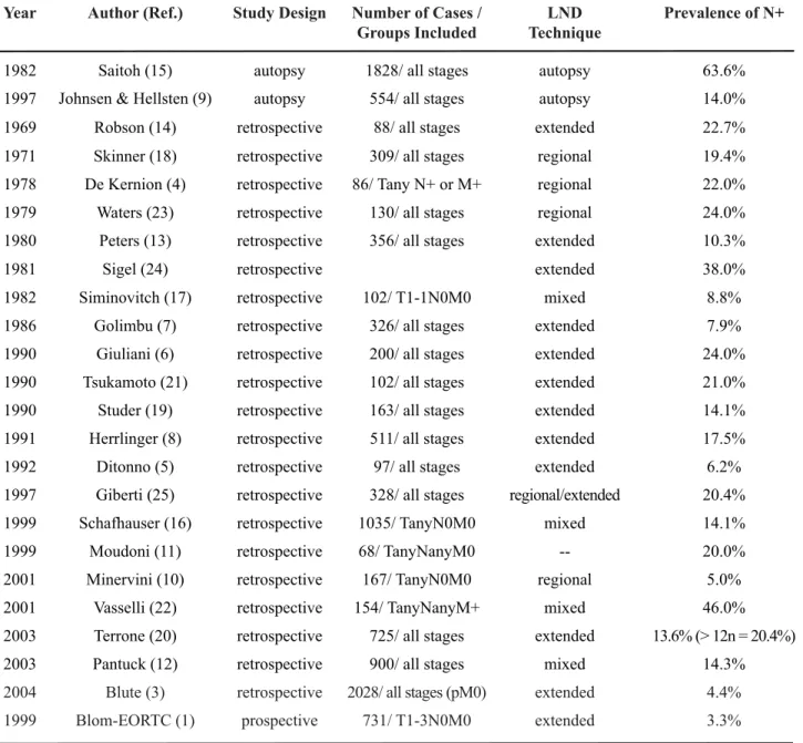
The reported overall incidence of lymph node metastasis in renal cancer in surgical and autopsy
VHULHVLVDSSUR[LPDWHO\EXWWKHUHLVDVLJQL¿ -cant variability reported in the literature (2). As seen
LQ7DEOHWKHLQFLGHQFHRILGHQWL¿HGO\PSKQRGH
Year Author (Ref.) Study Design Number of Cases / Groups Included
LND Technique
Prevalence of N+
1982 Saitoh (15) autopsy 1828/ all stages autopsy 63.6%
1997 Johnsen & Hellsten (9) autopsy 554/ all stages autopsy 14.0%
1969 Robson (14) retrospective 88/ all stages extended 22.7%
1971 Skinner (18) retrospective 309/ all stages regional 19.4%
1978 De Kernion (4) retrospective 86/ Tany N+ or M+ regional 22.0%
1979 Waters (23) retrospective 130/ all stages regional 24.0%
1980 Peters (13) retrospective 356/ all stages extended 10.3%
1981 Sigel (24) retrospective extended 38.0%
1982 Siminovitch (17) retrospective 102/ T1-1N0M0 mixed 8.8%
1986 Golimbu (7) retrospective 326/ all stages extended 7.9%
1990 Giuliani (6) retrospective 200/ all stages extended 24.0%
1990 Tsukamoto (21) retrospective 102/ all stages extended 21.0%
1990 Studer (19) retrospective 163/ all stages extended 14.1%
1991 Herrlinger (8) retrospective 511/ all stages extended 17.5%
1992 Ditonno (5) retrospective 97/ all stages extended 6.2%
1997 Giberti (25) retrospective 328/ all stages regional/extended 20.4%
1999 Schafhauser (16) retrospective 1035/ TanyN0M0 mixed 14.1%
1999 Moudoni (11) retrospective 68/ TanyNanyM0 -- 20.0%
2001 Minervini (10) retrospective 167/ TanyN0M0 regional 5.0%
2001 Vasselli (22) retrospective 154/ TanyNanyM+ mixed 46.0%
2003 Terrone (20) retrospective 725/ all stages extended 13.6% (> 12n = 20.4%)
2003 Pantuck (12) retrospective 900/ all stages mixed 14.3%
2004 Blute (3) retrospective 2028/ all stages (pM0) extended 4.4%
1999 %ORP(257& prospective 731/ T1-3N0M0 extended 3.3%
metastasis ranges from 3% in surgical series, up to 63.6% in autopsy series (1,3-25). The wide variation may be explained by the differences in patient selec-tion, the extent of LND, and the presence or absence of distant metastases.
Clinical stage and pathological grade of the tumor is highly predictive of the prevalence of lymph node metastases (6,12,26). When excluding patients with metastatic disease undergoing cytoreductive nephrectomy, the incidence of lymph node metastasis in surgical series decreases to 3-10% (1,6,8). The relationship of stage and lymph node metastasis has been demonstrated by several authors. Giuliani and colleagues reported a prevalence of nodal involvement of 13.2% in stages pT1-2 and 36.1% in stage pT3-4 tumors (6). Pantuck and colleagues reported a nodal involvement of 5.2% versus 23.4% for T1-2 and T3-4, respectively (12). Similarly, higher tumor grade is also associated with higher rates of positive nodes (5,6,20). Pantuck et al. reported nodal metastasis in 33% of Fuhrman grade 1-2 tumors (27), as compared with a rate of 68% in those with grade 3-4 tumors (12).
The incidence of lymph node metastasis at the time of clinical presentation has been steadily decreasing over time as evidenced by longitudinal analysis. When comparing the incidence of positive lymph nodes in Robson et al’s early series of radical nephrectomy and the more recent treatment arm of
WKH(257&DGURSIURPWRZDV
observed(1,14).
Another important factor contributing to the lower incidence of positive nodes is the downward stage migration in renal cancer seen with the increased incidental detection of the disease. Studying a series of 309 patients between 1935 and 1965, Skinner et al. found a 7% prevalence of incidental renal masses (18). More recently, the prevalence of incidental renal tumors reported in a series of 131 patients studied by Jayson and Sanders in 1998 was 61% (28). Konnak and Grossman also reported on the change in inciden-tal detection of renal tumors over time, from 13% to 48%, comparing two series of patients treated between 1961 and 1973, and between 1980 and 1984, respec-tively (29). In addition a simultaneous decline in the stage at the time of diagnosis was also observed, once
DJDLQFRQ¿UPLQJWKHVWDJHPLJUDWLRQSKHQRPHQRQ
(29). Since one of the most important risk factors
for the presence of lymph node metastasis is clinical stage, the increasing detection of incidental small renal tumors is presumably a major etiology for the observed decreasing prevalence of nodal metastasis at the time of presentation.
The importance of the discussion regarding nodal involvement in renal cell carcinoma is based
RQWKHIDFWWKDWWKHFDQFHUVSHFL¿FVXUYLYDOLVJUHDWO\
impacted by the presence of lymph node
metasta-VLV(DUO\VHULHVUHSRUWHGWKH\HDUFDQFHUVSHFL¿F
survival rate associated with lymph node metastasis for RCC ranging from 21% to 35% (14,30). More recently, Pantuck et al. and Blute et al. reported in a contemporary series an overall 5-year survival rate of 23% and 20.9%, respectively (3,12). They also noted on a multivariate analysis that the chance of dying from RCC was 7.87-fold higher with lymph node involvement than without (3), and that patients who did not undergo LND were three times more likely to die than those who did, regardless of the extent of the dissection (12). Despite all controversies about the necessity and extent of the dissection, the presence of lymph node involvement in RCC undoubtedly deserves attention, since the reported poor survival
UDWHVFDQGH¿QLWHO\EHLPSURYHGZLWK/1'LQVHOHFWHG
cases. The great challenge is to properly identify those
FDVHVWKDWZRXOGPRVWEHQH¿WIURPWKLVSUDFWLFH
Renal Lymphatic Drainage
Another factor that adds controversy to the indication of performing LND during radical ne-phrectomy is the unpredictability of renal lymphatic drainage.
The pathways of renal lymphatic drainage were initially described by Parker in 1935, during anatomical studies of the posterior lymphatic chan-nels of the abdomen (31). He noted that the lymphatic
GUDLQDJHRIWKHNLGQH\ZDVQHLWKHUXQLTXHQRUVSHFL¿F
im-portance of the vascular nature of RCC and hence the predilection for early hematogenic dissemination (32). Clinical series mimic these results (6,22). Vasselli et al. reported an incidence of 53% of distant metastasis without lymph node invasion (22) and Giuliani and colleagues also observed the extremely poor negative predictive value of regional LND in predicting disease progression (6). It was postulated that the neovascu-larization of RCC distorts the normal anatomy and renders the lymphatic drainage unpredictable (33).
7KHUHIRUH GH¿QLQJ WKH UROH RI O\PSKDGHQHFWRP\
during the surgical treatment of these tumors remains
DGLI¿FXOWWDVNDQGDEDODQFHEHWZHHQWKHPRUELGLW\ RIWKHSURFHGXUHDQGWKHEHQH¿WVRILWVSUDFWLFHPXVW
always be sought.
MORBIDITY VERSUS BENEFITS
Technique and Extent of LND
The recommended extent of LND has varied from an author to another. The extended dissection,
¿UVW SURSRVHG E\ 5REVRQ HW DO LQ LQFOXGHG
“all para-aortic and para-caval lymph nodes, from bifurcation of the aorta to the crus of the diaphragm” (14). Later, new limits were described dependent on laterality. Templates proposed for tumors on the right included the hilar, para-caval, pre-caval, retro-caval, interaortocaval and pre-aortic lymph nodes, whereas for left-sided tumors, inclusion of the hilar, para-aor-tic, aorpara-aor-tic, retro-aorpara-aor-tic, interaortocaval and pre-caval nodes was recommended (34). It is important to note that the primary renal lymphatic drainage on the right is to the interaortocaval lymph nodes, and on the left to the para-aortic nodes.
In practice, many surgeons attempt to de-crease morbidity by limiting the extent of dissection. Therefore, a limited regional dissection has been rec-ommended, involving only the para-caval, pre-caval and hilar nodes on the right side, and para-aortic, pre-aortic and hilar nodes on the left side, particularly in the setting of laparoscopically treated patients where extended node dissection would be technically
dif-¿FXOW$OVRGXHWRXQFHUWDLQWLHVDERXWWKHEHQH¿WRI
LND, disagreement persists about the ideal limits of LND (3,6,20). Given the distribution of lymphatic drainage, the use of limited node dissection in pa-tients with substantial risk of lymph node metastases
is likely to result in understaging, particularly on the right side.
Some authors have tried to overcome the divergences in the templates and to improve the stag-ing role of the procedure by analyzstag-ing the same issue from a different perspective. Terrone and colleagues reported on the impact of analyzing the number of dissected nodes, instead of anatomical extension of the LND and have found that a minimum number of 13 nodes should be retrieved, in order to properly stage and estimate the prognosis of these patients.
7KLVFXWRIIUHVXOWHGLQDVLJQL¿FDQWLQFUHDVHLQWKHUDWH
of lymph node metastasis found (20). Thus, based on this data, the best approach to effectively yield an ad-equate lymphadenectomy and optimize staging, would suggest a dissection of a nodal package extending be-tween the regional and the extended pattern, to assure that the proper number of nodes would be retrieved,
LUUHVSHFWLYHRIWKHVSHFL¿FWHPSODWHOLPLWV
Morbidity
It seems intuitive that an increased extent of lymph node dissection would also increase the mor-bidity of the procedure. However, when compared to nephrectomy alone, nephrectomy associated with LND, did not show increased morbidity based on retrospective and prospective data (1,35). Addition-ally, a direct comparison of various dissection patterns was performed by Siminovitch et al. who reported a group of N0M0 patients, who underwent extended, regional or hilar LND templates. They also failed to demonstrate any difference in the morbidity or sur-vival rates among these groups (17). Several other large retrospective series have likewise failed to dem-onstrate any difference in morbidity rates, as related to the extent of dissection (2,8,12,16,23,30,36).
The most common complications related to the surgical management of RCC are lymphocele, chylous ascites (36), bleeding from lumbar or great vessels, and injury to adjacent organs. However, it
LVGLI¿FXOWWRHVWDEOLVKWKHGLUHFWFRUUHODWLRQRIWKHVH HYHQWVZLWKWKH/1'SHUVH7KH(257&DOVR
addressed this issue and did not show any difference in complications rates, but an increased blood loss was observed among those undergoing LND (1).
complex procedure and because it carries a great potential risk for serious intraoperative life-threaten-ing complications, it should be performed only by skilled, well-trained surgeons, who are familiar with retroperitoneal dissections. In addition to providing surgical expertise, urologists should carefully identify
RQO\WKRVHFDQGLGDWHVLQZKRPWKHUHLVDFOHDUEHQH¿W
in performing the LND.
%HQH¿WV
Throughout the years, three potential
ben-H¿WVRI/1'DWWKHWLPHRIQHSKUHFWRP\KDYHEHHQ
evaluated: 1) improved staging and prognostication; 2) improved survival following surgery; and most recently 3) improved response to systemic therapy. Given the limitations of the inconsistent lymphatic anatomy described above, the accuracy of staging and the therapeutic value of the procedure in the set-ting of radiologically normal lymph nodes are highly dependent upon the rigor of dissection utilized and the pathological features of the disease. Recently the practice of LND in localized renal tumors has not
VKRZHG VLJQL¿FDQW EHQH¿W ,W LV DOVR TXHVWLRQDEOH
whether improved staging accuracy is important
JLYHQWKHDEVHQFHRIHI¿FDFLRXVDGMXYDQWWKHUDSLHV
for the disease (37,38). The recent approval in the United States of novel tyrosine kinase inhibitors for the treatment of advanced renal cancer will likely offer an opportunity for the adjuvant treatment of high risk pathology and for a rebirth of LND as staging and/or cytoreductive procedure (39-41).
Imaging Techniques and Staging role of LND
Although imaging advancements allow detection of nodules as small as 0.5 cm in the ret-roperitoneum, there is no imaging method that can
FRQ¿GHQWO\GLIIHUHQWLDWHHQODUJHGLQÀDPPDWRU\QRGHV
from metastatic ones in RCC (20,42). Studer et al. reported an incidence of only 42% of histologically positive nodes in his series of patients with preopera-tively enlarged nodes seen on computed tomography (CT) scan (19). The sensitivity of CT for enlarged nodal masses greater than 1 cc is higher than 95%
EXWWKHORZVSHFL¿FLW\RIWKLV¿QGLQJDQGWKHSRRU
predictive value of the method could argue both in favor and against routine LND in these patients. In
fact, Studer et al. found that nodes detected by CT, that measured between 1 cm and 2.2 cm were more
OLNHO\WREHLQÀDPPDWRU\%HFDXVHFRQYHQWLRQDO
imaging is unable to reliably discern lymph node me-tastasis from non-malignant lymph node enlargement, routine LND is recommended for any individual with
UDGLRORJLFDOO\LGHQWL¿HGO\PSKDGHQRSDWK\
%HQH¿WVIRU3DWLHQWVZLWK/RFDOL]HG7XPRUV The practice of LND in localized renal tumors
KDVQRWVKRZQVLJQL¿FDQWEHQH¿W7KHORZLQFLGHQFH
of positive nodes, reported to range from 0.4% in the UCLA data, up to 3.3% in other series (1,10,12,43), and the lack of survival advantage demonstrated in randomized trial (1) have favored the omission of routine LND in localized tumors with no suspicious nodes in the preoperative imaging. Moreover, the increasing popularity and the successful oncological outcomes of minimally invasive methods and neph-ron-sparing techniques have also contributed to the decreased enthusiasm for LND in early stages of the disease.
The staging role of LND is also questionable given the absence of effective adjuvant therapies for RCC (37,38). As data regarding adjuvant strategies continues to improve, offering routine LND to high risk patients, defined as those with large tumors (particularly clinical stage = T3), high nuclear grade (Fuhrman’s grade) (27), symptoms, and poor perfor-mance status (44,45), remains a topic of debate. These individuals have a reported incidence of positive nodes approaching 10% (46) and thus these patients warrant further attention in future clinical trials.
7KHDSSOLFDWLRQRIDULVNFODVVL¿FDWLRQVWUDWH -gy, according to the presence of predictive risk factors, has been proposed as a means of identifying those pa-tients at a higher risk of regional lymph node
involve-PHQWWKDWDUHPRVWOLNHO\WREHQH¿WIURP/1'7KH
only study that brought insight into such risk factors was published by Blute et al.(3). Using a multivariate model to identify pathologic features of the primary tumor that were independent predictors of increased risk of having positive nodes in non-metastatic RCC,
WKH\LGHQWL¿HGULVNIDFWRUVFOLQLFDOVWDJH77
more of these factors was associated with a 15-fold higher incidence of regional lymph node involvement. While provocative, the limitation of this approach is the lack of pre-treatment factors for segregating risk. In the absence of good pathologic support, its
applica-WLRQFRXOGEHGLI¿FXOW
%HQH¿WVIRU3DWLHQWVZLWK1RGDO0HWDVWDVLV Only
Little controversy exists about recommending LND in those with isolated positive nodes without distant metastasis. Although this situation is usually found in between 0.9% and 10% of the cases, as shown in Table-2 (1,6,9,10,12,17,25), it may reach rates up to 20.4%, as demonstrated by Terrone et al., using extended templates of dissection and retriev-ing more than 12 nodes (20). Additionally, in spite of the vast majority of patients who have positive nodes also present with concurrent distant metastasis (58-95% of cases), and exclusive nodal disease is a
VLWXDWLRQGLI¿FXOWWRGHWHFWWKHVXUYLYDORIWKLVJURXS
when treated with LND associated with radical ne-phrectomy is superior to that of nene-phrectomy alone (2). Moreover, the survival of this subset of patients who undergo LND associated with radical nephrec-tomy is far superior to those with distant metastasis, and more closely approximates that of those in the T3N0M0 stage disease (6,13). Data from Giuliani et
al. shows that survival rates of this group of patients are 47.9% and 31.9%, at 5 and 10 years of follow-up, respectively, in comparison to the 7% 5-year survival rate of patients with distant metastases (6). Peters and Brown also demonstrated an improved survival as-sociating LND with nephrectomy, which increased the survival rates from 56.5% to 87.5% at 1 year and from 25.79% to 43.75% at 5 years follow-up (13). In this situation, there is little controversy among the experts, and the LND must be performed with curative intent, therefore using an extended template (8). The chal-lenge lies in identifying those cases preoperatively. Perhaps the lack of the proven morbidity associated with the LND, allows for a more liberal indication for
WKHGLVVHFWLRQRIDOOVXVSLFLRXVQRGHVLGHQWL¿HGEHIRUH RUGXULQJVXUJHU\0RUHRYHUWKHLQFLGHQWDO¿QGLQJRI
suspicious nodes at radical nephrectomy should also be managed according to this same rationale, since
WKH(257&WULDOGHPRQVWUDWHGWKDWGHVSLWHRQO\
of nodes removed due to suspicious palpation were
SRVLWLYHLWZDV\HWVLJQL¿FDQWO\PRUHWKDQWKH
found to be positive, in those patients who underwent routine dissection (1).
%HQH¿WVIRU3DWLHQWVZLWK6\VWHPLF Metastasis
With the recent advances in systemic thera-pies using cytokines and tyrosine kinase inhibitors,
Table 2 – Prevalence of N+M0 disease.
LND = lymph node dissection; N+ = Node positive disease.
Year Author (Ref.) Study Design Number of Cases / Groups Included
LND Technique Prevalence of N+
1982 Siminovitch (17) retrospective 102/ T1-2N0M0 mixed 08.8%
1990 Giuliani (6) retrospective 200/ all stages extended 10.0%
1997 Johnsen & Hellsten (9) autopsy 554/ all stages autopsy 00.9%
1997 Giberti (25) retrospective 328/ all stages regional / extended 07.0%
1999 Blom (1) prospective 731/ T1-3N0M0 extended 03.3%
2001 Minervini (10) retrospective 167/ TanyN0M0 regional 05.0%
WKHYDOXHRIWKHVWDJLQJDQGWKHUDSHXWLFEHQH¿WRI
LND has been increasingly discussed. Although im-munotherapy using cytokines such as interleukin-2 and interferon-D, alone or in combination, have
EHHQZLGHO\XVHGDV¿UVWOLQHWUHDWPHQWRIPHWDVWDWLF
RCC, response rates are usually low (5-20%) with median survival rates ranging 12-17.9 months (or lower in the presence of adverse prognostic factors) and with substantial toxicity (47-51). It has been previously shown that systemic therapy with cyto-kines improves survival after radical nephrectomy and that lymph node metastases typically have a poor response to chemotherapy and immunotherapy (12,22,52). Vasseli et al. observed that survival rates were longer in patients with systemic disease without retroperitoneal nodal metastasis (median survival of 14.7 months), and that the preoperative presence of retroperitoneal lymphadenopathy predicted a short-ened survival. However, when the lymph nodes were completely resected during surgery, overall survival rates of these patients were similar to those without retroperitoneal lymphadenopathy (22). Although
WKHUHZDVQREHQH¿WLQVXUYLYDOUDWHVEHWZHHQJURXSV
receiving immunotherapy, this data supports the cy-toreductive role of the LND during the management of metastatic RCC with lymph node involvement. A more aggressive surgical approach could positively impact the outcomes of the systemic therapy, reduc-ing the burden of the disease and eliminatreduc-ing the metastatic tissue that is less susceptible to the therapy agents (22). Therefore, the extent of the dissection should always be guided by the rationale of clearing the most as possible the grossly involved nodes and its regional packages. However, as this strategy is still considered palliative, extended (more morbid) templates should be avoided, since these patients
EHQH¿WIURPDUDSLGDQGXQHYHQWIXOSRVWRSHUDWLYH
recovery allowing them to receive systemic therapy as soon as possible.
EVIDENCE-BASED RECOMMENDATIONS
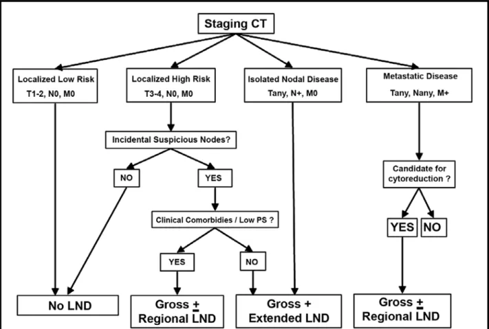
Based upon the data reviewed above, we propose the following approach for each patient group (Figure-1).
/RFDOL]HG'LVHDVH/RZ5LVN710 In the contemporary age of decreasing inci-dence of nodal metastasis, lymph node dissection is of little value in this group. Despite the low morbidity rates, the majority of practitioners agree that there is no indication for LND in these patients, as no survival
EHQH¿WKDVEHHQGHPRQVWUDWHGIRU/1'ZLWKORFDOL]HG
RCC.
/RFDOL]HG'LVHDVH+LJK5LVN710 In this subset of tumors lies the greatest con-troversy. Most urologists generally do not perform LND in this situation because of the substantial risk of concomitant hematogenic metastasis regardless of the state of the lymph nodes, as well as due to the lack of validated criteria to predict nodal metastasis. How-ever, with the poor negative predictive value of the imaging examinations and the promising emerging targeted therapies, the staging role of the LND again becomes a reasonable argument. We recommend regional LND in those patients that present with the risk factors aforementioned, including the hilar and para-aortic nodes for tumors on the left side, and the hilar, para-caval, retro-caval and interaortocaval nodes on the right side, from the crus of the diaphragm until the emergence of the inferior mesenteric artery. In the absence of any risk factors or incidental suspicious lymphadenopathy, probably any LND beyond the
KLODUQRGHVOHYHOLVQRWMXVWL¿HG
Nodal Metastasis Only (Tany, N+0
Metastatic Disease (Tany, Nany, M+)
In this group of patients, the cytoreductive
DSSURDFKLVEHQH¿FLDODQGZLOOOLNHO\LPSURYHRXW -comes of systemic therapies. The surgical procedure should include a radical nephrectomy and regional lymphadenectomy including only the grossly posi-tive nodes. As nodal disease shows poor response to immunotherapy, trying to extirpate all visible gross nodal metastasis is always a good practice in selected patients with good performance status, who are likely to tolerate the surgery and recover to receive systemic therapies. Since there is no curative
inten-WLRQH[WHQGHGSDWWHUQVRI/1'DUHQRWMXVWL¿HG
CONCLUSIONS
Lymph node dissection has become an uncommon procedure during the surgical treatment of renal tumors, in the era of small, incidental and early stage renal masses. We have seen an increased rate of surveillance strategies, minimally invasive and nephron-sparing approaches substituting for the classic radical nephrectomy as described by Robson et al. (14). LND in high-risk cases has not shown
DQ\SURYHQVXUYLYDOEHQH¿WEXWLQWKHIXWXUHPD\EH
genetic/molecular events in the carcinogenesis of RCC. To date, there are only two situations in which
/1'GH¿QLWHO\EHQH¿WVSDWLHQWVLQWKHSUHVHQFHRI
nodal involvement without distant metastasis, and as part of a cytoreductive approach. In the next few years, with advancements in novel targeted therapies, further
SURVSHFWLYHVWXGLHVZLOOEHZDUUDQWHGWRUHGH¿QHWKH
therapeutic/staging roles of LND in the management of renal tumors.
ACKNOWLEDGMENT
Dr. Guilherme Godoy is recipient of a Grant from Bruce and Cynthia Sherman Fellowship in
8URORJLF2QFRORJ\
CONFLICT OF INTEREST
None declared.
REFERENCES
1. Blom JH, van Poppel H, Marechal JM, Jacqmin D, Sylvester R, Schröder FH, et al.: Radical nephrectomy with and without lymph node dissection: preliminary UHVXOWVRIWKH(257&UDQGRPL]HGSKDVH,,,SURWRFRO (257&*HQLWRXULQDU\*URXS(XU8URO 36: 570-5.
2. Freedland SJ, Dekernion JB: Role of lymphadenec-tomy for patients undergoing radical nephreclymphadenec-tomy for renal cell carcinoma. Rev Urol. 2003; 5: 191-5. 3. Blute ML, Leibovich BC, Cheville JC, Lohse CM,
Zincke H: A protocol for performing extended lymph node dissection using primary tumor pathological features for patients treated with radical nephrectomy for clear cell renal cell carcinoma. J Urol. 2004; 172: 465-9.
4. Dekernion JB, Ramming KP, Smith RB: The natural history of metastatic renal cell carcinoma: a computer analysis. J Urol. 1978; 120: 148-52.
5. Ditonno P, Traficante A, Battaglia M, Grossi FS, Selvaggi FP: Role of lymphadenectomy in renal cell carcinoma. Prog Clin Biol Res. 1992; 378: 169-74. 6. Giuliani L, Giberti C, Martorana G, Rovida S: Radical
extensive surgery for renal cell carcinoma: long-term results and prognostic factors. J Urol. 1990; 143: 468-73; discussion 473-4.
7. Golimbu M, Joshi P, Sperber A, Tessler A, Al-Askari S, Morales P: Renal cell carcinoma: survival and prognostic factors. Urology. 1986; 27: 291-301. 8. Herrlinger A, Schrott KM, Schott G, Sigel A: What are
WKHEHQH¿WVRIH[WHQGHGGLVVHFWLRQRIWKHUHJLRQDOUHQDO lymph nodes in the therapy of renal cell carcinoma. J Urol. 1991; 146: 1224-7.
9. Johnsen JA, Hellsten S: Lymphatogenous spread of renal cell carcinoma: an autopsy study. J Urol. 1997; 157: 450-3.
10. Minervini A, Lilas L, Morelli G, Traversi C, Battaglia S, Cristofani R, et al.: Regional lymph node dissection in the treatment of renal cell carcinoma: is it useful in patients with no suspected adenopathy before or during surgery? BJU Int. 2001; 88: 169-72.BJU Int. 2001; 88: 169-72.
0RXGRXQL607D]L0RNKD.1RXUL0/UKRU¿0+ Koutani A, Iben Attya Andaloussi A, et al.: Renal can-cer in adults. Review of 68 cases. Ann Urol (Paris).Review of 68 cases. Ann Urol (Paris). 1999; 33: 395-401.
12. Pantuck AJ, Zisman A, Dorey F, Chao DH, Han KR, Said J, et al.: Renal cell carcinoma with retroperitoneal lymph nodes: role of lymph node dissection. J Urol. 2003; 169: 2076-83.
13. Peters PC, Brown GL: The role of lymphadenectomy in the management of renal cell carcinoma. Urol Clin North Am. 1980; 7: 705-9.
14. Robson CJ, Churchill BM, Anderson W: The results of radical nephrectomy for renal cell carcinoma. J Urol. 1969; 101: 297-301.
15. Saitoh H, Nakayama M, Nakamura K, Satoh T: Distant metastasis of renal adenocarcinoma in nephrectomized cases. J Urol. 1982; 127: 1092-5.
16. Schafhauser W, Ebert A, Brod J, Petsch S, Schrott KM: Lymph node involvement in renal cell carcinoma and survival chance by systematic lymphadenectomy. Anticancer Res. 1999; 19: 1573-8.
17. Siminovitch JP, Montie JE, Straffon RA: Lymphad-enectomy in renal adenocarcinoma. J Urol. 1982; 127: 1090-1.
6NLQQHU'*&ROYLQ5%9HUPLOOLRQ&'3¿VWHU5& Leadbetter WF: Diagnosis and management of renal cell carcinoma. A clinical and pathologic study of 309 cases. Cancer. 1971; 28: 1165-77.
19. Studer UE, Scherz S, Scheidegger J, Kraft R, Sonntag R, Ackermann D, et al.: Enlargement of regional lymph nodes in renal cell carcinoma is often not due to metastases. J Urol. 1990; 144: 243-5.
21. Tsukamoto T, Kumamoto Y, Miyao N, Yamazaki K, Takahashi A, Satoh M: Regional lymph node metasta-sis in renal cell carcinoma: incidence, distribution and LWVUHODWLRQWRRWKHUSDWKRORJLFDO¿QGLQJV(XU8URO 1990; 18: 88-93.
22. Vasselli JR, Yang JC, Linehan WM, White DE, Rosenberg SA, Walther MM: Lack of retroperitoneal lymphadenopathy predicts survival of patients with metastatic renal cell carcinoma. J Urol. 2001; 166: 68-72.
23. Waters WB, Richie JP: Aggressive surgical approach to renal cell carcinoma: review of 130 cases. J Urol. 1979; 122: 306-9.
24. Sigel A, Chlepas S, Schrott KM, Hermanek P: Surgery of the kidney tumor. Chirurg. 1981; 52: 545-53. *LEHUWL&2QHWR)0DUWRUDQD*5RYLGD6&DUPLJ
-nani G: Radical nephrectomy for renal cell carcinoma: long-term results and prognostic factors on a series of 328 cases. Eur Urol. 1997; 31: 40-8.
26. Phillips CK, Taneja SS: The role of lymphadenectomy in the surgical management of renal cell carcinoma. 8URO2QFROGLVFXVVLRQ 27. Fuhrman SA, Lasky LC, Limas C: Prognostic
sig-QL¿FDQFH RI PRUSKRORJLF SDUDPHWHUV LQ UHQDO FHOO carcinoma. Am J Surg Pathol. 1982; 6: 655-63. 28. Jayson M, Sanders H: Increased incidence of
seren-dipitously discovered renal cell carcinoma. Urology. 1998; 51: 203-5.
29. Konnak JW, Grossman HB: Renal cell carcinoma as DQLQFLGHQWDO¿QGLQJ-8URO 30. Skinner DG, Vermillion CD, Colvin RB: The surgical
management of renal cell carcinoma. J Urol. 1972; 107: 705-10.
31. Parker AE: Studies on the main posterior lymph chan-nels of the abdomen and their connections with the lymphatics of the genitourinary system. Am J Anat.Am J Anat.. 1935; 56: 409.
32. Saitoh H: Distant metastasis of renal adenocarcinoma in patients with a tumor thrombus in the renal vein and/or vena cava. J Urol. 1982; 127: 652-3.
33. DeKernion JB: Lymphadenectomy for renal cell carci-noma. Therapeutic implications. Urol Clin North Am. 1980; 7: 697-703.
34. Wood DP Jr: Role of lymphadenectomy in renal cell carcinoma. Urol Clin North Am. 1991; 18: 421-6.
35. Carmignani G, Belgrano E, Puppo P, Gilberti C, Cicherro A: Lymphadenectomy in renal cancer. In: Cancer of the Prostate and Kidney (ed.), Pavone-Ma-caluso M. Smith PH. New York, Plenum Press. 1983, pp. 645-50.
36. Ferrigni RG, Novicki DE: Chylous ascites complicat-ing genitourinary oncological surgery. J Urol. 1985; 134: 774-6.
1DLWR6.XPD]DZD-2PRWR7,JXFKL$6DJL\DPD . 2VDGD< HW DO 3RVWRSHUDWLYH 8)7 DGMXYDQW and the risk factors for recurrence in renal cell carcinoma: a long-term follow-up study. Kyushu 8QLYHUVLW\8URORJLFDO2QFRORJ\*URXS,QW-8URO 1997; 4: 8-12.
38. Pizzocaro G, Piva L, Colavita M, Ferri S, Artusi R, Boracchi P, et al.: Interferon adjuvant to radical nephrectomy in Robson stages II and III renal cell carcinoma: a multicentric randomized study. J Clin 2QFRO
(VFXGLHU%(LVHQ76WDGOHU:06]F]\OLN&2XGDUG S, Siebels M, et al.: Sorafenib in advanced clear-cell renal-cell carcinoma. N Engl J Med. 2007; 356: 125-34. Erratum in: N Engl J Med. 2007; 357: 203. 40. Hudes G, Carducci M, Tomczak P, Dutcher J, Figlin
R, Kapoor A, et al.: Temsirolimus, interferon alfa, or both for advanced renal-cell carcinoma. N Engl J Med. 2007; 356: 2271-81.
41. Motzer RJ, Hutson TE, Tomczak P, Michaelson MD, %XNRZVNL505L[H2HWDO6XQLWLQLEYHUVXVLQWHU -feron alfa in metastatic renal-cell carcinoma. N Engl J Med. 2007; 356: 115-24.
42. Bechtold RE, Zagoria RJ: Imaging approach to staging of renal cell carcinoma. Urol Clin North Am. 1997; 24: 507-22.
43. Mickisch GH: Lymphatic metastases in renal cell car-cinoma. What is the value of operation and adjuvant therapy? Urologe A. 1999; 38: 326-31.
44. Zisman A, Pantuck AJ, Dorey F, Chao DH, Gitlitz BJ, Moldawer N, et al.: Mathematical model to predict in-dividual survival for patients with renal cell carcinoma. -&OLQ2QFRO
45. Zisman A, Pantuck AJ, Wieder J, Chao DH, Dorey F, Said JW, et al.: Risk group assessment and clinical outcome algorithm to predict the natural history of patients with surgically resected renal cell carcinoma. -&OLQ2QFRO
46. Zisman A, Pantuck AJ, Belldegrun AS: Lymph Node Dissection in Renal and Adrenal Tumors: Biology and 0DQDJHPHQW1HZ<RUN2[IRUG8QLYHUVLW\3UHVV 2003; p. 318.
47. Fisher RI, Rosenberg SA, Fyfe G: Long-term survival update for high-dose recombinant interleukin-2 in patients with renal cell carcinoma. Cancer J Sci Am. 2000; 6 (Suppl 1): S55-7.
trial of high-dose interleukin-2 versus subcutaneous interleukin-2 and interferon in patients with metastatic UHQDOFHOOFDUFLQRPD-&OLQ2QFRO (UUDWXPLQ-&OLQ2QFRO
49. Motzer RJ, Murphy BA, Bacik J, Schwartz LH, Nanus DM, Mariani T, et al.: Phase III trial of interferon alfa-2a with or without 13-cis-retinoic acid for patients with DGYDQFHGUHQDOFHOOFDUFLQRPD-&OLQ2QFRO 2972-80.
50. Négrier S, Escudier B, Gomez F, Douillard JY, Ravaud A, Chevreau C, et al.: Prognostic factors of survival and rapid progression in 782 patients with metastatic renal carcinomas treated by cytokines: a report from
WKH*URXSH)UDQoDLVG¶,PPXQRWKpUDSLH$QQ2QFRO 2002; 13: 1460-8.
51. Negrier S, Escudier B, Lasset C, Douillard JY, Savary J, Chevreau C, et al.: Recombinant human interleu-kin-2, recombinant human interferon alfa-2a, or both in metastatic renal-cell carcinoma. Groupe FrançaisGroupe Français d’Immunothérapie. N Engl J Med. 1998; 338: 1272-N Engl J Med. 1998; 338: 1272-8.
52. Flanigan RC, Salmon SE, Blumenstein BA, Bearman SI, Roy V, McGrath PC, et al.: Nephrectomy followed by interferon alfa-2b compared with interferon alfa-2b alone for metastatic renal-cell cancer. N Engl J Med. 2001; 345: 1655-9.
Accepted: April 4, 2008
Correspondence address:
Dr. Samir S. Taneja Department of Urology New York School of Medicine (DVWQGVWUHHWQGÀRRU New York, NY, 10016, USA Fax: + 1 646 825-6394