OR
IGI
N
A
L
R
E
S
E
A
R
C
H
Corresponding address: Bruna Felix Apoloni – Rua Osaka, 311, Jardim Imperial II, Maringá (PR) – Zip code: 87023-100 – Phone: (44) 3305-7542 – E-mail: [email protected] – Finance source: CNPQ, Finep e Seti-PR – Conlict of interest: Nothing to declare – Presentation: Dec 1, 2016 – Accepted for publication: July 1, 2017 – Approved by the Human Research Ethics Committee (COPEP) of Universidade Estadual de Maringá (UEM) of opinion no. 434/2008.
Study developed in the Biomechanics and Motor Behavior Laboratory of the Department of Physical Education of the Universidade Estadual de Maringá – Maringá (PR), Brazil.
1Master’s Degree Candidate of the Graduate Program in Physical Education UEM/UEL of the Universidade Estadual de Maringá (UEM) – Maringá (PR), Brazil.
2PhD, Professor of the Graduate Program in Physical Education UEM/UEL of the Universidade Estadual de Maringá (UEM) – Maringá (PR), Brazil.
ABSTRACT | Disabilities in the gait motor pattern have been commonly found in individuals with Down Syndrome. This study evaluated the knee angle behavior of children with Down Syndrome for 24 months. The sample comprised 20 male and female children aged between 24 and 83 months. Participants had to walk straight in a speed of preference. We represented the biomechanical model by the external positioning of retrorelective markers in the greater trochanter of the femur, in the knee joint center, and the lateral ankle joint of the right hemibody. For registration and biomechanical analysis, we used two-dimensional kinematics. For data analysis, we used descriptive and comparative analysis of One-Way ANOVA and Kruskal-Wallis tests. There were no signiicant diferences in the knee angle values between diferent ages. The examined children showed regular values for knee maximum lexion at initial contact and knee maximum lexion at the swing phase, and excessive lexion over time.
Keywords | Down syndrome; Gait; Knee.
RESUMO | Desordens no padrão motor da marcha têm sido comumente encontradas em indivíduos com síndrome de Down. O presente estudo avaliou o comportamento angular do joelho de crianças com síndrome de Down ao longo de vinte e quatro meses de acompanhamento. A amostra foi constituída por 20 crianças, de ambos os sexos, com idade entre 24 e 83 meses. A tarefa proposta foi caminhar em linha reta, na velocidade autosselecionada. O
232
modelo biomecânico foi representado pelo posicionamento externo de marcadores retrorreletivos nas articulações trôcanter maior do fêmur, centro articular do joelho e maléolo lateral do hemicorpo direito. Para registro e análise biomecânica utilizou-se a cinemetria bidimensional. Para análise dos dados utilizou-se análise descritiva e os testes comparativos Anova One-Way e Kruskal-Wallis. Não foram encontradas diferenças signiicativas nos valores angulares do joelho entre diferentes faixas etárias. As crianças analisadas apresentaram valores regulares para a lexão máxima do joelho no contato inicial e a lexão máxima do joelho na fase de balanço apresentou lexão excessiva ao longo do tempo.
Descritores | Síndrome de Down; Marcha; Joelho.
RESUMEN | Desordenes en el patrón motor de la marcha están siendo comúnmente encontrados en los individuos con síndrome de Down. El presente estudio evaluó el comportamiento angular de la rodilla de niños con síndrome de Down a lo largo de veinticuatro meses de acompañamiento. La muestra fue constituida por 20 niños, de ambos sexos, con la edad entre 24 y 83 meses. La tarea propuesta fue la de caminar en línea recta, en la velocidad autoseleccionada. El modelo biomecánico fue representado por el posicionamiento externo de los marcadores retrorreflectantes en las articulaciones trocánter mayor del fémur, centro articular de la rodilla y maléolo lateral del hemicuerpo derecho. Para el registro
Kinematics pattern of knees in the gait of children
with Down Syndrome according to age
Padrão cinemático do joelho durante a marcha de crianças com síndrome de Down por
classiicação etária
Estándar cinemático de la rodilla durante la marcha de los niños con síndrome de Down por
clasiicación por edades
y el análisis biomecánico se utilizó la cinemetría bidimensional. Para el análisis de los datos se utilizó el análisis descriptivo y las pruebas comparativas Anova One-Way y Kruskal-Wallis. No fueron encontradas diferencias significativas en los valores angulares de la rodilla entre distintas franjas de edad. Los
niños analizados presentaron valores regulares para la flexión máxima de la rodilla en el contacto inicial y la flexión máxima de la rodilla en la etapa de balanceado que presentó flexión excesiva a lo largo del tiempo.
Palabras clave | Síndrome de Down; Marcha; Rodilla.
INTRODUCTION
Down Syndrome is a genetic abnormality mostly caused by the irregular cell division of the chromosome 211. Individuals with this syndrome may develop learning disabilities, heart problems, joint instability, and the weakening of muscle strength and muscle tone. Regarding the gait, literature has reported that motor disabilities have been found in individuals with Down Syndrome2.
Gait is the most basic form of human locomotion and also the most comfortable and economical one, being characterized by smooth and repetitive movements of the joints3. During the gait, knee joint kinematics comprises alternate lexion and extension movements. At the initial contact of the foot with the loor, as known as the support phase, the knee joint can show maximum lexion of 18°. At the swing phase, with lower limb transposition (from 40 to 70% of gait cycle), the knee lexes up to 70°4.
he acquisition of independent gait in children without disabilities develops their concept of space by stimulating the exploration of the environment. However, children such as the ones with Down Syndrome, those who have a motor disability, or who are moved over space can have limited development in addition to limited capacities to explore the environment. To achieve stable gait patterns, there is need for time, practice, and adequate stimuli5.
In the literature, we observed some studies on the gait of children with Down Syndrome2,6-8. However, we found no studies analyzing the knee angle behavior of children with Down syndrome over time. It is worth highlighting that the gait changes with motor development, thus requiring the frequent observation to monitor its progress9.
Wu et al.6 evaluated the inluence of generalized treadmill interventions of low intensity and individual
treadmill interventions of high intensity in the gait of children with Down Syndrome. Individual treadmill interventions of high intensity inluenced positively the kinetics pattern in hip, knee and ankle joints. he authors described that during the development of the motor pattern, and space/time and joint features, these children were diferent when compared with the ones with typical development.
Galli et al.7 studied the gait of children with Down Syndrome and with typical development. he results showed that children with the syndrome had a higher lexion of the hip and knee joints at support phase and higher plantar lexion at initial contact with the loor.
Cimolin et al.2 compared the kinematics variables during the gait of adult patients with the Prader-Willi syndrome, adult patients with Down Syndrome, and a control group without genetic disorders. hey concluded that patients with the two syndromes presented diferent gait patterns when compared with the control group.
Rodenbusch et al.8 analyzed the gait of 16 children with Down Syndrome in an inclined treadmill. he results showed that an inclination of 10% inluenced variables such as frequency, time of swing phase, and angle behavior of hips, knees and ankles.
Valentin-Gudiol et al.5 did a systematic review considering studies on the efectiveness of treadmill interventions in the motor development of children under six years old and with risk of neuromotor delay. Authors reported that research with treadmill intervention had positive results, because they developed the children’s independent gait. However, the few studies based on this methodology used an adequate control group in their research.
METHODOLOGY
A total of 20 male and female children with Down Syndrome, aged between 24 and 83 months, and that attended classes in an educational institution participated in this study. Convenience sampling was performed, followed by the selection process. he selection process of the sample was carried out considering whether the time of access to participants would be enough to describe the possible changes in knee angle behavior over time.
During the monitoring period of 24 months, ive evaluations were conducted, each of them comprising anthropometrics and kinematics analysis of the gait. After data collection, children were grouped by age totaling 7 children aged between 24 to 35 months with average weight of 11.50±1.19kg and average height of 81.71±5.16cm, 8 from 36 to 47 months (12.12± 1.86kg and 91.28±14.86), 8 from 48 to 59 months (14.58±2.06kg and 106.93±17.40cm), 7 from 60 to 71 months (17.14±1.67kg and 112.54±11.13cm), and 10 from 72 to 83 months (19.65±3.16kg and 124.35±16.64cm). Considering the selection criterion, the number of children was based on their availability in collection periods.
For such evaluation, a laboratory provided by the educational institution was used. he parents and/or legal guardians of the children were informed on the procedures of the survey and agreed to participate by signing an informed consent form. he study was approved by the Human Research Ethics Committee.
In anthropometric evaluations, a Camry BR9010® scale was used to measure body mass (Kg), and a Cardiomed® stadiometer was used to measure the height (cm).
In gait analysis, children with Down Syndrome, wearing comfortable clothing, walked in a straight line, in the preferred speed, thus performing the gait movement for three consecutive times. he laboratory environment for data collection was previously organized in a two-dimensional reference system.
he biomechanical model of the study was represented by the positioning of retrorelective markers in the following anatomical points of the lower limb: the greater trochanter of the femur, the knee joint center, and the lateral ankle joint. he coordinates of the positioned markers in the greater trochanter of the femur and the knee joint center deined the representative vector of the
thigh. he coordinates of the knee joint center and the lateral ankle joint deined the leg vector.
To record the gait movements, a camcorder camera (Panasonic NV-GS180®) with acquisition frequency 30Hz (frame/s) and manual shutter at 1/500s was placed perpendicularly to the reference system. After registration, images were scanned and deinterlaced, generating sequences of 60Hz.
The Dvideow10 software was used in processes of calibration, deinterlacing and two-dimensional reconstruction.
he gait cycle was deined by the successive contact of the heel of the right food with the loor. he children’s gait was carried out adjacently to the reference system, thus excluding the possible interferences from parallax errors. he analysis of the knee angle behavior during the gait was conducted in the children’s right hemibody. Initially, two-dimensional coordinates from the markers were iltered by a 4th order Butterworth ilter with cutof frequency of 5 Hz.
he relative angle of the knee was measured by the scalar product between thigh and leg vectors. For each gait cycle, a knee angle behavior value was obtained. For the analysis, an average relative value was selected to each child in the three gait cycles.
he normality of the data was veriied using the Shapiro Wilk test. Mean and standard deviation were chosen to present the data on maximum lexion angles (peak) at the initial contact of the foot with the loor, at the swing phase and percentages of occurrence of movements in the gait cycle.
After identifying the distribution type, statistical inference tests were carried out. To compare age groups regarding maximum lexion variables at initial contact, at the swing phase, and on the percentage of occurrence at initial contact, the One-Way Anova test was used. When it comes to the percentage of occurrence at swing phase, the Kruskal-Wallis test was used. he t test was used to compare knee maximum lexion at initial contact with the reference value described in the literature for all ages.
RESULTS
right foot with the loor for all the analyzed ages and the reference values described in the literature4.
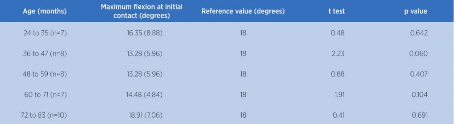
We found no signiicant diferences when comparing the maximum lexion values of knee at initial contact of the foot with the loor for all the analyzed ages and the reference values (18°) described in the literature2.
Table 1. Comparison between the angles of knee lexion at initial contact according to age and reference values
Age (months) Maximum flexion at initial
contact (degrees) Reference value (degrees) t test p value
24 to 35 (n=7) 16.35 (8.88) 18 0.48 0.642
36 to 47 (n=8) 13.28 (5.96) 18 2.23 0.060
48 to 59 (n=8) 13.28 (5.96) 18 0.88 0.407
60 to 71 (n=7) 14.48 (4.84) 18 1.91 0.104
72 to 83 (n=10) 18.91 (7.06) 18 0.41 0.691
Statistical test: t test for a group. p<0.05
Table 2. Comparison between knee angle behavior during the gait of children with Down Syndrome according to age
Age (months) Maximum flexion at initial
contact (degrees)
Percentage of occurrence at initial contact (%)
Maximum swing phase flexion (degrees)
Percentage of occurrence at swing phase (%)
24 to 35 (n=7) 16.35 (8.88) 10.79 (4.07) 61.90 (16.84) 74.22 (2.72)
36 to 47 (n=8) 13.28 (5.96) 11.06 (3.89) 58.97 (12.96) 73.30 (3.07)
48 to 59 (n=8) 21.67 (11.80) 11.00 (5.84) 64.43 (10.26) 74.26 (2.38)
60 to 71 (n=7) 14.48 (4.84) 11.65 (3.90) 56.80 (10.77) 75.90 (4.27)
72 to 83 (n=10) 18.91 (7.06) 13.42 (4.40) 56.50 (10.78) 73.22 (3.76)
Statistical test: One–way Anova for variables such as maximum lexion at initial contact and at swing phase, and occurrence percentage at initial contact; Kruskal-Wallis (p<0.05) for the variable occurrence percentage at swing phase
Table 2 presents data on knee angle behavior based on mean and standard deviation values during the gait of children with Down Syndrome according to age.
We found no signiicant diferences when comparing the analyzed variables between ages.
DISCUSSION
his study aims at describing the knee angle behavior in children with Down Syndrome, for 24 months, according to their age.
Regarding knee angle behavior at initial contact of the foot with the loor, the researched literature describes that at this moment the articulation can lex up to 18°. In this study, we found no signiicant diferences when comparing the maximum knee lexion angle for each age and reference values. However, from the described results (Table 1), it is possible to infer that between 24 and 35 months, 36 and 47 months and 60 to 71 months, the analyzed group of children showed
an average below the reference value. In ages between 48 and 59 months and 72 to 83 months, the maximum lexion value was higher than the value normally used for analysis. From these results, it is possible to note that at this point of the gait, children of all ages who participated in this study presented average values within the limits described by the literature4.
he analysis of corporal angles that directly or indirectly inluence the gait could also contribute to this study.
CONCLUSION
From the obtained results, it is possible to infer that the participants of this study presented knee angle behavior values at the initial contact that corroborated reference values by the reviewed literature for all ages. In the transposition of the lower limb at the swing phase, knee maximum lexion values were higher than reference values for all ages.
he main indings of this study were that the analyzed children showed regular values for maximum knee lexion at the irst contact, i.e., the lexion of this articulation was stable over time and corroborated the values of the literature on the matter when knee maximum lexion at swing phase showed to be excessive over time.
REFERENCES
1. Thabet NS, Kamal HM. Modulation of balance and gait in children with Down syndrome via gravity force stimulation program training. Bull Fac Ph Th. [Internet]. 2011 [acesso em 9 ago. 2017];16(2):87-98. Disponível em: https://goo.gl/ DY2nJe
2. Cimolin V, Galli M, Grugni G, Vismara L, Albertini G, Rigoldi C, et al. Gait patterns in Prader-Willi and Down syndrome patients. J Neuroeng Rehabil. 2010;7:28. doi: 10.1186/1743-0003-7-28. 3. Pietraszewski B, Winiarski S, Jaroszczuk S. Three-dimensional
human gait pattern: reference data for normal men. Acta Bioeng Biomech. 2012;14(3):9-16. doi: 10.5277/abb120302. 4. Perry J. Análise de marcha: marcha normal. Vol. 1. São Paulo:
Manole; 2005.
5. Valentin-Gudiol M, Bagur-Calafat C, Girabent-Farrés M, Hadders-Algra M, Mattern-Baxter K, Angulo-Barroso R. Treadmill interventions with partial body weight support in children under six years of age at risk of neuromotor delay: a report of a Cochrane systematic review and meta-analysis. Eur J Phys Rehabil Med. 2013;49(1):67-91.
6. Wu J, Looper J, Ulrich DA, Angulo-Barroso RM. Efects of various treadmill interventions on the development of joint kinematics in infants with Down syndrome. Phys Ther. 2010;90(9):1265-76. doi: 10.2522/ptj.20090281.
7. Galli M, Rigoldi C, Brunner R, Virji-Babul N, Giorgio A. Joint stifness and gait pattern evaluation in children with Down syndrome. Gait Posture. 2008;28(3):502-6. doi: 10.1016/j. gaitpost.2008.03.001.
8. Rodenbusch TL, Ribeiro TS, Simão CR, Britto HM, Tudella E, Lindquist AR. Efects of treadmill inclination on the gait of
discrepancies in the length of the dominant lower limb or because of an excessive movement of the hips, knees and arms to compensate the decrease in plantar lexion and the progress of the limb.
he knee angle behavior during the gait cycle can have a motion range between 0° to 70°, and factors such as individual characteristics, the biomechanical model used in data collection, and the gait speed can lead to diferences in lexion and extension values at each moment of the gait4. In this study, knee angle behavior showed motion range between 2.24° and 88.02° in all age groups.
At the swing phase, with lower limb transposition (swing phase), we found no signiicant diferences when comparing the maximum knee lexion value between diferent ages. At this point of the gait cycle, the knee can lex up to 70° and between 40 to 70% of the cycle. In the analyzed sample, maximum lexion values were higher than the value described in the literature for all ages.
Cimolin et al.2 found an average value for maximum knee lexion of 41.06°±10.68° in the second moment (swing phase) in adults with Down Syndrome.Wu et al.6 investigated the diference between two kinds of treadmill interventions in the development of thirteen children with Down Syndrome. We monitored the development of the gait over a year and at the end of this period, the group subjected to a general and low intensity intervention showed average value for the knee angle at the second lexion moment of 74.2° (±3.6°) and 70.4° (±4.0°), respectively. he group already subjected to high intensity intervention showed average value of 73.7° (4.3°) in the pre-intervention period and 69.5° (3.2°) in the post-intervention period.
In this study, children with Down Syndrome that walked on a treadmill with 0% of inclination showed an average knee lexion value of 15.59° (6.71°) at initial contact and a maximum lexion value at swing phase of 43.09° (6.26°).
In this study, only the children of aged between 48 to 59 months showed average knee lexion value at initial contact relatively higher than in the study previously described. While in maximum lexion at swing stage, all ages showed average values higher than those by Rodenbusch et al.8.
children with Down syndrome. Res Dev Disabil. 2013;34(7):2185-90. doi: 10.1016/j.ridd.2013.02.014.
9. Gufey K, Regier M, Mancinelli C, Pergami P. Gait parameters associated with balance in healthy 2-4 year-old children. Gait Posture. 2016;43:165-9. doi: 10.1016/j.gaitpost.2015.09.017.
10. Figueroa PJ, Leite NJ, Barros RML. A lexible software for tracking of markers used in human motion analysis. Comput methods programs biomed. 2003;72(2):155-65. doi: 10.1016/ S0169-2607(02)00122-0.