Supra-auricular versus Sinusectomy Approaches
for Preauricular Sinuses
Mohammad Waheed El-Anwar
1Ahmed Shaker ElAassar
11Department of Otorhinolaryngology - Head and Neck Surgery, School
of Medicine, Zagazig University, Zagazig, Egypt
Int Arch Otorhinolaryngol 2016;20:390–393.
Address for correspondence Mohammad Waheed El-Anwar, MD, Department of Otorhinolaryngology - Head and Neck Surgery, School of Medicine, Zagazig University, Zagazig, Zip code 0020552309843, Egypt (e-mail: [email protected]).
Introduction
Preauricular sinuses (pits) are common congenital abnormal-ities that werefirst described in 1864 by Heusinger.1,2The malformation is associated with either a defect in thefirst branchial arch development during the sixth week of gesta-tion3due to incomplete fusion of the six auditory hillocks of His, or with the sinus developing during embryonal auricular development from an isolated ectodermal folding, a less accepted hypothesis.2
Classically, a preauricular sinus presents as a small open-ing, usually near the anterior limb of the ascending helix, although most preauricular sinuses are found anterior to the external auditory canal.2 A small percentage has been reported and located in other areas such as the
superopos-terior edge of the helix, the tragus, the lobule, the ascending helix crus, supra-auricular area, and the postauricular area.4–8
Preauricular sinuses are usually asymptomatic, isolated, and require no treatment. However, if infected, these sinuses become painfully swollen with offensive discharge. Given that preauricular sinuses may be associated with hearing and renal anomalies, auditory testing and renal ultrasound are useful in patients presenting associated syndromes.9–11
Complete excision of the sinus sac orfistula is ideal in treatment.1,6 However, even if excision is performed by experienced surgeons, recurrence can still occur after exci-sion.4Several surgical techniques have been used for total excision of preauricular sinuses to avoid recurrence:
Keywords
►
recurrence
►
preauricular
fi
stulae
►
facial nerve
Abstract
Introduction
Several surgical techniques and modi
fi
cations have been described to
reduce the high recurrence rate after excision of preauricular sinus.
Objectives
The aim of this study is to review the literature regarding surgical
approaches for preauricular sinus.
Data Synthesis
We performed searches in the LILACS, MEDLINE, SciELO, PubMed
databases and Cochrane Library in September, 2015, and the key words used in the
search were
“
preauricular sinus,
” “
sinusectomy,
” “
supra-auricular approach,
” “
methy-lene blue,
”
and/or
“
recurrence.
”
We revised the results of 17 studies, including 1270
preauricular sinuses that were surgically excised by sinusectomy in 937 ears and by
supra-auricular approach in 333 ears. Recurrence with supra-auricular was 4 (1.3%) while
sinusectomy was 76 (8.1%) with signi
fi
cant difference (
p
<
0.0001). There were no
reported facial nerve paresis or paralysis in any of the approaches. The sinusectomy
approach showed signi
fi
cantly more complications (
p
¼
0.0048).
Conclusion
Supra-auricular approach had signi
fi
cantly less recurrence rate than tract
sinusectomy approaches. Thus, it could be regularly chosen as the standard procedure
for preauricular sinus excision. As such, it would be helpful for surgeons to be familiar
with this approach.
received
October 26, 2015
accepted
February 1, 2016
published online
April 26, 2016
DOI http://dx.doi.org/ 10.1055/s-0036-1583305. ISSN 1809-9777.
Copyright © 2016 by Thieme Publicações Ltda, Rio de Janeiro, Brazil
Systematic Review
Sinusectomy Approaches
(a)Classic simple sinusectomy using lacrimal probe.Under general or local anesthesia, an elliptical incision is done parallel to the edge of the anterior helix including the sinus opening. Gentle probing with a blunt ended mal-leable probe is done first to delineate the extent and presence of multiple ramifications. All ramifications are meticulously dissected and totally excised.12
(b)Classic simple sinusectomy using methylene blue. El-liptical incision is done parallel to the edge of the anterior helix including the sinus opening. Methylene blue injec-tion is used as a guide for tracing and excising the complete sinus, including its surrounding soft tissue.13 (c) Classic simple sinusectomy using microscopy or
mag-nifying glasses (inside-out technique).This technique wasfirst described in 2005 by Baatenburg de Jong,14but wasfirst introduced by Jesma in Rottendam in the 1970s (not published at that time). This method involves a small elliptical incision around the sinus pit. Stay sutures are placed to facilitate dissection of the tract and the sinus is opened. The sinus tract and its branches are then followed from the inside and outside.15
Supra-auricular Approaches
(a)Parasad technique of supra-auricular approach. The elliptical incision used is extended down to the superior end of the tragus and up parallel to the anterior edge of the anterior helix. The incision is deepened till the temporalis fascia is identified as a medial limit of the dissection. The dissection continues over the cartilage of the anterior helix. The base of the sinus attached to the perichondrium of the anterior helix is excised with the perichondrium to ensure complete excision of the epi-thelial lining.12
(b)Fig. 8 incision with extendedfistulectomy.For cases with
fistulae formation, Huang et al13performed the Fig. 8 inci-sion with extendedfistulectomy under general anesthesia. This surgical method consists of two wedge incisions: one includes the sinus opening and the other includes the abscess openings and surrounding necrotic skin. The surgeon then elevates the skinflap and dissects along the perichondria of the ear down to the temporalis fascia and removes all of the inflamed tissue en bloc, including the sinus orfistula tract. The use of microscopy or glasses in this technique is an option. The Fig. 8 incision method can preserve more intact skin than the large wedge excision can, attaining a better cosmetic result.13
The aim of this study was to review, collect, and analyze the published results of each technique.
Review of Literature
We conducted a search in the LILACS, MEDLINE, SciELO, PubMed, and Cochrane Library databases in September 2015, and used“preauricular sinus,” “sinusectomy,” “supra-auricular approach,” “methylene blue,” and/or “recurrence.” We searched for studies published after 2001.
We collected all methods of preauricular tract(s) identifi -cation and excision and their modifications and referred to them as sinusectomy approaches. We also gathered studies that used the supra-auricular approach as described by Prasad et al, referring to them as supra-auricular approaches. We collected, tabulated, and analyzed the results. Then, we performed a statistical analysis and comparison using SPSS 14.0 statistical software for Windows (SPSS Inc., Chicago, IL). The significance level was set atpless than 0.05.
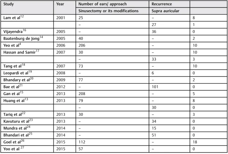
We revised seventeen studies and the results of 1270 surgi-cally excised preauricular sinuses: 937 by sinusectomy techni-ques and 303 by supra-auricular approach (►Table 1).4,12–27 Recurrence with supra-auricular was 4 (1.2%) and with sinus-ectomy was 76 (8.1%) with a highly significant difference (X2¼19.874;p<0.0001) (►Table 2).
None of the approaches reported major complications such as facial nerve paresis or paralysis. Of the sinusectomy approach cases, there were 4 dehiscent wounds (0.43%), 10 infections (1.06%), 14 bad scars (1.5%) documented, whereas only one operated preauricular sinus (0.3%) by supra-auricular approach reported infection with no scar or wounds. Total reported complications resulting from the sinusectomy approach was 28 ears (3%) and one ear (0.3%) from the supra-auricular approach. Therefore, the sinusectomy approach showed signifi -cantly more complications (X2¼7.955;p¼0.0048).
Discussion
Recurrence after excision of preauricular sinus is a result of incomplete excision of the sinus tract and presence of residual viable squamous epithelium.28
The real problem in the surgical removal of preauricular sinus is the high recurrence rate following sinusectomy techniques due to tortuous tract course12 and the high variability and number of sinus ramifications,12,24 particular-ly of the terminal ramifications, which are difficult for the surgeon to follow,12and especially upwards and medially.24 Furthermore, infectious episodes, possibly with abscess, can induce scars that further alter the sinus route and courses.18 Recurrences result from the difficulty during sinusectomy to follow the tract and its branches. Pre- and intraoperatory precautions are often not sufficient to guarantee there is no recurrence, which remains high.17
Although there are several tools and methods used for proper tract(s) identification, such as the use of methylene blue, probing, microscope, or magnifying glasses.14,15 None-theless, recurrence remains of significant concern.
Based on the theory that a preauricularfistula is almost always found in subcutaneous tissues between the temporalis fascia and perichondrium of the helical cartilage, the supra-auricular approach proposed by Prasad et al in 1990 is assumed to have a lower recurrence risk.17The supra-auric-ular technique is based on identification of the temporalis fascia (medial border of the dissection) and the cartilage of the helix and auditory canal (posterior border of the dissec-tion). Subsequently, the surgeon performs an en bloc resec-tion of the sinus,16removing all subcutaneous tissue between the temporalis fascia and the helix through a postauricular
International Archives of Otorhinolaryngology Vol. 20 No. 4/2016
extended incision. Thus, there is no need to identify the entire sinus tract and its branches.12,17
Thereafter, Lam et al,12in his comparative study, found a significant difference in recurrence rates between the classic sinusectomy technique and the supra-auricular approach (32% and 3.7%, respectively).
We analyzed the published operated preauricular cases since Lam et al12 and collectively found that recurrence rate was 4/333 (1.2%) with the supra-auricular approach, and 76/937 (8.1%) with various sinusectomy approaches with statistically significant difference in favor of the supra-auricular approach (►Table 2).
Even though, the sinusectomy relied on magnification,25 which was not employed in any previous study on the supra-auricular approach, recurrence was significantly minimized12,17 and even not encountered16,19,21,23–25after the supra-auricular approach. This demonstrates that the supra-auricular approach is highly effective and successful.
The supra-auricular approach was also described as a simple, less time consuming approach and shows fewer difficulties12,16,17,19,24,29because it does require the surgeon to isolate and follow the sinus branches, as in the sinusectomy technique and its modifications but simply identify a surgical plane such as the temporalis fascia.25Moreover, it carries a risk of injury of the facial nerve or any important structure, with a low risk of scar formation. That is why our statistical analysis of reported complications detected that the supra-auricular approach causes significantly (p¼0.0048) less complications (0.3%) than sinusectomy (3%).
Since this kind of surgery is often performed by relatively inexperienced surgeons, the supra-auricular approach may represent a further guarantee of preventing recurrences as it does not require a learning curve. It is less time consuming and can be done under local anesthesia
The supra-auricular approach is simple, effective, with negligible recurrence. Thus, it is better to be used regularly
Table 2 Statistical analysis of recurrence rate difference between supra-auricular and sinusectomy approaches
Approach Number of ears Recurrence Chi square test pvalue
Supra auricular 333 4 (1.2%) 19.874 <0.0001 HS
Sinusectomy or its modifications 937 76 (8.1%)
Abbreviations: HS; highly significant.
Table 1 Recurrence among different studies
Study Year Number of ears/ approach Recurrence
Sinusectomy or its modifications Supra auricular
Lam et al12 2001 25 – 8
– 27 1
Vijayendra16 2005 – 36 0
Baatenburg de Jong14 2005 40 – 2
Yeo et al4 2006 206 – 10
Hassan and Samir17 2007 30 – 10
– 33 3
Tang et al18 2007 73 – 10
Leopardi et al19 2008 – 6 0
Bhandary et al20 2009 77 – 2
Bae et al21 2012 – 101 0
Gan et al15 2013 208 – 5
Huang et al13 2013 79 – 8
– 30 0
Tariq et al22 2013 30 – 3
Kavuturu et al23 2013 – 34 0
Mundra et al24 2014 – 15 0
Bhandari et al25 2014 – 51 0
Goel et al26 2015 112 – 18
Yoo et al27 2015 57 – 0
International Archives of Otorhinolaryngology Vol. 20 No. 4/2016
as a standard procedure for preauricular sinus excision, especially because it has shown no significant complications and less post-operative scar formation18 with no need for extra tools as microscope and loops. Moreover, it is the ideal technique for recurrent cases or cases undergoing sinus-ectomy after abscess incision and drainage.
Studies to compare supra-auricular approach and sinus-ectomy approaches for preauricular sinus excision in bilateral cases by the same surgeon are still needed.
Final Comments
Supra-auricular approach had a significantly lower recurrence rate than tract sinusectomy approaches. Thus, it is a good option as a standard procedure for preauricular sinus excision, It is especially useful as an alternative in cases where the sinus-ectomy approaches are difficult to be performed. Therefore, it would be helpful for surgeons to be familiar with this approach.
Conflict of Interest and Financial Disclosure Statement The authors declare no financial support or conflict of interest in this study.
References
1 Scheinfeld NS, Silverberg NB, Weinberg JM, Nozad V. The preaur-icular sinus: a review of its clinical presentation, treatment, and associations. Pediatr Dermatol 2004;21(3):191–196
2 Tan T, Constantinides H, Mitchell TE. The preauricular sinus: A review of its aetiology, clinical presentation and management. Int J Pediatr Otorhinolaryngol 2005;69(11):1469–1474
3 Nofsinger YC, Tom LWC, LaRossa D, Wetmore RF, Handler SD. Periauricular cysts and sinuses. Laryngoscope 1997;107(7):883–887 4 Yeo SW, Jun BC, Park SN, et al. The preauricular sinus: factors contributing to recurrence after surgery. Am J Otolaryngol 2006; 27(6):396–400
5 Chami RG, Apesos J. Treatment of asymptomatic preauricular sinuses: challenging conventional wisdom. Ann Plast Surg 1989; 23(5):406–411
6 Choi SJ, Choung YH, Park K, Bae J, Park HY. The variant type of preauricular sinus: postauricular sinus. Laryngoscope 2007; 117(10):1798–1802
7 Minkowitz S, Minkowitz F. Congenital aural sinuses. Surg Gynecol Obstet 1964;118:801–806
8 Chang PH, Wu CM. An insidious preauricular sinus presenting as an infected postauricular cyst. Int J Clin Pract 2005;59(3):370–372 9 Kumar S, Marres HA, Cremers CW, Kimberling WJ. Autosomal-dominant branchio-otic (BO) syndrome is not allelic to the bran-chio-oto-renal (BOR) gene at 8q13. Am J Med Genet 1998;76(5): 395–401
10 Fraser FC, Aymé S, Halal F, Sproule J. Autosomal dominant dupli-cation of the renal collecting system, hearing loss, and external ear
anomalies: a new syndrome? Am J Med Genet 1983;14(3): 473–478
11 Clementi M, Mammi I, Tenconi R. Family with branchial arch anomalies, hearing loss, ear and commissural lip pits, and rib anomalies. A new autosomal recessive condition: branchio-oto-costal syndrome? Am J Med Genet 1997;68(1):91–93
12 Lam HCK, Soo G, Wormald PJ, Van Hasselt CA. Excision of the preauricular sinus: a comparison of two surgical techniques. Laryngoscope 2001;111(2):317–319
13 Huang WJ, Chu CH, Wang MC, Kuo CL, Shiao AS. Decision making in the choice of surgical management for preauricular sinuses with different severities. Otolaryngol Head Neck Surg 2013;148(6): 959–964
14 Baatenburg de Jong RJ. A new surgical technique for treatment of preauricular sinus. Surgery 2005;137(5):567–570
15 Gan EC, Anicete R, Tan HK, Balakrishnan A. Preauricular sinuses in the pediatric population: techniques and recurrence rates. Int J Pediatr Otorhinolaryngol 2013;77(3):372–378
16 Vijayendra H, Sangeetha R, Chetty KR. A safe and reliable technique in the management of preauricular sinus. Indian J Otolaryngol Head Neck Surg 2005;57(4):294–295
17 Mohamed EG. Hassan, Ayman Samir. Pre-Auricular sinus: com-parative study of two surgical techniques. Ann Pediatr Surg 2007; 3:139–143
18 Tang IP, Shashinder S, Kuljit S, Gopala KG. Outcome of patients presenting with preauricular sinus in a tertiary centre—afive year experience. Med J Malaysia 2007;62(1):53–55
19 Leopardi G, Chiarella G, Conti S, Cassandro E. Surgical treatment of recurring preauricular sinus: supra-auricular approach. Acta Otorhinolaryngol Ital 2008;28(6):302–305
20 Bhandary S, Singh RK, Karki P, Sharma SK, Chettri ST. Preauricular sinus: Prospective study of clinical course and associations. Gujarat Journal of Otorhinolaryngology and Head & Neck Surgery 2009;6(2):6–9
21 Bae SC, Yun SH, Park KH, et al. Preauricular sinus: advantage of the drainless minimal supra-auricular approach. Am J Otolaryngol 2012;33(4):427–431
22 Tariq M, Murtaza G, Akram A, Bashir T. Pre-Auricular sinus and its microsurgical excision. Esculapio 2013;9(3):123–125
23 Kumar Chowdary KV, Sateesh Chandra N, Karthik Madesh R. Preauricular sinus: a novel approach. Indian J Otolaryngol Head Neck Surg 2013;65(3):234–236
24 Mundra RK, Sinha R, Agrawal R. Supra-auricular approach: a simple recurrence-free technique for pre-auricular sinus. EJNSO 2014;1(1):11–15
25 Bhandari R, Limbu TR, Parajuli R, Thapa S. Auricular dissection method for treatment of preauricular sinus. Journal of Chitwan Medical College 2014;4(9):21–24
26 Goel AK, Sylonia SC, Garg A, Rattan K. Preauricular sinus: When to operate? Indian Journal of Otology 2011;17(2):63–65
27 Yoo H, Park DH, Lee J, Park MC. A Surgical Technique for congenital preauricular sinus. Arch Craniofac Surg 2015;16(2):63–66 28 Kumar KK, Narayanamurthy VB, Sumathi V, Vijay R. Preauricular
sinus: Operating microscope improves outcome. Indian J Otolar-yngol Head Neck Surg 2006;58(1):6–8
29 Prasad S, Grundfast K, Milmoe G. Management of congenital preauricular pit and sinus tract in children. Laryngoscope 1990; 100(3):320–321
International Archives of Otorhinolaryngology Vol. 20 No. 4/2016