35
TRAUMATIC RUPTURE OF ADRENAL PSEUDOCYST Case Report
International Braz J Urol
Official Journal of the Brazilian Society of Urology
Vol. 30 (1): 35-36, January - February, 2004
TRAUMATIC RUPTURE OF ADRENAL PSEUDOCYST LEADING TO
MASSIVE HEMORRHAGE IN RETROPERITO
NEUM
LUCIANO A. FAVORITO, FELIPE M. LOTT, ANDRÉ G. CAVALCANTE
Service of Urology, Souza Aguiar Municipal Hospital, Rio de Janeiro, RJ, Brazil
ABSTRACT
We present the case of a patient who had a large pseudocyst in the right adrenal gland, which was ruptured following blunt abdominal trauma, leading to a voluminous hemorrhage in retroperitoneum. A 29-year old female patient was admitted in the emergency room following a fall from stairs with trauma in right flank. She underwent a computerized tomography that evidenced a large retroperitoneal collection, with no apparent renal damage. She was submitted to surgery, where a large ruptured cyst was observed, originating from the upper portion of the right adrenal gland. Cystic diseases of adrenal gland are rare. Highly voluminous cysts can be damaged in cases of blunt trauma to the lumbar region leading to large hematomas in retroperitoneum.
Key words: adrenal glands; cysts; wounds and injuries; hemorrhage; retroperitoneal space Int Braz J Urol. 2004; 30: 35-6
INTRODUCTION
Cystic pathologies of the adrenal gland are rare, with an incidence of 0.06% in 1,400 autopsies (1). The cysts most frequently found in the adrenal gland are endothelial (45%), hematic pseudocyst (39%), epithelial (9%) and parasitic (7%) (2,3).
The objective of this work is to report the case of a female patient who had a voluminous pseudocyst in right adrenal gland that was ruptured following a blunt abdominal trauma, leading to an extensive hemorrhage in retroperitoneum.
CASE REPORT
A female, 29-year old patient was admitted to the emergency room reporting that 12 hours ear-lier she had fallen from the third step of a stair. She presented intense pain in right flank and a volumi-nous mass that occupied a large portion of the upper abdominal region. The patient did not present hemo-dynamic instability and denied previous pathologies.
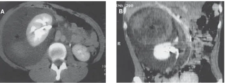
She underwent a computerized tomography that evidenced a large retroperitoneal collection, with preservation of the right kidney, compatible with extra-renal hematoma (Figure-1). The kidneys and the liver were apparently intact in the image scan. It was decided to perform an exploratory laparotomy.
During the surgery, the presence of a large retroperitoneal bulging was observed, without any lesion in liver or intestine. An exploration of the ret-roperitoneal hematoma was performed. A voluminous cyst was found, which was ruptured, originated from the upper portion of the right adrenal gland. It was performed the hematoma drainage and partial exer-esis of the region in the adrenal gland where the cyst was located. The pathological report showed it was a hematic adrenal pseudocyst.
COMMENTS
36
TRAUMATIC RUPTURE OF ADRENAL PSEUDOCYST
cases where there is some pathology that causes the gland’s enlargement, as in cases of adrenal cysts, tu-mors and congenital hyperplasia (1).
The majority of adrenal cystic pathologies are assymptomatic, being detected in autopsies; never-theless, large cysts have a tendency to develop com-plications such as intracystic hemorrhage and rup-ture, which can manifest as a surgical emergency (2). Symptoms of lumbar pain, nauseas, vomiting, in-creased abdominal volume and constipation occur only in cases of highly voluminous cysts. Usually, adrenal cysts are incidentally seen in routine exami-nations such as ultrasonography and computerized tomography of abdomen (3).
Among the adrenal cystic pathologies, the endothelial cysts and the hematic pseudocyst are the most frequent (2). The hematic pseudocyst ac-counts for 40% of all cystic lesions of the adrenal gland (2). The hematic pseudocysts are covered by a thin capsule that isolates the region from the re-maining normal adrenal parenchyma (2). They are generally unilocular and the liquid present inside them has a reddish color, and can reach large vol-umes, with reports of pseudocysts containing up to 1 liter inside (2). They usually present encapsu-lated residues of previous hemorrhages in the ad-renal gland (2).
Highly voluminous cysts can be damaged in cases of blunt trauma to the lumbar region, leading to voluminous hematomas in retroperitoneum.
REFERENCES
1. Pasciak RM, Cook WA: Massive retroperitoneal hem-orrhage owing to a ruptured adrenal cyst. J Urol. 1988; 139: 98-100.
2. Chew SP, Sim R, Teoh TA, Low CH: Haemorrhage into non-functioning adrenal cysts - report of two cases and review of the literature. Ann Acad Med Singapore. 1999; 28: 863-6
3. Cheema P, Cartagena R, Staubitz W: Adrenal Cysts: diagnosis and treatment. J Urol. 1981; 126: 396-9.
Received: August 27, 2003 Accepted after revision: November 14, 2003
Correspondence address: Dr. Luciano Alves Favorito Rua Professor Gabizo, 104 / 201 Rio de Janeiro, RJ, 20551-030, Brazil Fax: + 55 21 2587-6121
E-mail: [email protected]
Figure 1 - A) Computerized tomography (CT) of abdomen with contrast medium evidencing a voluminous retroperitoneal hematoma in the peri-renal area (*). Cross section. B) Abdominal CT with contrast medium evidencing renal integrity and the presence of a voluminous hematoma in the peri-renal area, as well as the presence of a voluminous expansive lesion with cystic content in the region of the adrenal gland (*). Frontal section