175
C
ASER
EPORTReceived for publication 24/04/2014 - Accepted for publication 23/10/2014 The authors declare no conflicts of interest
R
ESUMOA
BSTRACTEvaluation of retinal nerve fiber layer thickness
in a patient with bilateral optic disc drusen
Avaliação da espessura da camada de fibras nervosas da retina
em um paciente com drusas do disco óptico bilateralmente
Alime Gunes, Seden Demirci, Serpil Demirci, Hasan Rifat Koyuncu
1 Departments of Ophthalmology, Süleyman Demirel University Faculty of Medicine, Isparta, Turkey. 2 Departments of Neurology, Süleyman Demirel University Faculty of Medicine, Isparta, Turkey.
As drusas do disco óptico (DDO) são depósitos de material hialino calcificado dentro da substância da cabeça do nervo óptico. Drusas do disco óptico, especialmente se for bilateral, podem apresentar o quadro clínico de edema de papila. Usualmente o espessamento da camada de fibras nervosas da retina (RCFN) podem estar presentes em DDO. Neste relato apresentamos o caso de um homem com 17 anos de idade que foi encaminhado por um outro centro, com o diagnóstico de edema do disco óptico. A acuidade visual do paciente, o exame de lâmpada de fenda e a pressão intraocular foram normais em ambos os olhos. No exame de fundo de olho havia discos elevados de forma irregular bilateralmente e os nervos ópticos com margens de disco nebulosas. Ele não tinha defeitos do campo visual em perimetria computadorizada. Drusas do disco óptico (DDO) bilateral foram identificados e confirmados pela ultrassonografia B-scan e tomografia de coerência óptica (TCO) que demonstraram 4 horas de relógio de RCFN com espessamento. As drusas do disco óptico podem ser diagnosticadas como papiledema. Assim, a suspeita clínica de DDO é importante a fim de evitar intervenções desne-cessárias. Embora a maioria dos olhos com DDO têm espessura normal ou thinner RCFN, alguns desses olhos podem ter camada mais grossa na RCFN.
Descritores: Drusas do disco óptico; Papiledema; Nervo óptico/patologia; Fibras nervosas/patologia
Optic disc drusen (ODD) is the accumulations of calcified hyaline-like material within the substance of the optic nerve head. Optic disc drusen, especially if it is bilateral, may mimic the clinical presentation of papilledema. Usually retinal nerve fiber layer (RNFL) thinning can be present in ODD. In this report we present uncommon RNFL changes in a patient with bilateral ODD. A 17-year-old male was referred by another center with a diagnosis of optic disc edema. The patient’s visual acuity, the slit-lamp examination and the intraocular pressures were normal in both eyes. On fundus examination, there were irregularly elevated discs bilaterally and the optic nerves appear with hazy disk margins. He did not have visual field defects in automated perimetry. Bilateral ODD were identified and confirmed by B-scan ultrasonography and optical coherence tomography (OCT) demonstrated 4 clock hours of RNFL thickening. Optic disc drusen may be misdiagnosed as papilledema. Thus, clinical suspicion of ODD is important in order to diagnose papilledema and prevents unnecessary interventions. Although most of eyes with ODD have normal or thinner RNFL thickness, some of these eyes can have thicker RNFL thickness.
Keywords: Optic disc drusen; Papilledema; Optic nerve/pathology; Nerve fiber/pathology
176 A Gunes, S Demirci, S Demirci, HR Koyuncu
Rev Bras Oftalmol. 2015; 74 (3): 175-7
I
NTRODUCTIONO
ptic disc drusen (ODD) is the accumulations of calcifiedhyaline-like material within the substance of the optic
nerve head(1). Compression of ganglion cells by calcified
hyaline structures results in alteration in axoplasmic flow and cell death and so retinal nerve fiber layer (RNFL) defects are
seen in ODD(2).
Patients with ODD are often asymptomatic, with the condition being found incidentally during fundus examination. Visual acuity is well preserved but the visual fields of these
patients can be abnormal and may deteriorate over time(3).
The mechanism of visual field loss from ODD is speculative, and there is no known treatment. Atrophy of the RNFL can be appreciated as RNFL thinning, therefore measurement of RNFL thickness can be used as an objective index of optic nerve damage(2).
In this report we present uncommon RNFL changes in a patient with bilateral ODD.
C
ASER
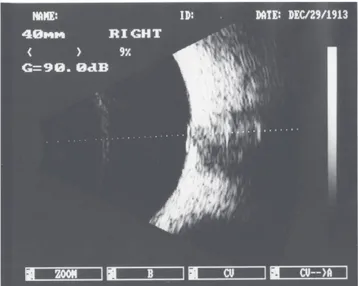
EPORTA 17-year-old male was referred by another center with a diagnosis of optic disc edema. The patient’s visual acuity, the slit-lamp examination and the intraocular pressures were normal in both eyes. On fundus examination, there were irregularly elevated discs bilaterally and the optic nerves appear with hazy disk margins (figures 1, 2). He did not have visual field defects in automated perimetry with the Humphrey Visual Field Analyzer (Zeiss Humphrey Systems, Dublin, California USA). Bilateral ODD were identified and confirmed by B-scan ultrasonography (BVI Compact) (figures 3, 4) and optical coherence tomography (OCT) (Spectral OCT SLO, OPKO/OTI Instrumentation, Miami, FL, USA) demonstrated 4 clock hours of RNFL thickening (fi-gure 5). The average RNFL thickness were 135 µm in right eye and 138 µm in left eye. His neurological examination was within normal limits except for the appearance of the optic discs. His cranial magnetic resonance imaging and Visual evoked potential showed no abnormality. Hence, the patient was diagnosed with bilateral ODD.
Figure 1. Fundus photography of the patient’s right eye reveal, irregularly elevated optic discs and the optic nerve appear with hazy disk margin
Figure 2. Fundus photography of the patient’s left eye reveal, irregularly elevated optic discs and the optic nerve appear with hazy disk margin
Figure 3. B-scan ultrasonography of the patient’s right eye identified the optic disc drusen
177
Corresponding author:
Alime Gunester, Assistant professor
Department of Ophthalmology, Süleyman Demirel University Faculty of Medicine, Isparta, Turkey
Phone: +905054828345
E-mail: [email protected]
Figure 5. Optical coherence tomography showing 4 clock hours of RNFL thickening
D
ISCUSSIONOptic disc drusen occurs in 0.3-2.0% of the population, is
bilateral in 75% of cases, and have no sex predilection(3).
Impairment of visual acuity is rare in ODD, but insignificant
vi-sual field defects may occur in up to half of cases(4).
Usually RNFL thinning can be present in ODD. Roh et
al. (5) reported that superior and inferior RNFL were
significantly thinner in the eyes with visible ODD, and OCT is sensitive and early indicator of RNFL thinning.
Noval et al.(6) studied 28 eyes with ODD in children and
classified as type 1 (buried), type 2 (ringed), and type 3 (superfici-al) ODD. They found that RNFL thickness was higher in type 1 and 2 ODD although RNFL thickness was lower in type 3 ODD. Similarly, our patient had buried ODD and had higher RNFL thickness in both eyes. Optic disc drusen can cause an increase in RNFL thickness but in the following years ODD can lead to decrease in RNFL thickness by damaging the optic nerve fibers. Optic disc drusen may be misdiagnosed as papilledema. Thus, clinical suspicion of ODD is important in order to diagnose papilledema and prevents unnecessary interventions. Although most of eyes with ODD have normal or thinner RNFL thickness, some of these eyes can have thicker RNFL thickness. Patient with ODD should be followed for further RNFL damage or visu-al field defects visu-although they have thicker RNFL.
R
EFERENCES1. Larentzen SE. Drusen of the optic disc: a clinical and genetic study. Acta Ophthalmol. 1966; Suppl 90:l-180.
2. Katz BJ, Pomeranz HD. Visual field defects and retinal nerve fiber layer defects in eyes with buried optic nerve drusen. Am J Ophthalmol. 2006;141(2):248-53.
3. Auw-Haedrich C, Staubach F, Witschel H. Optic disk drusen. Surv Ophthalmol. 2002;47(6):515-32.
4. Antcliff RJ, Spalton DJ. Are optic disc drusen inherited? Ophthalmol-ogy. 1999;106(7):1278-81.
5. Roh S, Noecker RJ, Schuman JS, Hedges TR 3rd, Weiter JJ, Mattox C. Effect of optic nerve head drusen on nerve fiber layer thickness. Oph-thalmology. 1998;105(5):878-85.
6. Noval S, Visa J, Contreras I. Visual field defects due to optic disk drusen in children. Graefes Arch Clin Exp Ophthalmol. 2013;251(10):2445– 50.
Evaluation of retinal nerve fiber layer thickness in a patient with bilateral optic disc drusen
Rev Bras Oftalmol. 2015; 74 (3): 175-7