ABSTRACT | Differentiating glaucomatous from nonglau comatous optic disc cupping remains challenging. We present a case of a 48yearold woman with an internal carotid aneurysm of approximately 3.5 mm × 6.5 mm that mimicked normaltension glaucoma. The patient had a 2year history of low vision acuity in her left eye and frontal oppressive headache. Owing to the carotid aneurysm, she developed an asymmetric vertical cuptodisc ratio above 0.2, and marked inferotemporal neu ronal rim loss and pallor of the residual rim were noted in the left disc. She also developed a visual field defect with an arcuate scotoma in the left eye. The patient was referred to a neurosurgeon and underwent endovascular aneurysm occlusion. This case highlights the diagnostic importance of recognizing that many neurological defects remain underdiagnosed.
Keywords: Glaucoma/diagnosis; Carotid artery, internal; Oph
thalmic artery/pathology; Aneurysm/diagnosis; Visual field; Diffe rential diagnosis
RESUMO | A diferenciação de escavações glaucomatosas e
não glaucomatosas ainda permanece um desafio ainda nos dias de hoje. Nos descrevemos um caso de aneurisma de carótida interna medindo 3.5mm x 6.5mm que simulava um glaucoma de pressão normal. O caso é sobre uma paciente feminino de 48 anos com história de 2 anos de baixa acuidade visual no olho esquerdo e cefaléia frontal. Devido ao aneurisma de carótida a paciente desenvolveu uma assimetria de escavação vertical maior que 0.2 no olho esquerdo em relação ao direito com defeito localizado da camada de fibras nervosas temporal inferior. Ela também apresentava um defeito arqueado temporal superior a
esquerda, cruzando a linha média vertical consistente. Após o diagnostico confirmado pela ressonância magnética funcional, a paciente foi enviada para o neurocirurgião para realização de uma oclusão endovascular do aneurisma. Esse caso nos alerta da importância de se lembrar que não apenas o glaucoma gera escavações suspeitas no disco óptico e que ainda muitos defeitos por causas neurológicas são subdiagnosticados.
Descritores: Glaucoma/diagnóstico; Carótida interna; Artéria
of tál mica/patologia; Aneurisma/diagnóstico; Campo visual; Diag nóstico diferencial
INTRODUCTION
Glaucoma is an optic neuropathy characterized by the progressive loss of retinal ganglion cells and associa ted morphological changes to the optic nerve and retinal nerve fiber layer(1,2). Among the various forms of glau
coma, normaltension glaucoma (NTG) is characterized by typical glaucomatous optic disc cupping associated with visual field loss, normal intraocular pressure (IOP), an open iridocorneal angle, and an absence of causative systemic or ocular disease(3). Prospective studies have
shown that NTG is more prevalent in woman, particu larly those with a recent history of migraine onset or changes in the pattern of migraines. NTG is also fre quently correlated with systemic findings of vasospasm, such as in Raynaud’s phenomenon, and low systemic blood pressure(3). A family history of glaucoma is also
fre quently reported(4,5).
The results of the Collaborative Normal Tension Glau coma Study and other studies have shown that compa red with patients without optic disc hemorrhage, those with hemorrhage have an increased risk of visual field progression(6,7). A diagnosis of NTG should be made after
excluding all causes of secondary glaucoma (use of ste roids, previous uveitis with angle closure, intermittent IOP elevation) and other causes of optic neuropathy(4,5).
Internal carotid artery aneurysm mimicking
normal-tension glaucoma
Aneurisma de carótida interna simulando glaucoma de pressão normal
Mário Pincelli Netto1,2, Pedro Vanalle Ferrari1,2, Bruno Torres Herrerias1,2, Flávio Eduardo Hirai1,2, Carolina Pelegrini Barbosa Gracitelli1,2
1. Department of Ophthalmology, Universidade Federal de São Paulo, São Paulo, SP, Brazil. 2. Glaucoma Unit, Ver Mais Oftalmologia, São Paulo, SP, Brazil.
Submitted for publication: May 22, 2017 Accepted for publication: November 16, 2017
Funding: No specific financial support was available for this study.
Disclosure of potential conflicts of interest: None of the authors have any potential conflict of interest to disclose.
Corresponding author: Carolina P. B. Gracitelli.
In this case report, we describe a patient with inter nal carotid aneurysm who presented with an asymmetric cuptodisc ratio and a consistent and corresponding vi sual field defect in the left eye. The case mimicked NTG; however, a careful evaluation ruled out this diagnosis.
CASE REPORT
A 48yearold African Brazilian woman presented to our clinic with a chief complaint of frontal oppressive headache of 2 years’ duration. The headache was cha racterized as intermittent, causing a moderate level of pain, and lasting from minutes to hours. The patient reported no irradiation and no typical period of mani festation or other neurological manifestations. She denied any remarkable personal, ophthalmological, or family history.
An initial examination revealed an IOP of 12 mmHg in both eyes (AU) without the administration of any to pical or systemic medication and a daily tension curve showing variation between 13 and 16 mmHg in the right eye (OD) and 12 and 15 mmHg in the left eye (OS). Her bestcorrected visual acuity was 20/20 (0 logMAR) AU. A color vision test was not performed during this examination. Anterior biomicroscopy showed no abnor ma lities, and the pupillary reflex was normal (AU). Go nios copy revealed an open angle of 360° to the scleral spur (AU). The results of a pachymetry exam were 542 µm OD and 535 µm OS. Fundoscopy showed a
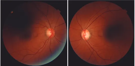
cuptodisc ratio of 0.5 OD and 0.7 OS. A lower tem poral retinal nerve fiber layer defect was detected in the OS (Figure 1). The visual field test was normal in
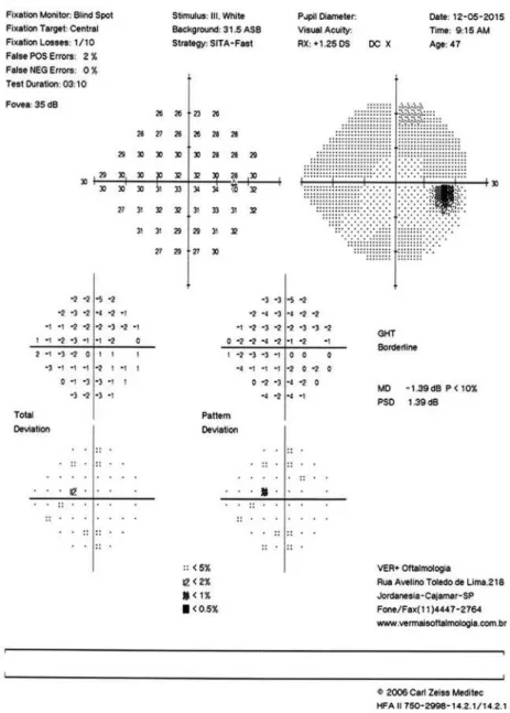
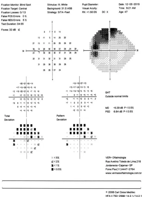
the OD (Figure 2); however, a temporal superior arched loss of sensitivity crossing the vertical midline was observed in the OS (Figure 3). Because the functional defect was compatible with the structural defect and IOP measurements were normal (although she had no epidemiologic findings for NTG), topical bimatoprost 0.03% AU was initiated for the treatment of NTG.
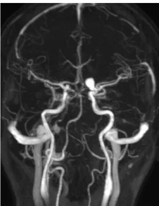
The patient returned 45 days later reporting the onset of dyschromatopsia. The ophthalmologic findings did not differ from those recorded at her initial visit. A nu clear magnetic resonance examination was requested and showed an internal carotid aneurysm of approxi mately 3.5 mm × 6.5 mm near the emergence of the ophthalmic artery. This aneurysm compressed the optic nerve (Figure 4). The patient was referred to a neurosur geon and underwent endovascular aneurysm occlusion. Three months after surgery, she reported improvement in the headache symptoms, but a visual field test revea led a diffuse loss of sensitivity.
DISCUSSION
The details of our case emphasize that clinicians must make the diagnosis of NTG with extreme care. Many neurological diseases can have anatomic and functional defects similar to those of NTG. These diseases include ischemic optic and hereditary optic neuropathies, de myelinating optic neuritis, multiple sclerosis, trauma, intraocular infections, and intraorbital or intracranial masses with compressive effects on the optic nerve or chiasm, all of which are reportedly characterized by disc
Figure 2. Visual field test of the right eye performed with the SITA 24-2 strategy. The image shows a relative depression of the upper temporal field despite the normal results of the glau-coma hemifield test. This test does not meet Anderson’s criteria for a diagnosis of glauglau-coma.
cupping and visual field defects(8) but do not present
with elevated IOP(9).
Our patient presented with the chief complaint of headache. This presenting symptom combined with the fun ctional and anatomical defects typical of glaucomaspecifically, a visual field defect crossing the vertical midline and nerve fiber layer loss (tem poral inferior retinal nerve fiber layer defect)initially suggested that the neurological abnormality was caused by the migraine. One study reported that the risk of lowtension glaucoma is several times higher in patients who have a history of headache, although
migraine is not thought to be a frequent cause of lowten sion glaucoma because migraine is too common and lowtension glaucoma is too rare(10). Only after the pa
tient noticed the onset of another neurological defect (dyschromatopsia) was a secondary cause considered and confirmed diagnostically as an aneurysm. Two cases similar to ours have appeared in the literature(11,12).
Figure 3. Visual field test of the left eye performed with the SITA 24-2 strategy. The image shows a deep relative supero-nasal scotoma from 10 (zero dB) to 20 (26 dB). The scotoma does not extend beyond the nasal hemifield. The results of the glaucoma hemifield test are abnormal. This test meets Anderson’s criteria for a diagnosis of glaucoma.
The differentiation between glaucomatous and nonglaucomatous cupping remains a challenge. The evidence does not support a recommendation that all patients with suspected NTG receive routine radiologic examination(4,10); therefore, clinicians should consider
predictive factors such as visual acuity worse than 20/40, visual field defects involving the vertical midline, age younger than 50 years, and pallor disproportionate to the disc cupping(4) in the differential diagnosis. Our
pa tient met only one of these criteria at the first visit to our clinic.
The carotidophthalmic artery includes the early in tracranial portion of the carotid from which the ophthalmic artery originates(11). Aneurysms in the carotid
portion of this artery account for 5.4% of all intracra nial aneurysms(12). Orbital and brain masses can also
cause the visual field defects and optic disc excavation typical of glaucoma by compressing components of the visual system at various levels(13). The physiopathology
Figure 4. Preoperative T1-weighted contrast magnetic reso-nance images showing a hyperintense aneurysm in the ophthal-mic segment of the right internal carotid artery.
com pression of the optic nerve by the lesion, or optic pathway ischemia(13).
The main clinical implication of our findings is that clinicians must recognize that many neurological defects are underdiagnosed and that glaucoma is not the only condition that generates suspicious cupping of the optic disc. A careful history combined with detailed clinical examination is crucial in reaching an accurate diagnosis.
In summary, patients with suspected NTG should undergo a careful evaluation. NTG is a diagnosis of ex clusion, and there is no evidence that all patients with
suspected NTG should undergo neuroimaging.(10) There
fore, consideration of the predictive factors mentioned above should be routine(4).
REFERENCES
1. Weinreb RN, Khaw PT. Primary openangle glaucoma. Lancet. 2004;363(9422):171120.
2. Marvasti AH, Tatham AJ, Zangwill LM, Girkin CA, Liebmann JM, Weinreb RN, et al. The relationship between visual field index and estimated number of retinal ganglion cells in glaucoma. PLoS One. 2013;8(10):e76590.
3. Mudumbai RC. Clinical update on normal tension glaucoma. Semin Ophthalmol. 2013;28(3):1739.
4. Greenfield DS, Siatkowski RM, Glaser JS, Schatz NJ, Parrish RK 2nd. The cupped disc. Who needs neuroimaging? Ophthalmology. 1998;105(10):186674.
5. Choudhari NS, Neog A, Fudnawala V, George R. Cupped disc with normal intraocular pressure: the long road to avoid misdiagnosis. Indian J Ophthalmol. 2011;59(6):4917.
6. Bengtsson B, Leske MC, Yang Z, Heijl A; EMGT Group. Disc he morrhages and treatment in the early manifest glaucoma trial. Ophthalmology. 2008;115(11):20448.
7. Gracitelli CP, Tatham AJ, Zangwill LM, Weinreb RN, Liu T, Medeiros FA. Estimated rates of retinal ganglion cell loss in glaucomatous eyes with and without optic disc hemorrhages. PLoS One. 2014; 9(8):e105611.
8. O’Neill EC, DaneshMeyer HV, Connell PP, Trounce IA, Coote MA, Mackey DA, et al. The optic nerve head in acquired optic neuropathies. Nat Rev Neurol. 2010;6(4):22136.
9. Esporcatte BL, Tavares IM. Normaltension glaucoma: an update. Arq Bras Oftalmol. 2016;79(4):2706.
10. Phelps CD, Corbett JJ. Migraine and lowtension glaucoma. A casecontrol study. Invest Ophthalmol Vis Sci. 1985;26(8):11058. 11. Kulkarni AA, Beale DJ, Byrne JV. Glaucoma mimicked by carotidoph thalmic artery aneurysm: a rare insidious cause of progressive visual loss. Eye (Lond). 2000;14(Pt 3A):4056.
12. Nucci C, Aiello F, Giuliano M, Colosimo C, Mancino R. Ophthalmic segment of internal carotid artery aneurysm mimicking normal tension glaucoma. International Ophthalmology. 2016;36(6):90714. 13. Lai LT, Morgan MK. Outcomes for unruptured ophthalmic segment