Objective: To report a rare case of dorsal brainstem syndrome in an infant after hypoxic-ischemic episode due to severe sepsis and the use of neurally adjusted ventilatory assist (NAVA) to aid in diagnosis and in the removal of mechanical ventilation.
Case description: A 2-month-old male infant, previously healthy, presented with severe sepsis that evolved to dorsal brainstem syndrome, which usually occurs after hypoxic-ischemic injury in neonates and infants, and is related to very speciic magnetic resonance images. Due to neurological lesions, thei nfant remained in mechanical ventilation. A NAVA module was installed to keep track of phrenic nerve conduction to the diaphragm, having successfully showed neural conduction and helped removing mechanical ventilation.
Comments: Dorsal brainstem syndrome is a rare condition that should be considered after hypoxic-ischemic episode in infants.
Keywords: Encephalitis; Critical care; Neurally adjusted ventilatory assist.
Objetivo: Relatar um caso raro de síndrome posterior do tronco cerebral em um lactente após um episódio hipóxico-isquêmico devido a sepse grave, e o uso da ventilação assistida ajustada neuralmente no auxílio diagnóstico e no desmame da ventilação mecânica.
Descrição do caso: Lactente masculino de 2 meses de idade, previamente hígido, apresentou sepse grave que evoluiu para síndrome posterior do tronco encefálico, entidade que pode ocorrer após lesão hipóxico-isquêmica em neonatos e lactentes e que apresenta imagens de ressonância magnética muito particulares. Devido à lesão neurológica, permaneceu em ventilação mecânica. Optou-se por iniciar ventilação assistida ajustada neuralmente para veriicar a patência da condução do nervo frênico ao diafragma e auxiliar no desmame da ventilação mecânica.
Comentários: A síndrome posterior do tronco cerebral é uma entidade rara que deve ser considerada em lactentes após evento hipóxico-isquêmico.
Palavras-chave: Encefalite; Terapia intensiva; Suporte ventilatório interativo.
ABSTRACT
RESUMO
*Corresponding author. E-mail: [email protected] (J. Colleti Junior).
aHospital Santa Catarina, São Paulo, SP, Brazil. bUniversity of São Paulo, São Paulo, SP, Brazil.
Received on October 24, 2016; approved on March 26, 2017; available online on September 14, 2017.
DORSAL BRAINSTEM SYNDROME AND THE
USE OF NEURALLY ADJUSTED VENTILATORY
ASSIST (NAVA) IN AN INFANT
Síndrome posterior do tronco cerebral e o uso de ventilação
assistida ajustada neuralmente (NAVA) em lactente
José Colleti Junior
a,*, Walter Koga
a, Werther Brunow de Carvalho
bDorsal brainstem syndrome in an infant
110
Rev Paul Pediatr. 2018;36(1):109-112
INTRODUCTION
Dorsal brainstem syndrome (DBSS) is a rare condition that afects children with hypoxic-ischemic encephalopathy. It may occur after severe sepsis, and Magnetic Resonance (MR) imag-ing is the method of choice for assessimag-ing this kind of injury in neonates and infants.1 We report a case of an infant who presented myocardiopathy, and develope DBSS after men-ingitis-related septic shock. he child became dependent of mechanical ventilation (MV). Neurally adjusted ventilatory assist (NAVA) was successfully used to diagnose the neural conduction through the phrenic nerve, and also helped in MV removal.2,3
CASE DESCRIPTION
A two-month-old male infant weighing 6.9 kg was admitted to the pediatric ICU with respiratory distress, tachycardia, hypotonia, cold extremities, and fever. About three days ear-lier, the infant started with fever (37.8ºC to 38.5ºC), vomit-ing, apathy and eventually progressed to respiratory distress and cardic arrest. No relevant facts about his previous health history were available.
At admission, the infant was hypotonic, with moderate subdiaphragmatic and intercostal retractions; the skin was pale and cold. Breathing rate was 80 incursions/minute and heart rate was 180 bpm. It was interpreted as circulatory collapse and the infant was put in orotracheal intubation. he nurses tried a peripheral venous access, but dehydration made it dii-cult. An intraosseous access was achieved. A total of 40 mL/ kg of saline solution was infused in 20 minutes. After that, a peripheral venous line was inserted and another 40 mL/ kg of saline solution was administered to the patient in 60 min-utes. Perfusion was regular and the mean arterial pressure (MAP) was low (30 mmHg). Milrinone (0.5 mcg/kg/min) and norepinephrine (0.2 mcg/kg/min) was started and per-fusion improved 30 minutes later. MAP raised to 50 mmHg. Ceftriaxone 100 mg/ kg/ day and acyclovir (45 mg/kg/day) were administered in the irst hour of admission, as per institutional sepsis protocol. A lumbar puncture was performed to collect cerebrospinal luid (CSF) and study the hypothesis of central nervous system virus infection.
Peripheral blood count showed 21,780 leucocytes (58% neutrophils, 4% bands, and 54% segmented), 12.6 g/dL hemoglobin, 34.9% hematocrit, and 482,000/mm3 plate-lets. C-protein reaction (CPR) level was 2.48 mg/dL (normal <0.6 mg/dL). Procalcitonin concentration was 7.6 ng/ mL (normal <0.5 ng/mL). Lactate level was 50 mg/dL (nor-mal <11.3 mg/dL). Troponin I was 1.67 ng/mL (nor(nor-mal <0.16 ng/ mL). Creatine kinase (CK) level was 926 U/L
(normal: 38-174 U/L) and creatine kinase MB (CKMB) was 24.1 ng/mL (normal <5 ng/mL). CSF analysis showed white blood cells (WBC) count of 174/mm3 (60% polymorphonu-clear cells), protein at 100 mg/dL, glucose at 92 mg/dL, and lactate at 33 mg/dL. Diagnosis of viral meningoencephalitis was considered, but CSF cultures for bacteria, fungi, herpes virus, and enterovirus resulted negative. Acyclovir was then suspended. A doppler echocardiography showed moderate left ventricular dysfunction (ejection fraction: 45%) and mild pulmonary hypertension (35 mmHg) while in milri-none (0.5 mcg/kg/min).
Seven days after admission, hemodynamics and infec-tious status improved. However, there was no sign of neu-rological improvement. he patient had no respiratory drive or cough relex, and Ramsay scale was 6 even after sedation was removed.
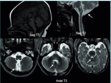
MR showed: “T1-D1 hypointense, T2/Flair hyperin-tense images in difusion sequence at the dorsal medulla oblongata and pons, with involvement of VI cranial pair” (Figure 1). Hyperintense lesion was seen in the difusion sequence, afecting the corpus callosum. hese alterations were compatible with the Dorsal Brainstem Syndrome (DBSS), a condition occurring after hypoxic-ischemic injury in neo-nates and infants. he pattern of brain injury relates to the pathogenetic mechanism and provides information about outcomes and prognosis.
Two weeks after admission, vasoactive drugs started being removed, the cardiovascular function had improved, and CSF was normal. On week 3, we started weaning the patient from MV. However, the procedure was not successful and pCO2 would raise every time we tried to wean MV. Hence, the patient
Figure 1 MRI: Sagittal T1, Sagittal T2 and Axial T2 views
showing well-deined hypointense (T1) and hyperintense
Colleti Junior J et al.
111
Rev Paul Pediatr. 2018;36(1):109-112
remained dependent of MV with no improvement in respi-ratory drive, although his neurological status improved and he started moving the limbs, doing eye contact, and reacting to painful stimuli. Tracheostomy and gastrostomy were per-formed at this time.
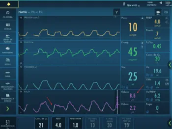
Since the MV weaning was challenging and the medical staf were in doubt about neural conduction integrity, we decided to use neurally-adjusted ventilatory assist (NAVA). After adequate eletrode positioning, Edi signal was detected (8 mV) and conirmed neural conduction (Figure 2). After that, a program of MV weaning using NAVA was proposed; each day the patient remained four hours in NAVA, switching to pressure support for another four hours and synchronized intermitent mandatory ventilation (SIMV) during the night. he patient was discharged from the hospital three months after admission, and is currently in homecare, receiving neb-ulization during the day, and bilevel positive airway pressure (BiPAP) overnight.
DISCUSSION
DBSS is a rare entity but it should be considered whenever a neonate or infant develops a hypoxic-ischemic injury. Located
in the watershed area between the paramedian and circumscrib-ing branches of the basilar artery, the brainstem tegmentum is vulnerable to ischemic distress.4 According to Sugama et al., “lesions in this area can be seen as changes in signal intensity at MR in some cases”,5,6 but in other cases, they may not be visible on neuroimaging.7,8 hese can present with or without supratentorial lesions that often involve the thalamus, basal gan-glia, and periventricular white matter. MR imaging is helpful for both diagnosis and prognosis, depending on lesion exten-sion. Prompt and adequate treatment of the shock is primor-dial to prevent injury expansion, promoting a better outcome for these patients. In the case reported, despite the accurate clinical assistance and readily execution of sepsis protocol, the infant developed DBSS.
NAVA is a mode of mechanical ventilation that could help determine neural conduction in DBSS patients. It is a safe method of ventilation. Most studies have shown no signiicant adverse events in children cared for with NAVA, and no difer-ences in intraventricular hemorrhage or pneumothorax rates when compared to conventional ventilation.3 Besides that, in neural triggering, the electrical trigger coming from the brain through the vagal nerve stimulates diaphragm comcomitantly to the ventilator, therefore improving patient-ventilator syn-chrony, permitting breath-to-breath variability, and reducing sedation need.
We conclude that dorsal brainstem syndrome is a rare condition in infants. However, it should be considered in all young patients with hypoxic-ischemic lesions. To our knowledge, this was the irst time NAVA was used to check neural conduction in an infant with this king of neuro-logical injuries. Spreading the concept of dorsal brainstem syndrome would contribute to the recognition of similar patients and alert neurologists and pediatricians for better therapeutic approaches.
Funding
his study did not receive funding.
Conflict of interests
he authors declare no conlict of interests.
Figure 2 Ventilation SERVO-i with NAVA display showing Edi signal (arrows).
REFERENCES
1. Quattrocchi CC, Longo D, Delino LN, Cilio MR, Piersigilli F, Capua MD, et al. Dorsal Brain Stem Syndrome: MR Imaging Location of Brain Stem Tegmental Lesions in Neonates with Oral Motor Dysfunction. AJNR Am J Neuroradiol. 2010;31:1438-42.
Dorsal brainstem syndrome in an infant
112
Rev Paul Pediatr. 2018;36(1):109-112 3. Narchi H, Chedid F. Neurally adjusted ventilator assist in very
low birth weight infants: Current status. World J Methodol. 2015;5:62-7.
4. Sarnat HB. Watershed infarcts in the fetal and neonatal brainstem. An aetiology of central hypoventilation, dysphagia, Moibius syndrome and micrognathia. Eur J Paediatr Neurol. 2004;8:71-87.
5. Sugama S, Ariga M, Hoashi E, Eto Y. Brainstem cranial-nerve lesions in an infant with hypoxic cerebral injury. Pediatr Neurol. 2003;29:256-9.
6. Saito Y, Kawashima Y, Kondo A, Chikumaru Y, Matsui A, Nagata I, et al. Dysphagia-gastroesophageal reflux
complex: complications due to dysfunction of solitary tract nucleusmediated vago-vagal relex.Neuropediatrics.
2006;37:115-20.
7. Natsume J, Watanabe K, Kuno K, Hayakawa F, Hashizume Y. Clinical, neurophysiologic, and neuropathological features of an infant with brain damage of total asphyxia type (Myers). Pediatr Neurol. 1995;13:61-4.
8. Roig M, Gratacòs M, Vazquez E, Toro M, Foguet A, Ferrer I, et al. Brainstem dysgenesis: report of five patients with congenital hypotonia, multiple cranial nerve involvement, and ocular motor apraxia. Dev Med Child Neurol. 2003;45:489-93.