Current clinical approach to patients with disorders of
consciousness
ROBSON LUIS OLIVEIRADE AMORIM1, MARCIA MITIE NAGUMO2*, WELLINGSON SILVA PAIVA3, ALMIR FERREIRADE ANDRADE3,
MANOEL JACOBSEN TEIXEIRA4
1PhD – Assistant Physician of the Neurosurgical Emergency Unit, Division of Neurosurgery, Hospital das Clínicas, Faculdade de Medicina, Universidade de São Paulo (FMUSP), São Paulo, SP, Brazil 2Nurse – MSc Student at the Neurosurgical Emergency Unit, Division of Neurosurgery, Hospital das Clínicas, FMUSP, São Paulo, SP, Brazil
3Habilitation (BR: Livre-docência) – Professor of the Neurosurgical Emergency Unit, Division of Neurosurgery, Hospital das Clínicas, FMUSP, São Paulo, SP, Brazil 4Habilitation (BR: Livre-docência) – Full Professor of the Division of Neurosurgery, Hospital das Clínicas, FMUSP, São Paulo, SP, Brazil
S
UMMARYStudy conducted at Hospital das Clínicas, Faculdade de Medicina, Universidade de São Paulo (FMUSP), São Paulo, SP, Brazil
Article received: 1/28/2015
Accepted for publication: 5/4/2015
*Correspondence:
Address: Av. Dr. Enéas de Carvalho Aguiar, 255, Cerqueira César São Paulo, SP – Brazil Postal code: 05403-000 [email protected]
http://dx.doi.org/10.1590/1806-9282.62.04.377
In clinical practice, hospital admission of patients with altered level of conscious-ness, sleepy or in a non-responsive state is extremely common. This clinical con-dition requires an effective investigation and early treatment. Performing a fo-cused and objective evaluation is critical, with quality history taking and physical examination capable to locate the lesion and define conducts. Imaging and laboratory exams have played an increasingly important role in supporting clinical research. In this review, the main types of changes in consciousness are discussed as well as the essential points that should be evaluated in the clinical management of these patients.
Keywords: coma, neurology, emergency medicine, state of consciousness.
I
NTRODUCTIONConsciousness is the individual’s ability to have perception of themselves and their surrounding environment. It is im-portant to distinguish the level and the content of con-sciousness. The first refers to the degree of alertness and the second is related to the quality and coherence of the indi-vidual’s thought, cognition and attitude. To be conscious one must be awake; however, being awake does not neces-sarily mean that one is conscious. Anatomically and func-tionally, consciousness depends on the interaction and ac-tivity of the cerebral cortex, brainstem and thalamus.¹ The activity of these three brain regions has fundamental im-portance on an individual’s capacity to open their eyes, re-spond to verbal and motor commands and also be aware of themselves and the environment through the interrelation of the five senses (sight, smell, touch, taste, and hearing).
The most common causes of brain injury are trau-matic, ischemic/hemorrhagic and metabolic (drugs, ex-cess insulin, diabetes, alcohol)2 in nature.
Altered levels of conscience can range from temporo-spatial disorientation to a state of deep coma.3 Therefore,
know how to correctly classify the patient’s type of alter-ation is as important as defining appropriate medical conduct for the clinical symptoms.
The different stages that may precede coma will be described below. Drowsiness or lethargy is considered a state of decreased level of consciousness in which the pa-tient can be woken up with mild stimuli and obey com-mands. In the state of torpor, the patient wakes up after mechanical stimulation, responds to simple requests, has self-psychic orientation, while generally suffering allo-psychic disorientation.4 A patient with clouding of
con-sciousness oscillates between states of drowsiness and ag-itation. Stupor is considered a deeper state of drowsiness in that the individual needs to receive vigorous and re-peated stimuli to be awakened.
his-tory of recent trauma, personal hishis-tory, life habits and medications in use are valuable for clinical management.3
Among the states that are characterized by altered content of consciousness are delirium and dementia. De-lirium is characterized by disorientation, attention defi-cit, a feeling of fear, irritability and changes in the per-ception of sensory stimuli, such as visual hallucinations.2
In dementia, there is the progressive and permanent loss of cognitive function over months or years, without chang-es in alertnchang-ess or level of consciousnchang-ess.
The following behavioral states can be commonly confused with coma: persistent vegetative state, minimal-ly conscious state, abulia, catatonia, locked-in syndrome, akinetic mutism and psychogenic passivity (Table 1). The first two can be states subsequent to coma.
TABLE 1 Types of consciousness disorder; -(absent), +(present); UWS: unresponsive wakefulness syndrome.
State Alert Consciousness
Coma -
-Persistent vegetative state/UWS +++ -Minimally conscious state +++ +
Delirium ++ ++
Abulia ++ ++
Catatonia ++ +
Akinetic mutism +++ +
Locked in syndrome +++ +++ Psychogenic passivity +++ +++
The clinical definition of the vegetative state, most recent-ly described as unresponsive wakefulness syndrome (UWS)5,8,10 the clinical condition of complete
unconscious-ness of self and the environment. However, the sleep-wake cycle is maintained and the autonomic functions of the hypothalamus and brainstem are completely or partially preserved.6,8 The patient usually presents reflexes, eye
opening and spontaneous breathing and may perform spontaneous movements like chewing and swallowing, emitting unintelligible sounds, and demonstrating cer-tain spontaneous reactions such as smiling and crying. The main causes are TBI and cardiorespiratory arrest. These symptoms must be present for more than 4 weeks
after the event that led to brain injury.6
In a minimally conscious state, the patient may pos-sess some degree of consciousness, obey verbal commands sporadically, and try to communicate; however, this is most often unintelligible, and they may cry or smile in response to affective stimuli, as well as track moving ob-jects, voices or people.
Abulia is a behavior in which there is serious indiffer-ence with a reduction or absindiffer-ence of emotional or mental behavior, in which the patient does not speak or move spontaneously, although alert and recognizing stimuli from the environment.3 This usually occurs in patients
with bilateral frontal lesions. Akinetic mutism presents symptoms similar to those of abulia but less severe; the pa-tient shows an unwillingness/extreme difficulty to move or speak, with the level and content of consciousness pre-served, and eyes following the observer or sound although the patient does not obey commands. Muscle tone and re-flexes are usually intact. In the case of locked-in syndrome, the patient also has the level and content of consciousness preserved; however, the patient presents complete paraly-sis, preventing any type of movement or verbal communi-cation. In some cases, eye movement may be present. This syndrome usually occurs in patients with basilar artery thrombosis and ischemic infarction of the base of the pons and must be differentiated from coma and persistent veg-etative state. This condition may show symptoms similar to those of acute polyneuropathies, myasthenia gravis and acute use of neuromuscular blockers. Advances in the field of neuroimaging suggest a new diagnosis: Functional locked-in syndrome. In this condition, patients do not show any behavioral signs of consciousness and differ from patients who are in a vegetative state as only examinations such as functional magnetic resonance imaging, positron emission tomography or evoked potential tests are able to identify responses that suggest some degree of conscious-ness.10 Catatonia is a state in which the individual may
re-main mute and with a marked decrease of motor activity, usually associated with psychiatric symptoms, but it can also occur due to metabolic disorders or induced by drugs.3
The patient exhibits bizarre and repetitive behavior, pos-ture disturbances and rigidity.
Psychogenic passivity is also associated with psychi-atric conditions and includes preserved muscular tone, resistance to passive movement of the limbs, resistance to opening of the eyelids or forcibly closed eye, eyes fo-cused on the ground regardless of the side on which they are lying, or the presence of non-epileptic seizures.
H
ISTORY AND CLINICAL EXAMINATIONThe identification of the cause of the coma must be ini-tiated by gathering information from family members and people who may have witnessed the clinical progres-sion of the patient. It is very important to obtain infor-mation with respect to the start and progression of symp-toms (sudden or gradual), life habits (possible use of drugs or toxic substances), presence of fever, history of trauma, previous symptoms or diseases, personal and psychiatric history and medication in use.
The general clinical examination should seek evidence of systemic conditions that could lead to changes in the level of consciousness. Vital signs and the examination of the cardiovascular, pulmonary and gastrointestinal systems, skin and other systems may provide important data regarding the etiology of the coma.
The approach should be carried out in a systematic manner in order for it to be concise and efficient: check-ing if the airways are patent; checkcheck-ing if breathcheck-ing is pro-viding adequate oxygenation (observing the respiratory pattern, oxygen saturation). Oxygen should be provided in cases of hypoxemia or establishing airway and ventila-tory assistance if the GCS score is lower than nine points. Blood pressure should be measured, verifying the heart rate and obtaining intravenous access, as well as checking blood glucose to exclude the suspicion of hypoglycemia. Signs of trauma, evidence of recent epilepsy crises (tongue lesions, sphincter release) should be sought, evaluating the skin, ophthalmoscopy, evidence of exogenous poisoning (needle marks suggesting drug use), evaluation of the breath, presence of neck stiffness, and temperature assessment.
Fever is most common in systemic infections such as pneumonia, bacterial meningitis or viral encephalitis. An excessively high temperature is related to burns and poi-sonings by drugs with anticholinergic effects. Hypother-mia can occur in intoxicated patients, those in shock, in barbiturate poisoning and myxedema.
An abnormally low pulse suggests a heart block by medications such as tricyclic antidepressants or anticon-vulsants. Severe hypertension is observed in intracranial hemorrhage and hypertensive encephalopathy.
Cherry-red skin coloration is typical of carbon mon-oxide poisoning. Abrasions, epistaxis and otorrhagia, and hematoma in the cephalic region suggest a traumatic cause of coma. Hyperemia of the face and conjunctiva is a common finding in alcoholics. Maculopapular rash sug-gests meningococcemia, staphylococcal endocarditis and typhoid fever. Excessive sweating is found in hypoglyce-mic patients or those in shock. Excessively dry skin is found in diabetic ketoacidosis and uremia.
The simple assessment of breath odor can provide the diagnosis of the cause of the coma. The odor of alco-hol is easily recognized and in patients with symptoms such as uremia, hepatic coma, diabetic ketoacidosis and cyanide poisoning, typical odors can be distinguished by an acute sense of smell.
N
EUROLOGICAL EXAMINATIONThe most important data from the neurological exami-nation for the location and prognosis include: level of consciousness, size and pupillary response to light, spon-taneous or reflex ocular motility, skeletal motor response and breathing pattern.
Level of consciousness
The assessment of the level of consciousness should in-clude a description of the patient’s alertness, and response to verbal and painful stimuli. The goal is to determine the degree of alteration to the level of consciousness and have a clinical parameter for evolution and prognosis. It should be carried out serially and following similar crite-ria and standards among the examiners for comparative purposes. It should begin with verbal stimulus and in the absence of a response, followed by painful stimulus. The painful stimulus can be applied to the supraorbital re-gion, nail bed or sternum. Asymmetric motor responses are suggestive of focal hemispheric damage.
The GCS is a standardized scale used to assess the level of consciousness originally designed for patients suffering traumatic injury, which, however, can also be used to assess any disturbance of consciousness in the acute phase.
Pupil size and reactivity to light
Pupil reactions have fundamental importance in the ex-amination as well as the shape, size and symmetry pre-sented by the pupils. A unilateral pupillary increase (>5.5 mm) is an early indicator of compression or stretching of the oculomotor nerve (cranial nerve III) as a side effect of an expansive unilateral process. Initially, there is a de-crease in reaction to unilateral light.
parasympathetic pathways, the effect on pupil size, mio-sis or mydriamio-sis, will depend on the action of the system affected the least or still intact. The pupillary reflex con-sists of the pupillary contraction after light stimulation via the parasympathetic pathway.
As the areas related to consciousness control are ad-jacent to these pathways, pupil changes can help us in the differentiation and location of possible causes of coma. Hypothalamic lesions, especially in the posterior and ventrolateral region, can produce ipsilateral pupillary constriction, usually associated with ptosis and anhidro-sis (Horner syndrome). Thalamus lesions can lead to small, reactive pupils, known as diencephalic pupils.
Mesencephalic lesions can produce three types of pu-pil changes depending on the location of the damage: dorsal tectal region lesions, lesions that disrupt pupillary reaction to light, pupils with medium or little dilation (5 to 6 mm), and fixed pupils with preservation of the ac-commodation reflex. Pupillary size oscillations may oc-cur (hippus) alongside maintenance of the ciliospinal re-flex; mesencephalic nuclear lesions, usually affect both the sympathetic and parasympathetic pathways, leading to medium-fixed pupils (4 to 5 mm), generally a little ir-regular, and generally related to transtentorial hernia-tion; lesions of bilateral cranial nerve III lead to paralyt-ic mydriasis, usually associated with uncal herniation. Pontine lesions lead to extreme bilateral miosis (<1 mm), with preservation of the pupillary light reflex, which is very hard to perceive. Pinpoint pupils due to pontine hem-orrhages may result from sympathetic pathway lesions and irritation of parasympathetic pathways.
In patients in a coma related to toxic-metabolic chang-es, in general the pupils are isochoric and reactive to light, except in the terminal stage. The actions of drugs wheth-er local or systemic can lead to pupil changes due to their effect on the sympathetic or parasympathetic pathways. Anticholinergic drugs such as atropine or scopolamine, can lead to pupillary dilation. Opiates (heroin and mor-phine) can lead to miosis, similar to pontine hemorrhage. Barbiturates may lead to fixed pupils. Data from the clin-ical history and other data from the neurologclin-ical exam can help us in the differentiation of these cases. Post-ic-tal patients usually present mydriatic but reactive pupils.
Ocular motility
Ocular motility depends on the integrity of structures lo-cated in the brain, cerebellum and brainstem. Since we cannot evaluate voluntary ocular motility in unrespon-sive patients, we must evaluate the integrity of reflex path-ways located in the brainstem. The absence of changes in
ocular motility means that the region located between the vestibular nuclei, in the medulla oblongata-pons junction, up to the oculomotor nuclei in the midbrain are intact.
In coma patients, we must evaluate three aspects with respect to ocular motility: (1) primary gaze at rest, to as-sess the presence of deviations; (2) observe the presence of spontaneous eye movements and (3) test eye movements. The presence of deviations in the rest position can provide us with evidence of cranial nerve palsies. Conju-gate deviations of the lateral gaze may be the result of le-sions from the cortex to the contralateral pontine retic-ular formation, which is common in strokes. Disconjugate deviations of the lateral gaze may reflect abducens and oculomotor nerve paralysis, or internuclear ophthalmo-plegia. The presence of downward eye deviations can mean brainstem lesions, compression of the mesencephalic tec-tum, which may be present in hydrocephalus. Thalamic and subthalamic lesions can lead to conjugate deviation both upward and downward. Other conditions that can lead to upward deviation of the eyes are sleep, epileptic seizure, syncope, apnea in Cheyne-Stokes respiration, bleeding in the cerebellar vermis, ischemia or brainstem encephalitis. Skew deviation is a vertical misalignment of the eyes, usually corresponding to brainstem or cerebel-lar lesions.
The observation of spontaneous eye movements can offer important information. The presence of roving movements, consisting of slow and spontaneous conju-gate movements of the lateral gaze, can indicate the in-tegrity of the oculomotor pathways and their connec-tions. The presence of nystagmus in comatose patients can be indicative of an irritative supratentorial focus. Oc-ular bobbing is characterized by fast conjugate move-ments downwards followed by a slow return to the pri-mary position, occurring generally in brainstem lesions, especially in the pons.
The evaluation of reflex eye movements can be done through the oculo-cephalic maneuver or the caloric test (vestibular-ocular). The oculo-cephalic maneuver is car-ried out by vertical or lateral rotation of the head and ob-servation of the eye movement. This test is not conduct-ed in patients with suspicion of trauma, due to the possibility of cervical lesion. In patients where this reflex is preserved, the eyes move in unison and in the opposite direction to the movement of the head.
conju-gate gaze to the stimulated side and a fast corrective move-ment or nystagmus in the contralateral direction. Loss of the fast phase is observed in comatose patients. This is a good examination in cases of suspected psychogenic or-igin of the coma. Obviously, a brainstem injury may lead to absent response by interrupting this reflex.
Through these maneuvers we can observe the integ-rity of oculomotor pathways in the brainstem and clear-ly show the palsy of isolated cranial nerves. The absence of a bilateral reflex response can indicate the presence of extensive lesions in the brainstem.
Motor response
The motor examination of comatose patients is carried out initially through observation of posture at rest, the presence of spontaneous movement or response to ver-bal or painful stimuli. The motor response should be an-alyzed in comparison to the opposite side and after sym-metrical stimulation, in the four limbs.
Head and eye deviation to one side and contralater-al hemiplegia indicate supratentoricontralater-al lesion, while devi-ation to the same side of the hemiplegia may indicate a brainstem lesion. External rotation of the lower limb may be indicative of hemiplegia or hip dislocation.
Decerebrate posturing consists of bilateral extension of the lower limbs and adduction and internal rotation of the shoulders and extension of the elbows and wrists. This generally means a bilateral mesencephalic and pons lesion but can appear in severe metabolic encephalopa-thies and supratentorial lesions involving the bilateral corticospinal tract. Decorticate posturing consists of bending the elbows and wrists, adduction of the shoul-ders and extension of the lower limbs. Although not a posture with a good topographic correlation, it usually indicates injury above the brainstem. These abnormal postures can be observed spontaneously or after painful stimuli, and their presence might suggest a brainstem herniation syndrome.
Other movements that can be observed are: tonic-clonic seizures in epileptic seizures; myoclonus, observed frequently in post-anoxic encephalopathy and other met-abolic comas; reflex responses, such as the triple flexion response of the lower limbs and the plantar reflex (Babin-ski sign).
Respiratory pattern
In comatose patients, the respiratory pattern can help us with the topographic diagnosis of the lesion. These may include: Cheyne-Stokes respiration (bilateral hemi-spheric lesions, severe heart failure), central neurogenic
hyperventilation (brainstem lesions), apneusis (lesions in the dorsal-medial region of the lower half of the pons), Biot’s respiration (lower pontine tegmental lesion) and ataxic respiration (generally damage to the medulla ob-longata).
L
ABORATORY/
IMAGING EXAMSThe requested exams should include blood tests, electro-lytes and biochemistry (sodium, calcium, phosphorus, magnesium, chlorine, glucose), renal, hepatic and thyroid functions, coagulogram, and blood gas. In selected cases, quantify the serum level of certain drugs and toxicolog-ical screening.
If there is suspicion of poisoning, aspiration and anal-ysis of gastric contents can contribute to the diagnosis. According to the availability of the service, the serum
con-centration of anticonvulsants, opioids, diazepinic agents, barbiturates, alcohol and a large number of toxic sub-stances can be measured.
White blood cell count can identify neutrophilic leu-kocytosis, present in bacterial infections such as menin-gitis, where it is usually greater than 12,000/mm3.
In some cases thyroid hormones and serum cortisol may be requested, such as in suspected myxedema coma and Addisonian crisis, respectively. One must mind that water and sodium disorders such as hypo- or hypernatre-mia can be results of brain disease that will directly or in-directly affect the hypothalamic-pituitary axis.
Head tomography
This is essential upon suspicion of intracranial lesion and must be the first image exam requested. It is performed in a few seconds, is available in most emergency services and has good sensitivity to detect bleeding (subarachnoid hemorrhage, subdural hematoma, epidural hematoma, or intraparenchymal hematoma), hydrocephalus, tumors and extensive brain infarcts.6
brain-stem is a real possibility in the presence of intracranial hypertension. Structural causes of coma may be related to subtle changes or even a normal CT scan (e.g. unilat-eral chronic isodense subdural hematomas, subarach-noid hemorrhage, deep vein thrombosis, herpes simplex encephalitis, and meningitis).
Cerebrospinal luid
Lumbar puncture is carried out upon suspicion of infec-tion of the central nervous system or on suspicion of sub-arachnoid hemorrhage with a normal head CT scan.
Magnetic resonance imaging
This is not an examination carried out routinely in the emergency department. However, it may provide valuable information on the number, characteristics and severity of the lesions,10 as well as allow the identification of
in-juries not visible to the head CT, as in cases of herpes sim-plex encephalitis or diffuse axonal injury.7 Functional
magnetic resonance imaging and positron emission to-mography (PET-SCAN) have proved promising and can help in the differential diagnosis of patients in a mini-mally conscious or vegetative state.9
The latest technologies include diffusion-weighted im-aging (DWI) and diffusion-tensor imim-aging (DTI), which are tests using methods similar to MRI, but with a more advanced technology. They provide in vivo images of
bio-logical tissues, and allow the observation of molecular wa-ter displacement as well as more accurate data about dam-age occurring in specific structures of the nervous system.10
Electroencephalogram (EEG)
This is most recommended upon suspicion of non-con-vulsive seizures, especially in patients with worsening of the level of consciousness that have a known underlying neurological disease. Some studies have shown promis-ing results in the integration of transcranial magnetic stimulation (TMS) with simultaneous EEG for assess-ment of brain connectivity and cortical interactions.11,12
T
REATMENT OF COMATOSE PATIENTSThe initial care of a comatose patient, the most serious altered state of consciousness, should emphasize basic precautions to maintain the airways and adequate venti-lation, hemodynamic stability and other measures to min-imize damage to the brain and other vital organs. We should exclude causes requiring urgent surgical interven-tion and medical causes that need immediate treatment. In general, the patient has no ability to protect the airways and the presence of hypoxia or hypoventilation
requires endotracheal intubation. At the same time, blood is collected for determination of glucose, electrolytes and renal and liver function tests. In patients with hypogly-cemia, infusion of 25 to 50 mL of 50% glucose should al-ways be supplemented with thiamine.
Gastric aspiration and lavage with saline solution can be a diagnostic and therapeutic measure upon suspicion of coma due to ingestion of drugs. Salicylates, opiates and anticholinergic drugs induce gastric atony and can be removed several hours after ingestion. Caustic mate-rials should not be washed out because of the risk of gas-trointestinal perforation. The administration of activat-ed charcoal is indicatactivat-ed in certain cases of poisoning. This is not effective in poisoning caused by heavy metals, cya-nides and alcohol.
In patients with intracranial hypertension that are getting worse neurologically (worsening of the GCS score, anisocoria, decerebrate or decorticate posturing), proceed with rapid infusion of 20% 1g/kg mannitol or hyperton-ic saline solution (NaCl 20% 1 mL/kg), associated with hyperventilation in order to maintain pCO2 at around 30
mmHg. In lesions with expansive effects suggestive of brain tumors, a bolus of 20 mg dexamethasone may be given. In selected cases, intracranial pressure monitoring may be appropriate. Lumbar puncture should be per-formed upon suspicion of meningitis and preferably af-ter a CT scan of the head.
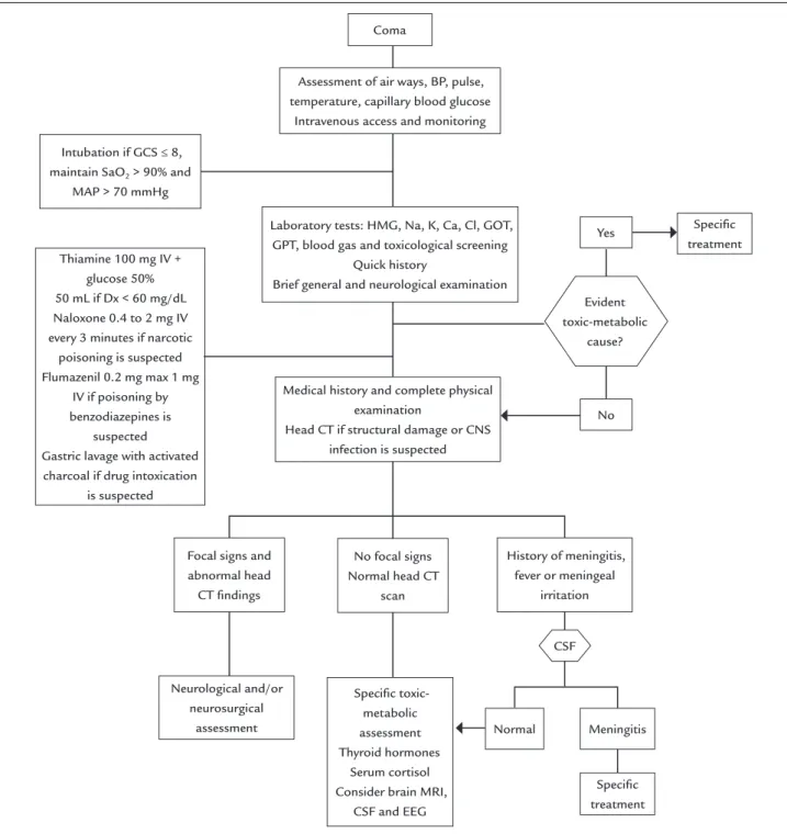
Extreme hyperthermia or hypothermia must be avoid-ed. To prevent bladder distension, a catheter should be inserted. Aspiration pneumonia is prevented by proper positioning, restriction of oral fluids, gastric tube and orotracheal tube. As comatose patients have an increased risk of deep vein thrombosis, elastic compression stock-ings in the lower limbs and subcutaneous application of unfractionated heparin 5000u every eight hours are to be used (Figure 1).
C
ONCLUSIONFIGURE 1 Algorithm of the initial approach to the patient in a coma.
BP: blood pressure; GCS: Glasgow Coma Scale; MAP: mean arterial pressure; HMG-CoA: 3-hydroxy-3-methyl-glutaryl coenzyme A; GOT: glutamic-oxaloacetic transaminase; GPT: glutamate-pyru-vate transaminase; CT: computed tomography; CNS: central nervous system; CSF: cerebrospinal luid; MRI: magnetic resonance imaging; EEG: electroencephalogram.
Yes
No
Normal
Speciic treatment
Speciic treatment Thiamine 100 mg IV +
glucose 50% 50 mL if Dx < 60 mg/dL Naloxone 0.4 to 2 mg IV every 3 minutes if narcotic
poisoning is suspected Flumazenil 0.2 mg max 1 mg
IV if poisoning by benzodiazepines is
suspected Gastric lavage with activated charcoal if drug intoxication
is suspected Intubation if GCS ≤ 8, maintain SaO2 > 90% and
MAP > 70 mmHg
Coma
Assessment of air ways, BP, pulse, temperature, capillary blood glucose Intravenous access and monitoring
Laboratory tests: HMG, Na, K, Ca, Cl, GOT, GPT, blood gas and toxicological screening
Quick history
Brief general and neurological examination
Speciic toxic-metabolic assessment Thyroid hormones
Serum cortisol Consider brain MRI,
CSF and EEG
Medical history and complete physical examination
Head CT if structural damage or CNS infection is suspected
CSF Focal signs and
abnormal head CT indings
Neurological and/or neurosurgical
assessment
History of meningitis, fever or meningeal
irritation No focal signs
Normal head CT scan
Meningitis Evident toxic-metabolic
cause?
that could be essential for localization and etiology of the patient’s symptoms. Basic laboratory investigations and imaging tests are of utmost importance for differential diagnosis.
The prognosis is extremely variable and depends on the cause, duration and extent of the affected site. How-ever, effective initial care and introduction of proper
treat-ment are crucial in order to prevent the occurrence of sec-ondary damage and to speed up patient recovery.
R
ESUMONa prática clínica é extremamente comum a admissão hospitalar de pacientes com nível de consciência alterado, sonolentos ou em estado não responsivo. Essa condição clínica demanda uma investigação eficaz e um tratamen-to precoce. É fundamental a realização de uma avaliação focada e objetiva, com a realização de anamnese e exame físico de qualidade para localizar a lesão e definir condu-tas. Exames de imagem e laboratoriais têm desempenha-do papéis cada vez mais relevantes no suporte à investi-gação clínica. Nesta revisão, são discutidos os principais tipos de alterações de consciência e os pontos imprescin-díveis que devem ser avaliados na abordagem clínica des-ses pacientes.
Palavras-chave: coma, neurologia, emergências, estado de consciência.
R
EFERENCES1. Gosseries O, Vanhaudenhuyse A, Bruno MA, Demertzi A, Schnakers C, Boly MM, et al. Disorders of consciousness: coma, vegetative and minimally conscious states. In: Cvetkovic D, Cosic I (eds.). States of consciousness – Experimental insights into meditation, waking, sleeping and dreams. The Frontiers Collection. Berlin: Springer, 2011. p.29-55.
2. Bonsignore LT, Macrì S, Orsi P, Chiarotti F, Alleva E. Coma and vegetative states: state of the art and proposal of a novel approach combining existing coma scales. Ann Ist Super Sanita. 2014; 50(3):241-8.
3. Andrade AF, Carvalho RC, Amorim RLO, Paiva WS, Figueiredo EG, Teixeira MJ. Coma e outros estados de consciência. Rev Med (São Paulo). 2007; 86(3):123-31.
4. Shinosaki JSM, Baiense RF. Manual de Neurologia. Manual do Residente da Universidade Federal de São Paulo – UNIFESP. São Paulo: Roca, 2009.
5. Von Wild K, Laureys ST, Gerstenbrand F, Dolce G, Onose G. The vegetative state – a syndrome in search of a name. J Med Life. 2012; 5(1):3-15. 6. Practice parameters: assessment and management of patients in the
persistent vegetative state (summary statement). The Quality Standards Subcommittee of the American Academy of Neurology. Neurology. 1995; 45(5):1015-8.
7. Moore SA, Wijdicks EF. The acutely comatose patient: clinical approach and diagnosis. Semin Neurol. 2013; 33(2):110-20.
8. Laureys S, Celesia GG, Cohadon F, Lavrijsen J, León-Carrión J, Sannita WG, et al.; European Task Force on Disorders of Consciousness. Unresponsive wakefulness syndrome: a new name for the vegetative state or apallic syndrome. BMC Med. 2010; 8:68.
9. Stender J, Gosseries O, Bruno MA, Charland-Verville V, Vanhaudenhuyse A, Demertzi A, et al. Diagnostic precision of PET imaging and functional MRI in disorders of consciousness: a clinical validation study. Lancet. 2014; 384(9942):514-22.
10. Bodart O, Laureys S, Grosseries O. Coma and disorders of consciousness: scientific advances and practical considerations for clinicians. Semin Neurol. 2013; 33(2):83-90.
11. Di Perri C, Thibaut A, Heine L, Soddu A, Demertzi A, Laureys S. Measuring consciousness in coma and related states. World J Radiol. 2014; 6(8):589-97. 12. Ilmoniemi, RJ, Kicic D. Methodology for combined TMS and EEG. Brain