* Post-graduation program in Human Movement Sciences, Study Group in Biochemistry and Exercise Physiology, Exercise Research Laborato-ry – Physical Education School, UFRGS, Porto Alegre, Brazil.
Received in 21/3/04. 2nd version received in 22/5/04. Approved in 25/5/04. Correspondence to: Rua Felizardo, 750 – 90690-200 – Porto Alegre, RS. E-mail: [email protected]/[email protected]
Oxygen free radicals and exercise: mechanisms
of synthesis and adaptation to the physical training
*
Cláudia Dornelles Schneider and Alvaro Reischak de OliveiraR
EVIEWA
RTICLEKey words: Exercise. Oxygen free radicals oxidative stress. Antioxidants. Training. ENGLISH VERSION
ABSTRACT
The interest about the mechanisms of generation and adapta-tion of oxygen free radicals (OFR) to exercise has increased signif-icantly from the demonstration of its relation with the oxygen in-take. The OFR are formed through the incomplete reduction of oxygen, generating species presenting high reactivity to other bio-molecules, especially lipids and proteins of the cell membranes and even DNA. The injuries caused by the oxidative stress present accumulative effects, being related to several diseases such as cancer, arteriosclerosis and diabetes. The acute physical exercise furthers the increase on the formation of OFR in function of the increment on the oxygen intake. However, the physical training generates adaptations able to soften the harmful effects caused by OFR. These adaptations are related to several systems, among which the most important are the enzymatic system, composed by the superoxide dysmutase, catalase and glutathione peroxidase; and the non-enzymatic system, composed by the ceruloplasmine, the sexual hormones, co-enzyme Q, uric acid, thermal shock pro-teins, among others. Such adaptations, despite the controversies about the mechanisms involved, further a higher tissue resistance and oxidative challenges such as those provided by long-duration high intensity exercises. The evaluations techniques of the oxida-tive stress, most times are not able to detect injuries in short-dura-tion exercises. Thus, studies of physical efforts performed for long periods or until exhaustion have been conducted. New lesion mark-ers by OFR action have been discovered and new techniques for its determination have been created. The objective of this work is discuss the formation mechanisms of OFR and the adaptations to the chronic oxidative stress caused by physical training.
INTRODUCTION
The increase on the oxygen intake as well as the activation of specific metabolic paths during physical exercise results in the for-mation of oxygen free radicals, substances simply known as free
radicals(1-3). These molecules are increased in high-intensity(4,5) and
exhausting(6) exercises and from the 80’s decade were related to a
large number of diseases such as pulmonary emphysema,
inflam-matory diseases, arteriosclerosis, cancer and aging(7,8). On the
oth-er hand, it is known that psychical activity is a source of stress, and the chronic exposition to this source of stress, called as phys-ical training, is able to release adaptations in responses to a higher production of these free radicals. In this context, the newest stud-ies establish the role of the physical activity in the prevention and control of several diseases such as colon cancer and possibly breast
and prostate cancers(9), diabetes and hypertension(10), dislipidemy
and arteriosclerosis(11), among others. This paper provides a
review-ing with regard to the mechanisms of the free radicals generation through exercise as well as the adaptive processes and the re-spective consequences induced by physical training.
FREE RADICALS
The oxygen free radicals (OFR) are naturally produced in our or-ganism through oxidative metabolic processes, many times being extremely useful as in situations where the activation of the im-munologic system is required (for example, macrophages use the hydrogen peroxide to destroy bacteria and other strange elements); in drugs detoxification and in the production of the endothelium-derived relaxing factor, the nitric oxide, extremely important in
pro-cesses that unchain the relaxation of the blood vessels(12).
According to Halliwell(1), the oxygen (O
2) that we breathe is
me-tabolized in our organism as follows: approximately 85 to 90% is used by mitochondria through the electrons transportation chain and the 10 to 15% remaining is used by several oxidase and deox-idase enzymes as well as by direct oxidation chemical reactions. At the terminal part of the electrons transportation chain, the en-zyme cytochrome oxidase (reaction 1) removes one electron from each one of the four cytochrome reduced molecules, oxidizing them
and add the four electrons to the O2 in order to form water (around
95 to 98% from the 85 to 90% mentioned above). The 2 to 5% remaining is univalently reduced into metabolites called reactive species of oxygen.
Reaction 1 – tetravalent oxygen reduction
O2 + 4e- + 4H+→ 2 H
2O + energy
FORMATION OF REACTIVE OXYGEN SPECIES
Due to the electronic configuration, the oxygen presents strong tendency of receiving one electron at the time. The univalent con-version of oxygen into water is given as follows:
(a) The addition of one electron to one oxygen molecule in its fundamental state generates the formation of the superoxide
rad-ical (O2•-) (reaction 2).
Reaction 2: O2 + e-→ O
2
•-(b) The superoxide that receives more than one electron and
two hydrogen ions forms hydrogen peroxide (H2O2) through the
process called as dismutation(13). This reaction is catalyzed by the
enzyme superoxide dismutase (SOD) found in high amounts in the
mammals cells and accelerates the reaction up to 104 times the
frequency for spontaneous dismutation in physiological pH (reac-tion 3).
Reaction 3: 2 O2•- + 2H+
→
SOD
H2O2
(c) When H2O2 receives more than one electron and one
hydro-gen ion, the hydroxyl radical is formed (OH•), which is the most
any near cellular structure, thus influencing enzymes, membranes
or nucleic acids(6).
The hydroxyl radical may be formed when H2O2 reacts with iron
or copper ions (reaction 4). This reaction is known as Fenton Reac-tion.
Reaction 4: Fe2+/Cu+ + H
2O2→ OH
• + OH- + Fe3+/Cu2+
The ions of transition metals may as well catalyze the reaction
between H2O2 and superoxide, leading to the production of
hy-droxyl radical (reaction 5), the called Haber-Weiss Reaction.
Reaction 5: H2O2 + O2
•-
→
Fe/CuOH• + OH- + O
2
The superoxide and hydroxyl radicals present unpaired electrons in its outer orbit and, therefore, called as free radicals. The hydro-gen peroxide is not a free radical, however, it represents a partly reduced oxygen metabolite. Other reactive species of interest are the singlet oxygen, which are spin-altered forms of oxygen. These oxygen-derived metabolites, if considered as a whole, are called as oxygen reactive species (ERO) in function of their increased
reactivity with regard to biomolecules(14), that generally change the
size and shape of compounds that they interact with.
Besides, the superoxide radical may react directly with the nitric oxide (NO). A free radical centralized in the nitrogen, generating peroxinitrite. This compound may lead to the formation of an oxi-dant agent with hydroxyl radical features (reaction 6).
Reaction 6: O2•- + NO → ONOO-→ ONOO- + H+→ OH•
Each ERO has its own characteristics, showing different
reac-tivity and half-life times(13,15).
OXIDATIVE STRESS
The term oxidative stress is used under circumstances in which the “challenge” for free radicals results in tissue damage or in the production of compounds toxic or harmful to tissues. One may say that an organism is found under oxidative stress (OE) when an unbalance between the pro-oxidant and antioxidant system occur
in such way that the pro-oxidant system prevail(16). One of the main
lesion mechanisms is the lipoperoxidation (LPO), in other words, the oxidation of the cellular membrane lipidic layer. Besides, the EO may generate damage to protein and DNA, causing several alterations in the cellular function and consequently to tissue.
ANTIOXIDANT DEFENSE
As the ERO are continuously formed in small amounts by the metabolism regular processes, all cells present mechanisms to soften their harmful effects. It is worthy emphasizing that the com-position of the antioxidant defenses is distinguished from tissue to tissue, from type of cell to type of cell and possibly from cell to cell
of the same type in a given tissue(1).
The antioxidant defense system is divided into enzymatic and non-enzymatic. The first one included the enzymes superoxide dis-mutase (SOD), catalase (CAT) and glutathione peroxidase (GPx).
The catalase plays important role in the elimination of H2O2,
fur-thering its catalysis into water.
Reaction 7: H2O2 + H2O2
CAT →
O2 + 2H2O
The GPx also acts as mechanism of protection against the dative stress, converting the reduced glutathione (GSH) into
oxi-dized glutathione (GSSG), removing H2O2 and forming water
(reac-tion 8)(17).
Reaction 8: 2GSH + H2O2
GPx→
GSSG + 2H2O
Thus, both the CAT and the GPx avoid the accumulation of the superoxide radical and hydrogen peroxide so that the hydroxyl rad-ical is not produced, against which there is no defense enzymatic
system(13).
The perfect balance between the antioxidant enzymes (CuZn-SOD, Mn(CuZn-SOD, CAT, GPx) is of great importance for the mainte-nance of the cellular integrity.
The non-enzymatic system includes compounds synthesized by the human organism such as bilirubin, ceruloplasmine, sexual hor-mones, melatonin, co-enzyme Q, uric acid and others ingested through regular diet or through supplementation such as ascorbic
acid (vitamin C), α-tocopherol (vitamin E), β-carotene (precursor of
the vitamin A) and phenol groups of plants (flavonoids).
In studies developed in our laboratory(18,19) using hearts isolated
from rats in a coronary perfusion model (Langendorff), we demon-strated that both the vitamin A and the Trolox (vitamin E hydro soluble analogue) acted by reducing the lipoperoxidation levels and
the H2O2-induced inotropic, chronotropic and lusitropic negative
effects. This is due to the quencher capacity of the singlet oxygen of both vitamins. A quencher is a molecule that receives the exci-tation energy from the singlet oxygen to itself, leading it to the
fundamental state, becoming excited(1).
FORMATION MECHANISMS OF THE OXYGEN REACTIVE SPECIES
During muscular activity, the energy demand may exceed 35
times the rest demand(20). Thus, during the performance of the
muscular activity, the oxygen intake is greatly increased, mostly due to the increase of the muscular work. Because ERO are pro-duced through intermediate metabolism, the exercise causes in-creases on its production. As an example, suppose an adult man
with 70 kg in rest uses 3.5 mL O2.kg-1.min-1 or 352.8 L.d-1 or 14.7
mol.d-1. If 1% generates O
2•-, it means 0.147 mol. d-1 or 53.66
mol.year-1 or ≅ 1.7 kg.year-1 (of O
2•-). Now during physical exercise,
with the increase on the oxygen intake, it may increase up to ten
to fifteen times(1).
According to Viña et al.(21), the oxidative stress and muscular
damage degrees do not depend on the exercise absolute intensi-ty, but rather on the exhaustion degree of people who performs the exercise. Besides, knowing the free radicals’ formation mech-anisms with exercise is important in order to avoid the oxidative stress and the damage associated with exhaustive physical activi-ty. The mechanisms are the following:
(1) temporary interruptions of the calcium-dependent (Ca++) ATP
pumps lead to increases on the calcium intracellular concentra-tions, what may activate the path of the xanthine oxidase during exercises. Increased concentrations of intramuscular calcium dur-ing periods of high intensity exercises may activate the calcium-dependent proteases, which convert xanthine dehydrogenase into xanthine oxidase. The xanthine oxidase uses the molecular
oxy-gen instead of NAD+ as electrons acceptor, thus generating the
superoxide radical;
(2) periods of intense exercise may increase the oxidative stress due to the temporary hypoxia and re-oxygenation occurring in the exercised muscle in function of the contractions and relaxations cyclically established. During contraction, the vascular compres-sion establishes an ischemia condition and, therefore, hypoxia. In relaxation, the reperfusion occurs and consequently re-oxygenation. Under hypoxia condition, the reduced equivalent may accumulate within the mitochondria electrons transportation chain, resulting in a phenomenon known as reductive stress. In the re-oxygenation a burst of mono-electronic reductions may convert molecular oxy-gen into superoxide radical;
(4) increases on the Ca++ concentration may activate the
en-zyme phospholipase A2, which releases the arachidonic acid from
the phospholipids. The ciclooxygenase reacts with the arachidonic acid to form the hydroxyl radical;
(5) hypoxic conditions have also been shown in the increase on the nitric oxide synthase activity (NOS), leading to the formation of nitric oxide radicals. These radicals may perform a weak pro-oxi-dant effect by themselves or to combine with the superoxide to
form a more powerful oxidant agent, the peroxinitrite(22), as already
demonstrated (reaction 6).
Thus, during the aerobic metabolism, the possibility of oxidative lesions to occur in the tissues will depend on a precise balance between the generation of oxygen radicals and the efficiency of the antioxidant mechanisms.
PHYSICAL EXERCISE AND OXYGEN REACTIVE SPECIES
Several studies from the 80’s decade presented results in which repeated loads of exercise led to accelerated damage or aging of the muscle in individuals or guinea-pigs that exercised regularly.
However, Heath et al.(23), after following athletes during many years,
verified that the metabolic potential and the muscular functional
capacity of athletes were not impaired. Besides, Gutteridge et al.(24),
pointed as possibility of protective mechanism the fact of finding increments on the iron and copper levels in the sweat of athletes after exercise, speculating that the excretion of such metals in the sweat would decrease the extension of the oxidative damage caused by such metals. From these studies, the possibility that the regular exercise could further an adaptive increase of the skel-etal muscle defense mechanisms able to protect against lesions cause by ERO has been considered.
In 1982, Davies et al.(25) proposed that the formation of
exercise-induced free radicals could be the initial stimulus for the
mitochon-drial biogenesis in a chronic training situation. In this context, Ji(26)
demonstrated that in the skeletal muscle, an isolated load of ex-haustive work produced an increase on the LPO and that the activ-ity of the antioxidant enzymes glutathione reductase, GPx, SOD
and CAT was significantly increased. In the same way, Alessio(27)
showed increases on LPO in the fast and low muscular fibres of rats submitted to exercise loads, indicating an increase on the phys-ical activity-induced oxidative stress. This stress was better toler-ated by trained rats, suggesting an adaptation of the antioxidant systems.
Studying humans, Nies et al.(28), demonstrated the occurrence
of damage to DNA in the circulating leukocytes after exhaustive exercise in treadmill. For the first time it was demonstrated in trained individuals, but as the damage extension was not large, the authors suggest that the adaptation to the aerobic resistance training may reduce the EO effects such as the damage to DNA. In
the same year, the results of the work of Mills et al.(29) with
race-horses showed that the exercise may induce changes on the bio-chemical parameters indicative of the oxidative stress and that these parameters are exacerbated in the presence of high temperatures and humidity. In an interesting work involving a training overload
model, Palazzetti et al.(30) studied triathletes submitted to a work
load increment of 21% in swimming, 51% in cycling and 44% in running during four weeks. The simple fact that the athlete was submitted to training overload already caused significant elevation of urinary adrenaline and CK plasmatic activity in rest. However, the highest differences showed when the effects of a simulated duathlon (running and cycling) were evaluated. The athletes in train-ing overload condition presented higher lipoperoxidation indexes, evaluated through the level of substances reactive to the tiobarbi-turic acid (TBA-RS), CK-MB and plasmatic myoglobulin, muscular lesion markers and the drop of the GSH:GSSG relation, clearly indi-cating that this overload impairs the antioxidant defense mecha-nisms related to exercise-induced response.
Margaritis et al.(31) proposed that the improvement range of the
antioxidant defense system is dependant on the training loads. The
same authors also demonstrated the higher the VO2max in
triath-letes is, the higher the activity of the antioxidant enzyme GPx will be in the erythrocytes, protecting the organism from the damage
to the cellular membrane. Leewenburgh et al.(32) verified that the
exercise-induced oxidative stress may release adaptations in re-sponse to training and that such adaptations would be tissue-spe-cific, suggesting a complex regulatory mechanism. Besides, Leaf
et al.(33) suggest that in healthy individuals, the physical exercise
induces the lipidic peroxidation transitorily, and that the LPO prod-ucts are removed during the recovery phase. The work of Venditti
and De Meo(34) with adult rats submitted to a regular training
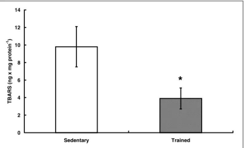
pro-gram with duration of one year, proved the hypothesis that such training lengthens the aerobic resistance capacity and increases the antioxidant defenses, thus limiting the tissue damage caused by free radicals. In the same way, we have demonstrated in our laboratory that the aerobic training in rats, performed through run-ning in treadmill walking belt, increases the myocardic capacity of
handling a perfusion challenge with H2O2, causing lower
contrac-tion and formacontrac-tion of TBA-RS(35) (figures 1 and 2). In the same
in-vestigation line, 11 weeks of aerobic training in aged rats, besides causing bradycardia and increase on the glucose/insulin index (an insulin resistance marker), was associated to a reduced response
to the H2O2-induced oxidative stress. In this study, we demonstrated
a positive correlation between basal FC and the TBA-RS levels (fig-ure 3), in other words, the higher the basal FC is, the higher the
radical lesion levels were(36). These effects may be partly explained
due to the higher SOD activity found in another study from our
group(37), where 21 days-old rats were submitted to training at 50%
Fig. 1 – Cardiac contracture (%) in different coronary perfusion times with H2O2 (256 mmolar.L-1) in trained and untrained rats
0 10 20 30 40 50 60 70 80
5 10 15 20
Time (min)
Card
iac c
o
n
tra
ctu
re (
%
)
Sedentary
Trained
*
*
Fig. 2 – TBARS in cardiac homogenized after 20 minutes of coronary perfu-sion with H2O2 (256 mmolar.L-1) in trained and untrained rats
0 2 4 6 8 10 12 14
Sedentary Trained
TBARS
(
n
g
x
m
g
pr
ot
ein
-1)
of the for 4 weeks. No increase on the TBA-RS or QL was verified in the heart of these animals, what suggests that compensatory adaptations in the tissue antioxidant system have occurred. In this
context, Ramires and Ji(38) demonstrated that the physical training
associated to the glutathione supplementation protects the heart of rats against oxidative lesions and depression of the cardiac func-tion caused by in vivo ischemia and reperfusion. The mechanism for this adaptation was the elevation on the myocardic glutathione content and on the antioxidant capacity through the increment of
the SOD, GPx, GSH reductase, and γ-glutamyl transpeptidase
ac-tivities in the myocardium.
Still using the animal model, Smolka et al.(39) analyzed the effect
of two different training protocols on the expression of HSP72 (Heat shock protein – 72 KDa), a stress protein with the function of mataining and repairing the protein conformation. This protein is in-volved in the protection of cells against different types of injuries. Some of these injuries, as the oxidative stress, thermic stress and low pH resulting from the lactate accumulation are generated dur-ing the exercise. Besides the adaptations already described such as the increment of the citrate synthase activity, catalase and mus-cular GSH reductase after training, an original finding of this study was the demonstration that the HSP72 induction caused by an exercise isolated load only occurs to the group kept inactive, sug-gesting that it acts as a complementary protective mechanism to exercise-induced oxidative stress. Furthermore, the group of ani-mals submitted to a short duration and high intensity protocol pre-sented higher susceptibility to the challenge provided by the acute exercise, if compared to the group trained through continuous
pro-tocol. In the study of Child et al.(40), observed, through the measure
of the total antioxidant capacity and uric acid in trained individuals submitted to a simulated half-marathon test, a higher scavenger ability (capacity of neutralizing free radicals forming less reactive compounds) on the free radical in the serum. Still, the exercise induced an increase on the malondialdehyde concentrations, sug-gesting that such responses were sufficient to prevent exercise-induced LPO.
Powers et al.(41), assert that the usual high-intensity training
re-quired for the elite competition level is able to increase the
antiox-idant defenses. In this context, Halliwell(1) suggests that athletes
present high ceruloplasmine concentrations in the plasma. The
ceruloplasmine is a α-globulin involved in the copper
transporta-tion and regulatransporta-tion, directly reducing oxygen without known inter-mediates, therefore participating in the extracellular antioxidant defense system.
In 2000, the study of Selamoglu et al.(42) presented adaptive
dif-ferences between aerobic and anaerobic exercises. The activity of the enzyme GPx in erythrocytes was found increased in long-dis-tance runners if compared to weight lifters. In the same way, Inal
et al.(43) analyzed the aerobic metabolism in acute swimming
exer-Fig. 3 – Correlation between FC and rest and the lipoperoxidation levels evaluated by TBARS in untrained aged rats and rats physically trained
R = 0,7 p < 0,05
0 0,2 0,4 0,6 0,8 1 1,2
275 300 325 350 375 400
HRbasal (bpm)
TBARS
(
n
mol x
mg
pr
ot
ei
n
-1)
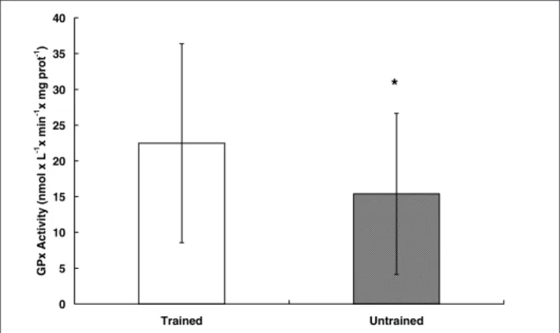
Fig. 4 – GPx activity in erythrocytes of trained (triathletes) and untrained individuals
0 5 10 15 20 25 30 35 40
Trained Untrained
G
P
x
Ac
ti
v
ity
(nm
o
l x
L
-1x m
in
-1x
mg
prot
-1)
*
Fig. 5 – Total plasma antioxidant capacity of trained (triathletes) and un-trained individuals after 40 minutes of exercise
0 50 100 150 200 250 300 350 400 450 500
Before After
TRAP
(
m
m
o
l x
L
-1 Tr
o
lox
)
*
cise and observed that the production of free radicals was higher
than the antioxidant capacity. On the other hand, Subudhi et al.(22)
evaluated elite alpine ski racers after intense training and observed no change on the oxidative stress markers, suggesting that these athletes suffered a positive adaptation in their antioxidant mecha-nisms with training.
Recently in our laboratory, Schneider(44), Schneider et al.(45) and
Oliveira et al.(46), found a higher erythrocyte activity of the enzyme
GPx in trained triathletes if compared to untrained individuals (fig-ure 4) and an increased total plasmatic antioxidant capacity (TRAP) after treadmill exercise in both groups (figure 5). The higher GPx activity is in agreement with several studies that show the
adapta-tion of the enzymatic defense system(32,41,47-49). The increased TRAP
was also observed in the study of Child et al.(38), and it might have
occurred due to a higher release of antioxidant substances such as
the uric acid, as observed in the work of Mastaloudis et al.(49).
FINAL CONSIDERATIONS
The physical training adaptive process is able to protect trained individuals in most situations of exercise exposition. A failure in detecting any change in the lipoperoxidation or in any other target of damage may suggest that some compensatory changes in the antioxidant system might have occurred. The results point to an up-regulation in relation to enzymes GPx and SOD in the skeletal muscle and erythrocytes, but with regard to the enzyme catalase, the results are conflicting. It is interesting observing that several works that evaluated the human antioxidant defense system ana-lyzed the glutathione system, the GPx and SOD, the total antioxi-dant capacity, but none of them included the activity of the en-zyme catalase.
Furthermore, the HSPs activation in acute and chronic exercise participates in the antioxidant protection process. This mechanism has deserved attention in the last years.
Finally, as alternatives of study, we could suggest the utilization of protocols that include long-duration exercises or until exhaus-tion associated to a diet rich in antioxidant nutrients or to the sup-plementation of vitamins and enzyme co-factors on the exercise-induced oxidative stress as well as the study of the genic expression of antioxidant enzymes, protein oxidation and DNA from more sen-sible techniques.
All the authors declared there is not any potential conflict of inter-ests regarding this article.
REFERENCES
1. Halliwell B, Gutteridge JMC. Free radicals in biology and medicine. 3rd ed. New York: Oxford, 1999.
2. Heunks LMA, Viña J, Van Herwaarden CLA, Folgering HTM, Gimeno A, Dekhuijzen PNR. Xanthine oxidase is involved in exercise-induced oxidative stress in chron-ic obstructive pulmonary disease. Am J Physiol 1999;277:R1697-704. 3. Rowlands DS, Downey B. Physiology of triathlon. In: Garrett WE Jr, Kirkendall
DT, editors. Exercise and sport science. Philadelphia: Lippincott Williams & Wilkins, 2000;921-2.
4. Lovlim R, Cottle W, Pyke I, Kavanagh M, Belcastro AN. Are indices of free radi-cal damage related to exercise intensity? Eur J Appl Physiol 1987;56:312-6. 5. Ebelling CB, Clarkson PM. Exercise-induced muscle damage and adaptation.
Sports Med 1989;7:207-34.
6. Jenkins RR. Free radical chemistry relationship to exercise. Sports Med 1988;5: 156-70.
7. Meneguini R. A toxicidade do oxigênio. Ciência Hoje 1987;5:28.
8. Southorn PA, Powis G. Free radicals in medicine II. Involvement in human dis-ease. Mayo Clin Proc 1988b;63:390-408.
9. Marret LD, Theis B, Ashbury FD. Workshop report: physical activity and cancer prevention. Chronic Dis Can 2000;21:143-9.
10. Albright A, Franz M, Hornsby G, Kriska A, Marrero D, Ullrich I, et al. American college of sports medicine position stand. Exercise and type 2 diabetes. Med Sci Sports Exerc 2000;32:1345-60.
11. Diretrizes Brasileiras Sobre Dislipidemias e Diretriz de Prevenção da Ateroscle-rose do Departamento de AteroscleAteroscle-rose da Sociedade Brasileira de Cardiologia, III. Arq Bras Cardiol 2001;77(Supl 3):1-48.
12. Moncada S, Higgs A. Nitric oxide: role in human disease. Encyclopedia of Life Sciences, 2001. Disponível em www.els.net em 20 dez 2001.
13. Pal Yu B. Cellular defenses against damage from reactive oxygen species. Phys-iol Rev 1994;74:139-62.
14. Fischer AB. Intracellular production of oxygen-derived free radicals. Proceed-ings of a Brook Lodge Symposium, Augusta, Apr. 1987;27-29:99-104. 15. Del Maestro RF. An approach to free radicals in medicine and biology. Acta Physiol
Scand 1980;492:153-68.
16. Sies H. Biochemistry of oxidative stress. Angew Chem Int Ed Ingl 1986;25:1058-71.
17. Ferrari R, Ceconi C, Curello S, Guarnieri C, Caldarera M, Albertini A, Visioli O. Oxygen-mediated myocardial damage during ischaemia and reperfusion: role of the cellular defenses against oxygen toxicity. J Mol Cell Cardiol 1985;17:937-45. 18. Belló-Klein A, Oliveira AR, Brunetto AF, Irigoyen MC, Llesuy S, Belló AA. Effect of vitamin A on cardiac contracture induced by hydrogen peroxide. Med Sci Res 1994;22:411-13.
19. Belló-Klein A, Oliveira AR, Miranda MFS, Irigoyen MC, Homem-de-Bittencourt Jr PI, Llesuy S, et al. Effect of trolox C on cardiac contracture induced by hydro-gen peroxide. Braz J Med Biol Res 1997;30:1337-42.
20. Åstrand PO, Rodhal K, Dahl HA, StrØmme SB. Textbook of work physiology. Physiological basis of exercise. 4th ed. Champaign: Human Kinetics, 2003.
21. Viña J, Gomez-Cabrera MC, Lloret A, Marquez R, Miñana JB, Pallardó FV, et al. Free radical in exhaustive physical exercise: mechanism of production, and pro-tection by antioxidants. IUBMB Life 2000;50:271-7.
22. Subudhi AW, Davis SL, Kipp RW, Askew EW. Antioxidant status and oxidative stress in elite alpine ski racers. Int J Sport Nutr Exerc Metab 2001;11:32-41. 23. Heath GW, Hagberg JM, Ehsani AA, Holloszy JO. A physiological comparison of
young and older endurance athletes. J Appl Physiol 1981;51:634-40. 24. Gutteridge JMC, Rowley DA, Halliwell B, Cooper DF, Heeley DM. Cooper and
iron complexes catalytic for oxygen radical reactions in sweat from human ath-letes. Clin Chim Acta 1985;145:267-73.
25. Davies KJA, Quintanilha AT, Brooks GA, Packer L. Free radicals and tissue dam-age produced by exercise. Biochem Biophys Res Commun 1982;107:1198-205. 26. Ji LL, Fu R. Responses of glutathione system and antioxidant enzymes to
ex-haustive exercise and hydroperoxide. J Appl Physiol 1992;72:549-54. 27. Alessio HM. Exercise-induced oxidative stress. Med Sci Sports Exerc 1993;25:
218-24.
28. Nies AM, Hartmann A, Grunert-Fuchs M, Poch B, Speit G. DNA damage after exhaustive treadmill running in trained and untrained men. Int J Sports Med 1996;17:397-403.
29. Mills PC, Smith NC, Casas I, Harris P, Harris RC, Marlin DJ. Effects of exercise intensity and environmental stress on indices of oxidative stress and iron ho-meostasis during exercise in the horse. Eur J Appl Physiol 1996;74:60-6. 30. Palazzetti S, Richard M-J, Favier A, Margaritis I. Overloaded training increases
exercise-induced oxidative stress and damage. Can J Appl Physiol 2003;28:588-604.
31. Margaritis I, Tessier F, M-J Richard, Marconnet P. No evidence of oxidative stress after a triathlon race in highly trained competitors. Int J Sports Med 1997;18: 186-90.
32. Leeuwenburgh C, Hollander J, Leichtweis S, Griffiths M, Gore M, Ji LL. Adapta-tions of glutathione antioxidant system to endurance training are tissue and muscle fiber specific. Am J Physiol 1997;272:R363-9.
33. Leaf DA, Kleinman MT, Hamilton M, Barstow TJ. The effect of exercise intensi-ty on lipid peroxidation. Med Sci Sports Exerc 1997;29:1036-9.
34. Venditti P, Di Meo S. Effect of training on antioxidant capacity, tissue damage, and endurance of adult male rats. Int J Sports Med 1997;18:497-502. 35. Belló AA, Belló-Klein A, Oliveira AR, Brunetto AF, Irigoyen MC, Bauermann LF,
et al. Hydrogen peroxide as a tool for studying oxidative stress in the heart. J Braz Assoc Adv Sci 1996;48:28-36.
36. De Angelis KLD, Oliveira AR, Werner A, Bock P, Bello-Klein A, Fernandes TG, et al. Exercise training in aging: hemodynamic, metabolic, and oxidative stress eval-uations. Hypertension 1997;30:767-71.
37. Belló-Klein A, Lagranha CJ, Bock P, Barp J, Araújo ASR, Llesuy S, et al. Submax-imal exercise training in postnatal rats: hemodynamic and oxidative stress chang-es. Exp Clin Cardiol 2000;5:149-53.
38. Ramires PR, Ji LL. Glutathione supplementation and training increases myocar-dial resistance to ischemia-reperfusion in vivo. Am J Physiol 2001;281:H679-88. 39. Smolka MB, Zoppi CC, Alves AA, Silveira LR, Marangoni S, Pereira-Da-Silva L, et al. HSP72 as a complementary protection against oxidative stress induced by exercise in the soleus muscle of rats. Am J Physiol 2000;279:R1539-45. 40. Child RB, Wilkinson DM, Fallowfield JL, Donnelly AE. Elevated serum
antioxi-dant capacity and plasma malondialdehyde concentration in response to a simu-lated half-marathon run. Med Sci Sports Exerc 1998;30:1603-7.
41. Powers SK, Ji LL, Leeuwenburgh C. Exercise training-induced alterations in skel-etal muscle antioxidant capacity: a brief review. Med Sci Sports Exerc 1999;31: 987-97.
42. Selamoglu S, Turgay F, Kayatekin BM, Günenc S, Yslegen C. Aerobic and anaer-obic training effects on the antioxidant enzymes of the blood. Acta Physiol Hung 2000;87:267-73.
43. Inal M, Akyüz F, Turgut A, Getsfrid WM. Effect of aerobic and anaerobic metabo-lism on free radical generation swimmers. Med Sci Sports Exerc 2001;33:564-7. 44. Schneider CD. Avaliação do estresse oxidativo em indivíduos submetidos a dife-rentes intensidades de exercício em esteira rolante [Dissertação apresentada para obtenção do título de mestre em ciências do movimento humano]. Porto Alegre: Escola de Educação Física da Universidade Federal do Rio Grande do Sul, 2002.
45. Schneider CD, Barp J, Ribeiro JL, Belló-Klein A, Oliveira AR. Oxidative stress after three different intensities of running. Can J Appl Physiol 2004 (submetido). 46. Oliveira AR, Schneider CD, Ribeiro JL, Deresz LF, Barp J, Belló-Klein A. Oxida-tive stress after three different intensities of running. Med Sci Sports Exerc 2003;35:S367.
47. Tessier F, Margaritis I, Richard M-J, Moynot C, Marconnet P. Selenium and train-ing effects on the glutathione system and aerobic performance. Med Sci Sports Exerc 1995;27:390-6.
48. Hellstein Y, Apple F, Sjodin B. Effect of sprint cycle training on activities of anti-oxidant enzymes in human skeletal muscle. J Appl Physiol 1996;81:1484-7. 49. Mastaloudis A, Leonard SW, Traber MG. Oxidative stress in athletes during