UNIVERSIDADE FEDERAL DE MINAS GERAIS INSTITUTO DE CIÊNCIAS BIOLÓGICAS
DEPARTAMENTO DE BIOQUÍMICA E IMUNOLOGIA LABORATÓRIO DE GNOTOBIOLOGIA E IMUNOLOGIA
THE ROLE OF GP91
PHOX-DERIVED REACTIVE OXYGEN
SPECIES IN THE INFECTION CAUSED BY
LEISHMANIA
AMAZONENSIS
: IMPLICATIONS RELATIVE TO THE
SITE OF INFECTION
Eric Henrique Roma de Lima
Orientadora: Prof. Dr. Leda Quercia Vieira
Co-orientadores: Dr. Nathan Peters Dr. David Sacks
UNIVERSIDADE FEDERAL DE MINAS GERAIS INSTITUTO DE CIÊNCIAS BIOLÓGICAS
DEPARTAMENTO DE BIOQUÍMICA E IMUNOLOGIA LABORATÓRIO DE GNOTOBIOLOGIA E IMUNOLOGIA
THE ROLE OF GP91
PHOX-DERIVED REACTIVE OXYGEN
SPECIES IN THE INFECTION CAUSED BY
LEISHMANIA
AMAZONENSIS
: IMPLICATIONS RELATIVE TO THE
SITE OF INFECTION
Tese apresentada para o programa de pós-graduação em Bioquímica e Imunologia do departamentos de Bioquímica e Imunologia do Instituto de Ciências Biológicas da Universidade Federal de Minas Gerais como pré-requisito para obtenção do título de doutorado em Imunologia.
Eric Henrique Roma de Lima
Orientadora: Prof. Dr. Leda Quercia Vieira
Co-orientadores: Dr. Nathan Peters
Dr. David Sacks
Dedicatória
Para minha mãe Lu, a qual devo tudo que sou hoje. Sempre ao meu lado com
apoio e carinho... não importando o caminho e sacrifícios realizados sempre me
ajudou a trilhar meus caminhos. Se estou aqui hoje é tudo devido a você. Terei por
você gratidão eterna, pois nunca conseguirei retribuir o que sempre fez por mim.
Para minha esposa Juliana, minha companheira há 9 anos... sempre me
apoiando tanto dentro como fora do laboratório. Com amor e carinho nas horas
difíceis me ajudou a ultrapassar obstáculos e é responsável por grande parte da minha
felicidade. Sem você ao meu lado, este trabalho seria bem mais difícil.
Para Leda, por me dar a oportunidade de realização deste trabalho e ter me
acolhido em seu laboratório. Por ter confiado em mim e me ajudado em tudo que
precisei. Por ter me dado a oportunidade de conhecer os EUA e vivenciar a ciência do
jeito que ela deveria ser no Brasil. Obrigado Leda, por todo apoio dado neste tempo.
Essa tese não existiria sem você.
Agradecimentos
Ao Dr. David Sacks por ter me aceitado em seu laboratório e ter fornecido todo
suporte necessário para realização de grande parte deste trabalho, além dos
ensinamentos sobre ciência os quais me ajudaram bastante a crescer como cientista.
Ao Dr. Nathan Peters, que acompanhou de perto meu trabalho nos EUA, com
grandes contribuições técnicas e intelectuais, sendo de fato muito importante para a
realização deste trabalho.
Ao NIH , por ter me dado a oportunidade de conhecer como é feito ciência de
ponta no maior instituto de pesquisa do mundo.
À UFMG por ter sido responsável por toda minha formação como cientista,
desde a graduação há dez anos atrás até os dias de hoje.
Aos integrantes do laboratório do Dr. David Sacks nos EUA, pelo apoio e a
ótima convivência.
A todos os integrantes do LAGI pela ótima convivência nestes anos. Lugar este
que com certeza foi um dos melhores ambientes nos quais vivi.
Aos amigos dos EUA, principalmente Helton e Ester, que me acolheram com
carinho em sua casa no início da minha jornada fazendo com que eu me sentisse mais
confortável mesmo longe da família.
Aos amigos da UFMG com os quais sempre pude contar. Sempre me trataram
com carinho e por isso me sinto muito bem quando estou nessa universidade.
Aos amigos de 2003/02 pelos bons momentos que passamos juntos durante
nossa graduação dentro e fora do ambiente acadêmico.
Aos meus familiares pelo incentivo, carinho e pelos momentos prazerosos que
passamos juntos.
Aos familiares da Juliana, especialmente seus pais Namira e Márcio os quais
também considero meus pais. Pelo acolhimento e por me tratarem como filho quando
estou junto a eles.
Aos meus cães Raica, Julie, Princesa e Sadam por seu amor incondicional.
Como disse Darwin: "O próprio homem não pode expressar o amor e humildade por
sinais externos, tão claramente como um cachorro, quando ele encontra seu amado
mestre". Por serem a válvula de escape de problemas através da demonstração do
carinho que sempre estão dispostos a fornecer. Sempre que estou com eles me sinto
bem e feliz. Afinal, você não precisa de psiquiatra se tem um cachorro.
V Table of Contents
List of Figures...IX List of Tables ... XVIII List of Abbreviations ... XIX 1. Resumo ... XXII 1. Abstract ... XXIV
2. Introduction ... 26
3. Literature review ... 29
3.1. Leishmania genus taxonomy ... 30
3.2. Epidemiology ... 31
3.3. Disease ... 34
3.3.1. Etiological agent and clinical aspects ... 34
3.3.2. Biological cycle ... 35
3.3.3. Immune response ... 38
3.3.4. Evasion of immune response by the parasite ... 42
3.4. NADPH oxidases, production of ROS and inflammation ... 43
3.5. ROS and Leishmania ... 48
3.5.1. Production of ROS by host cells in Leishmania infections ... 48
3.5.2. Importance of ROS in the direct killing of Leishmania ... 50
3.5.3. Subversive mechanisms used by Leishmania to avoid damage by ROS ... 51
3.6. Relevance of the work and explanation of thesis structure ... 52
4. Part one: subcutaneous inoculation with L. amazonensis in gp91phox-/- mice promote changes in inflammatory response to infection ... 55
4.1. Objectives ... 56
4.1.1. General objective ... 56
4.1.2. Specific objectives ... 56
4.2. Material and Methods ... 57
4.2.1. Parasites ... 57
4.2.2. Mice ... 57
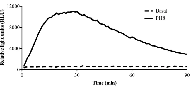
4.2.3. Luminometry assay ... 57
4.2.4. Infection with L. amazonensis ... 58
4.2.5. Euthanasia, collection of blood and organs ... 58
VI
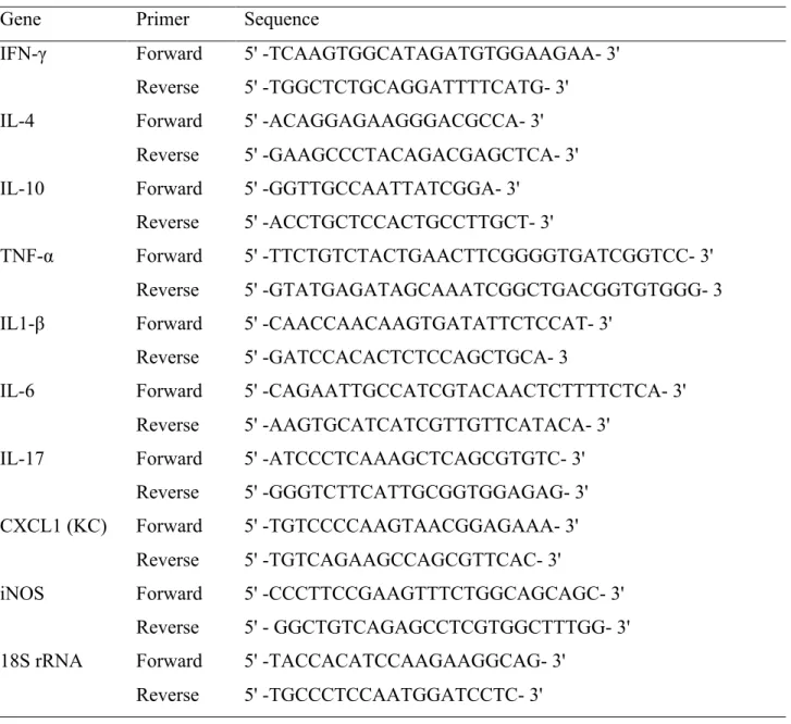
4.2.7. Real time PCR ... 59
4.2.8. Generation of L. amazonensis antigen of (LALa) ... 61
4.2.9. ELISA for cytokine measurements ... 61
4.2.10. ELISA for anti-Leishmania IgG1, IgG2a and total IgG ... 61
4.2.11. Flow cytometry ... 62
4.2.12. Mieloperoxidase assay for neutrophil numbers inference ... 63
4.2.13. Phagocytic and microbicidal activity of macrophages in infections invitro ... 64
4.2.14. Cytokine and chemokine production and nitrite measurements from peritoneal macrophages cultures ... 64
4.2.15. Statistical analysis ... 65
4.3. Results ... 65
4.3.1. L. amazonensis induces respiratory burst in macrophages from C57BL/6 mice ... 65
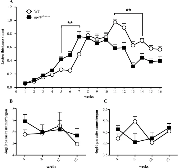
4.3.2. Gp91phox-/- mice show different lesion development, but present similar parasite loads in footpads and dLNs to those of WT mice. ... 66
4.3.3 WT mice express IL-1β in footpads at chronic phase of L. amazonensis infection when compared to gp91phox-/-. ... 68
4.3.4. Mice lacking gp91phox induces a higher Th17 response in acute phase of L. amazonensis infection ... 70
4.3.5. Deficiency in ROS production is not compensated by increase in iNOS expression ... 72
4.3.6. ROS alter the influx of innate and adaptive immune cells to the site of infection and the dynamics of dLNS expansion in mice subcutaneously infected with L. amazonensis ... 73
4.3.7. ROS increased the migration and decreased clearance of neutrophils immediately after infection with L. amazonensis ... 78
4.3.8. ROS influence anti-Leishmania IgG antibodies secretion during L. amazonensis infection ... 79
4.3.9. Activated gp91phox-/- macrophages secrete higher levels of inflammatory cytokines in L. amazonensis infection in vitro ... 81
4.3.10. ROS promotes increased parasite loads in macrophages infected with L. amazonensis in vitro ... 85
5. Part two: Site of L. major infection determines dominant host cell phenotype ... 87
5.1. Objectives ... 88
5.1.1. General objective ... 88
5.1.2. Specific objectives ... 88
5.2. Material and Methods (the following text is taken from a manuscript in preparation) 88 5.2.1. Mice ... 88
VII
5.2.3. Parasite preparation and inoculations ... 89
5.2.4. Processing of tissues ... 90
5.2.5. Immunolabeling and flow cytometry ... 90
5.2.6. Statistical analysis ... 91
5.3. Results ... 91
5.3.1. The patterns of cell migration changes according to the site of L. major infection ... 91
5.3.2. The site of inoculation determines the cells involved in the initial uptake of L. major ... 99
5.3.3. Infection at the intradermal site elicited higher cell activation and inflammatory response in the acute phase of L. major infection ... 105
6. Part three: Reactive oxygen species (ROS) control the inflammation in L. amazonensis intradermal infection impairing neutrophil necrosis ... 110
6.1. Objectives ... 111
6.1.1. General objective ... 111
6.1.2. Specific objectives ... 111
6.2. Material and Methods ... 112
6.2.1. Mice ... 112
6.2.2. Parasites ... 112
6.2.3. Infection with L. amazonensis ... 113
6.2.4. Euthanasia and tissue processing ... 114
6.2.5. Parasite load quantification ... 114
6.2.6. Quantitative Real time PCR ... 115
6.2.6. Generation of L. amazonensis antigen (LALa) ... 116
6.2.8. Flow cytometry ... 116
6.2.9. Apoptosis assessment ... 117
6.2.10. Statistical analysis ... 118
6.3. Results ... 118
6.3.1. Intradermally inoculated gp91phox-/- mice exhibit higher inflammation in L. amazonensis infection ... 118
6.3.2. ROS do not influence the killing of L. amazonensis in vivo at the site of infection, but is required to control parasite loads in dLNs ... 120
6.3.3. ROS impairs neutrophil accumulation at the site of inoculation with L. amazonensis .. 121
6.3.4. Gp91phox-/- mice do not presented with alterations in T lymphocytes during L. amazonensis infection ... 126
IX List of Figures
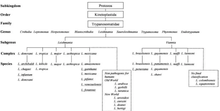
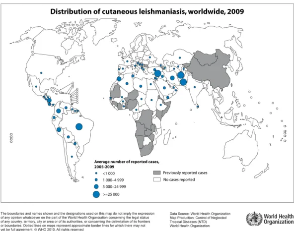
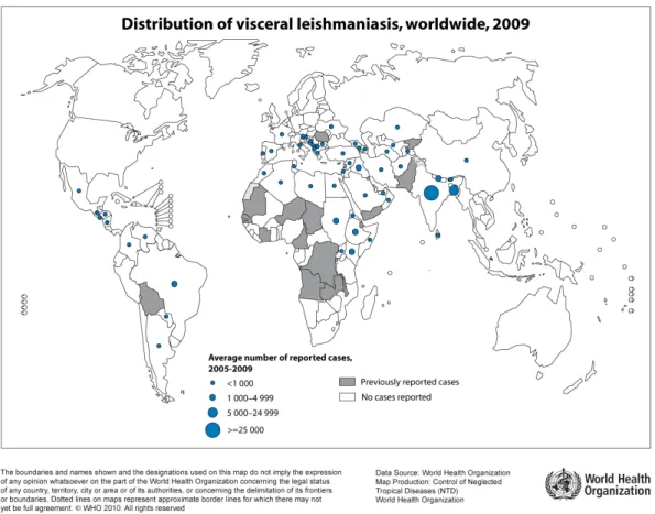
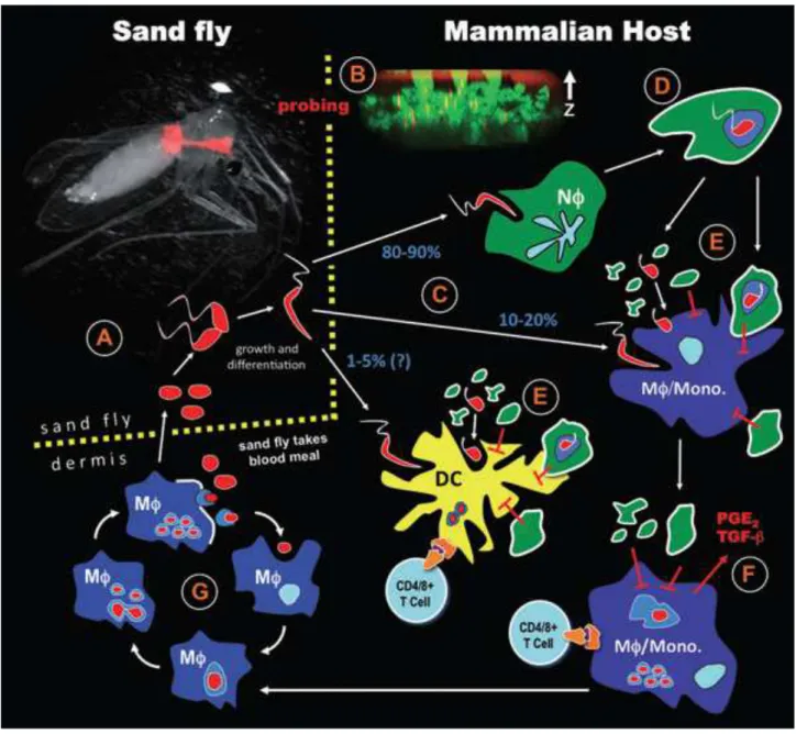
Figure 1: Taxonomy of Leishmania genus (Banuls et al., 2007). Underlined species have had their taxonomic classification questioned. Twenty species of the thirty known are pathogenic for humans. ... 31 Figure 2: World distribution of tegumentar form of leishmaniasis. The blue-dotted areas indicate endemic zones between 2005 to 2009. WHO/CNTD. Map: Control of Neglected Tropical Diseases of the World Health Organization, 2010. ... 32 Figure 3: World distribution of visceral form of leishmaniasis. The blue-dotted areas indicate endemic zones between 2005 to 2009. WHO/CNTD. Map: Control of Neglected Tropical Diseases of the World Health Organization, 2010. ... 33 Figure 4: Distribution of Leishmania species causing cutaneous leishmaniasis in the Brazilian territory. L. amazonensis and L. braziliensis are responsible for the majority of cutaneous leishmaniasis cases in Brazil. Map: Ministry of Health of Brazil, Manual of leishmaniasis vigilance, 2007. ... 34 Figure 5: Biological cycle of Leishmania major (Peters and Sacks, 2009). (A) The cycle starts with the vector feeding on the blood from infected host. With the blood, the vector ingests amastigotes that are inside macrophages in the skin. In the insect gut, the amastigotes are released by macrophage lysis and differentiate into promastigote forms. They attach to the gut wall and start to replicate. (B) In the new blood meal, these metacyclic forms are inoculated in the skin of the host and (C) they are phagocyted majorly by neutrophils. (D) The neutrophils fail to kill phagocyted parasites, start the apoptotic process and (E) release the parasites together with apoptotic bodies into the extracellular environment. (D) Phagocytosis by the macrophages/DCs of these apoptotic bodies and parasites (F) induce the production of anti-inflammatory mediators such as TGF-β, which favor the survival of the parasite in the newly infected cells. (F and G) The fusion of phagosome membrane containing parasites with lysosomes in the host cells creates favorable environment to differentiation of the promastigotes into amastigotes, which start cycles of replication inside of the infected cells. (G) The amastigotes replicate indefinitely inside of macrophages until their lysis. Thus, many amastigotes are released in the extracellular environment and then are phagocyted by others phagocytic cells, enhancing the replication cycles. (A) Eventually, the vector might feed on contaminated host blood, re-initiating the cycle. ...37 Figure 6: Models of immune responses in the infection caused by L. major (Gollob et al.,
X Figure 7: Many ways of inhibition of microbicidal activities used by Leishmania spp. to immune response evasion. Adapted (Peters and Sacks, 2006). Leishmania evade the microbicidal activities of macrophages in many ways. The phagocytosis of Leishmania by specific CR1 and CR3, manose receptors and phosphatidylserine receptors promotes, among other responses, release of IL-10. The uptake by these receptors and IL-10 production induce downregulation of oxidative defenses by macrophages. Inside of macrophages, the Leishmania
XI iNOS mRNA levels were normalized by 18S mRNA expression 4, 8, 12 and 16 weeks post infection. (B) Thioglycolate-elicited macrophages were harvested from WT or gp91phox-/- mice peritoneum 3 days after stimulation. Macrophages were infected with L. amazonensis
XII assay to estimate the neutrophil numbers. Data are shown as mean ± SD from one representative experiment of 2, n=5 for each experiment, *p<0.05. ... 79 Figure 21. Production of anti-Leishmania IgG in NADPH oxidase deficient mice infected with
L. amazonensis. Mice were infected with 1 x 10⁶ metacyclic promastigote forms of L. amazonensis in the right hind footpad and followed until 16 weeks. After 4, 8, 12 and 16 weeks of infection, the serum was collected and ELISA to detect anti-Leishmania IgG was performed. A, B and C represent the absorbance found for anti-Leishmania IgG, IgG1 and IgG2a, respectively. The dotted lines represent the absorbance of serum of non-infected mice in dilution of 1:200, the same dilution used for the serum of the infected mice. Data are shown as mean ± SD from one representative experiment of 2, n=6 for each experiment. ... 81 Figure 22. Production of cytokines and chemokines in gp91phox-/- macrophages infected with L. amazonensis. Thioglycolate-elicited macrophages were infected with stationary-phase L. amazonensis promastigotes at 10 parasites per macrophage. Macrophages were activated 50U/ml of IFN-γ plus 100ng/ml of LPS. After 72h of infection with L. amazonensis or activation the supernatants were collected and ELISAs were performed to quantify cytokines and chemokines. For TNF-α assays, aliquots from cell cultures were collected at 24h of infection. Controls without stimuli and infection were used to quantify the basal levels of cytokines. A, B, C, D, E and F represent the concentration showed in pg/ml of the IL-6, TNF-α, IL-17, CXCL-1, MCP-1 and IL-10 respectively. Data are shown as mean ± SD from one representative experiment of 2, n=6 for each experiment. ... 84 Figure 23. Quantification of parasite loads in macrophages treated with ROS and nitric oxide inhibitors in infection caused by L. amazonensis in vitro. Peritoneal macrophages were infected with metacyclic promastigote forms of L. amazonensis in proportion 10 parasites per macrophage for 4h. During the infection, the cells were treated with several inhibitors (WT macrophages treated with L-NMMA alone; apocynin alone; L-NMMA plus apocynin; L-NMMA plus SOD; L-NMMA plus catalase; WT infection without treatment; and gp91 phox-/-macrophages treated or not with L-NMMA. After 72h of infection the cells were stained counted under light microscope. ... 86 Figure 24. Flow cytometry gate strategy adopted to analyse the recruited cells after L. major
XIII cells, respectively. The data are represented as mean ±SD of one experiment representative of two performed, n=5 for each group. *=p<0.05, **=p<0.01 and ***=p<0.001. ... 96 Figure 26. Total cell numbers of subpopulations gated on CD11b+ recruited to the inoculation site after L. major infection. Mouse ears or footpads were infected with 5 x 10⁵ promastigote forms of L. major and 2h, 48h and 9 days post infection and the 0h time point represents the non-infected controls. The tissues were processed and stained to surface markers. A, B, C, D, E and F represent the kinetics of cell recruitment of neutrophils, inflammatory monocytes, macrophages, DCs, activated macrophages and CD11c+MHCII- cells, respectively. The data are represented as mean ±SD of one experiment representative of two, n=5 for each group. *=p<0.05, **=p<0.01 and ***=p<0.001. ... 98 Figure 27. Flow cytometry gate strategy adopted to analyse the infected cells after L. major
inoculation. Mouse ears or footpads were infected with 5 x 10⁵ promastigote forms of L. major
transfected with rfp gene and 2h, 48h and 9 days post infection the tissues were processed and stained for surface markers. The same gate strategy was used in both tissues and all times. The red arrows indicate the pathways to split populations and identify subsequent sub-populations. The gates represent one sample of ears 48h post infection. ... 99 Figure 28. Percentage of subpopulations gated on infected CD11b+rfp+ cells recruited to the L. major inoculation site. Mouse ears or footpads were infected with 5 x 10⁵ L. major
promastigote forms and 2h, 48h and 9 days post infection the tissues were processed and stained to surface markers. A, B, C, D, E and F represent the kinetics of phagocytic cells, neutrophils, inflammatory monocytes, macrophages, DCs and CD11c+MHCII- cells, respectively. The data are represented as mean ±SD of 1 experiment representative of 2, n=5 for each group. *=p<0.05, **=p<0.01 and ***=p<0.001. ... 102 Figure 29. Total numbers of infected CD11b+ cells recruited to the L. major inoculation site. Mouse ears or footpads were infected with 5 x 10⁵ promastigote forms of L. major and 2h, 48h and 9 days post infection the tissues were processed and stained for surface markers. A, B, C, D, E and F represent phagocytic cells, neutrophils, inflammatory monocytes, macrophages, DCs and CD11c+MHCII- cells, respectively. The data are represented as mean ±SD of 1 experiment representative of 2, n=5 for each group. *=p<0.05, **=p<0.01 and ***=p<0.001. ... 104 Figure 30. Mean fluorescence intensity (MFI) of MHCII on macrophages or DCs infected with L. major. Mouse ears or footpads were infected with 5 x 10⁵ promastigote forms of L. major and 2h, 48h and 9 days post infection and the 0h time point represents the non-infected controls. The tissues were processed and stained for surface markers. A, B, C and D represent the MFI of MHCII+ macrophages, DCs, infected macrophages and infected DCs, respectively. The data are represented as mean ±SD of 1 experiment representative of 2, n=5 for each group. **=p<0.01 and ***=p<0.001. ... 106 Figure 31. qRT-PCR of cytokines and chemokines expressed during acute phase of L. major
infection. Mouse ears or footpads were infected with 5 x 10⁵ promastigote forms of L. major
XIV the mRNA expression 2h, 48h and 9 days post infection of IL-1β, CXCL-1, IL-6, IL-10 and TNF-α, respectively and normalized by mRNA expression of the cytokines in naïve tissues. E and F represent the mRNA expression 2h, 48h and 9 days post infection of IFN-γ and IL-17, respectively and normalized by constitutive expression of 18S mRNA. N.D. indicates not determined. The data are represented as mean ±SD, n=5 for each group. *=p<0.05 and ***=p<0.001. ... 109 Figure 32. Lesion size of mice infected with L. amazonensis. Mice were infected with 5 x 10³ L. amazonensis metacyclic promastigotes in pinna ears and followed until 16 weeks. (A) The lesion diameter was measured week by week with a calliper, *p<0.05, **p<0.01 and ***p<0.001 Data are shown as mean ± standard deviation (± SD). (B) Percentage of mice that had some tissue loss from 7 to 16 weeks post infection. (C) Representative pictures of ears from mice infected with L. amazonesis 4, 8, 12 and 16 weeks post infection. The figure represents one representative experiment of 2, n=5 for each experiment. ... 120 Figure 33. Parasite loads found in L. Amazonensis-infected mice 4, 8, 12 and 16 weeks post infection. Mice were infected with 5 x 10³ L. amazonensis metacyclic promastigotes in pinna ears and followed for 16 weeks. (A) Parasite loads found in ears of infected mice 4, 8, 12 and 16 weeks post infection. (B) Parasite loads found in draining lymph nodes of infected mice 4, 8, 12 and 16 weeks post infection. Data are shown as mean ± SD from one representative experiment of 2, n=5 for each experiment. ... 121 Figure 34. Number of CD11b+ cells recruited to the site of inoculation with L. amazonensis 4, 8 and 12 weeks post infection. Mice were infected with 5 x 10³ L. amazonensis metacyclic promastigotes in pinna ears and followed for 16 weeks. The data represents the total numbers of CD11b+ cells in ears 4, 8 and 12 weeks post infection. Data are shown as mean ± SD from one representative experiment of 2, n=5 for each experiment. ... 122 Figure 35. CD11b+ subpopulations recruited to the site of inoculation with L. amazonensis 4, 8 and 12 weeks post infection. The mice were infected with 5 x 10³ of L. amazonensis
XV proliferating CD4 T cells and CD8 T cells in ears, respectively. T cell populations were analysed 4, 8 and 12 weeks post infection. Data are shown as mean ± SD from one representative experiment of 2, n=5 for each experiment. ... 127 Figure 38. Production of cytokines by CD4+ T cells at the site of L. amazonensis inoculation 4, 8 and 12 weeks post infection. Mice were infected with 5 x 10³ L. amazonensis metacyclic promastigotes in pinna ears and followed for 16 weeks. A represents CD4+ T cells producing IFN-γ, B represents CD4+ T cells producing TNF-α, C represents CD4+ T cells producing IFN-γ and TNF-α, D represents CD4+ T cells producing IL-2, E represents CD4+ T cells producing IFN-γ, TNF-α and IL-2, F represents CD4+ T cells producing IL-10 and G represents CD4+ T cells producing IL-17 in ears 4, 8 and 12 weeks post infection. Data are shown as mean ± SD from one representative experiment of 2, n=5 for each experiment. ... 129 Figure 39. Total number of cells in draining lymph nodes in L. amazonensis infected mice 4, 8, 12 and 16 weeks post infection. Mice were infected with 5 x 10³ L. amazonensis metacyclic promastigotes in pinna ears and followed for 16 weeks. At 4, 8, 12 and 16 the dLNs were harvested and cells counted. Data are shown as mean ± SD from one representative experiment of two, n=5 for each experiment. ... 130 Figure 40. Percentage phagocytic and T cells in draining lymph nodes in L. amazonensis
infected mice 4, 8, 12 and 16 weeks post infection. Mice were infected with 5 x 10³ L. amazonensis metacyclic promastigotes forms of in pinna ears and followed for 16 weeks. At 4, 8, 12 and 16 the dLNs were disrupted and the total cells counted. A, B and C represent the percentage of phagocytic, CD4+ and CD8+ T cells ranging from non-infected mice (week 0) until 16 weeks post infection. Data are shown as mean ± SD from one representative experiment of 2, n=5 for each experiment. ... 131 Figure 41. Percentage phagocytic subpopulations in draining lymph nodes in L. amazonensis
infected mice 4, 8, 12 and 16 weeks post infection. Mice were infected with 5 x 10³ L. amazonensis metacyclic promastigotes in pinna ears and followed for 16 weeks. At 4, 8, 12 and 16 the dLNs were harvested, disrupted and the total cells counted. A and B represent the percentage of neutrophils and inflammatory monocytes in lymph nodes from non-infected mice (week 0) until 16 weeks post infection. Data are shown as mean ± SD from one representative experiment of 2, n=5 for each experiment. ... 132 Figure 42. Percentage CD4+ T cell subpopulations in draining lymph nodes in L. amazonensis
XVI weeks. 4, 8, 12 and 16 the dLNs were disrupted and the total cells counted. A, B, C, D, E, F and G represent the percentage of IFN-γ, TNF-α, double (IFN-γ and TNF-α), IL-2, triple (IFN-γ, TNF-α and IL-2), IL-10 and IL-17 CD4+ T producing cells, respectively, ranging from non-infected mice (week 0) until 16 weeks post infection. Data are shown as mean ± SD from one representative experiment of 2, n=5 for each experiment. ... 136 Figure 44. mRNA expression levels of CXCL1, CXCL2 and IL-1β from L. amazonensis
infected ears measured by qPCR. Mice were infected with 5 x 10³ metacyclic promastigotes forms of L. amazonensis in pinna ears and followed until 16 weeks. A, B and C represent the mRNA expression of CXCL1, CXCL2 and IL-1β, respectively, normalized to mRNA expression in naïve mice 4 and 8 weeks post infection. Data are shown as mean ± SD from one representative experiment of 2, n=5 for each experiment. ... 138 Figure 45. Percentage and total number of neutrophils recruited to the site of inoculation in L. amazonensis 8 weeks post infection. Mice were infected with 5 x 10³ L. amazonensis
metacyclic promastigotes transfected with rfp gene in pinna ears and followed for 8 weeks. A represents the dot plots of subpopulations gated on Ly6G and Ly6C markers. B and C represent the percentage and total numbers of neutrophils, respectively. Data are shown as mean ± SD from one experiment, n=13. ... 140 Figure 46. Percentage and total number of infected neutrophils at site of L. amazonensis
inoculation 8 weeks post infection. Mice were infected with 5 x 10³ L. amazonensis
metacyclic promastigotes transfected with rfp gene in pinna ears and followed for 8 weeks. A represents the dot plots of the percentage of neutrophils gated on RFP. B and C represent the percentage and total numbers of infected neutrophils, respectively. Data are shown as mean ± SD from one experiment, n=13. ... 141 Figure 47. Percentage and total number of dying neutrophils at the site of inoculation with L. amazonensis 8 weeks post infection. Mice were infected with 5 x 10³ L. amazonensis
XVII present in ears 10h, 36h and 60h post infection. Data are shown as mean ± SD from one representative experiment of 3, n=5 for each experiment. ... 145 Figure 50. Percentage of infected CD11b+RFP+ subpopulations at the site of inoculation with
L. amazonensis 10h, 36h and 60h post infection. Mice were infected with 5 x 10⁵ metacyclic promastigotes forms of L. amazonensis transfected with rfp gene in pinna ears and followed since 10h to 60h post infection. A represents the dot plots of the percentage of infected CD11b+ cells subpopulations. B, C D and E represent the percentage of neutrophils, inflammatory monocytes, macrophages and DCs presented in ears. Data are shown as mean ± SD from one representative experiment of 3, n=5 for each experiment. ... 147 Figure 51. Percentage of dying neutrophils at the site of inoculation with L. amazonensis 36h and 60h post infection. Mice were infected with 5 x 10⁵ metacyclic promastigotes forms of L. amazonensis transfected with rfp gene in pinna ears and followed until 60h post infection. A represents the dot plots of the percentage of neutrophils in necrotic or apoptotic death process 36h and 60h post infection. B and C represent the percentage of apoptotic or necrotic cells, respectively, in total recruited neutrophils 36h or 60h post infection. D and E represent the percentage of apoptotic or necrotic cells, respectively, in infected neutrophils 36h or 60h post infection. Data are shown as mean ± SD from one representative experiment of 2, n=5 for each experiment. ... 149
XVIII
List of Tables
Table 1. Primers used in qPCR to quantify the expression of mRNA from footpads in the infection. ...60
XIX
List of Abbreviations
7-AAD 7-aminoactinomycin D
ANOVA Analysis of variance
APC Antigen presenting cell
BOD Biochemical Oxygen Demand
BSA Bovine serum albumin
cDNA Complementary DNA
CGD Chronic granulomatous disease
CR1 Complement receptor 1
CR3 Complement receptor 3
DAMPs Danger associated molecular patterns
DC Dendritic cells
DETC Diethyldithiocarbamate
dLNs draining Lymph Nodes
DMEM Dulbecco's Modified Eagle Medium
DNA Deoxyribonucleic acid
DTH Delayed type hypersensitivity
EDTA Ethylenediamine tetraacetic acid
ELISA Enzyme-Linked Immunosorbent Assay
FBS Fetal bovine serum
FcR Fc receptor
FCS Fetal calf serum
FITC Fluorescein isothiocyanate
GIPs Glycoinositolphospholipids
HEPES 4-(2-hydroxyethyl)-1-piperazineethanesulfonic acid
HMGB1 High mobility group box protein 1
HO-1 Heme oxygenase 1
HTAB Hexadecyltrimethylammonium bromide
IFN-β Interferon β
IFN-γ Interferon γ
IgG Immunoglobulin G
XX
IgG2a Immunoglobulin G subtype 2a
IL-10 Interleukin 10
IL-12 Interleukin 12
IL-13 Interleukin 13
IL-17 Interleukin 17
IL-1β Interleukin 1β
IL-4 Interleukin 4
IL-5 Interleukin 5
IL-6 Interleukin 6
IL-8 Interleukin 8
iNOS inducible nitric oxide synthase
L-NMMA L-NG-monomethyl Arginine citrate
LF Lactoferrin
LPG Lipophosphoglycan
LPS Lipopolysaccharide
MCP-1 Monocyte chemoattractant protein 1
MHCII Major histocompatibility complex class II
MPO Mieloperoxidase
mRNA messenger RNA
NAC N-acetylcysteine
NADPH Nicotinamide adenine dinucleotide phosphate-oxidase
OD Optical density
OPD o-phenylenediamine
PAMPs Pathogen-associated molecular patterns
PBS Phosphate buffer saline
PCR Polymerase chain reaction
PE Phycoerythrin
PEG-catalase Polyethylene glycol linked catalase
PEG-SOD Polyethylene glycol linked superoxide dismutase
PGE2 Prostaglandin E2
PIP3 Phosphatidylinositol (3,4,5)-triphosphate
PVs Parasitophorous vacuoles
qPCR quantitative PCR
XXI
RNS Reactive nitrogen species
ROS Reactive oxygen species
RPMI Roswell Park Memorial Institute Medium
rRNA ribosomal RNA
LA Leishmania antigen
LALa
LALm
Leishmania antigen of L. amazonensis Leishmania antigen of L. major
SOD Superoxide dismutase
TGF-β Transforming growth factor β
Th1 T helper response type 1
Th17A T helper response type 17A
Th2 T helper response type 2
Th3 T helper response type 3
TLRs Toll like receptor
TNF-α Tumor necrosis factor α
Tregs Regulatory T response
UCP2 Uncoupling protein 2
XXII
1. Resumo
As leishmanioses são um espectro de doenças causadas por parasitas do gênero Leishmania.
Por ser um patógeno intracelular obrigatório, infectando principalmente macrófagos e neutrófilos, a produção de radicais de oxigênio e nitrogênio poderia ser importante fator na eliminação de parasitas. No caso específico da infecção causada por Leishmania amazonensis, uma importante espécie causadora de leishmaniose cutânea no Brasil, não há dados na literatura demonstrando o efeito desses radicais em infecções in vivo.Dados do nosso grupo confirmaram que o óxido nítrico possui um importante papel na eliminação dos parasitas em infecções causadas por L. amazonensis in vivo, porém o ânion superóxido e o peróxido de hidrogênio não parecem estar associados com a morte dos parasitas in vivo. Neste trabalho, nós mostramos que animais C57BL/6 deficientes da subunidade gp91phox da NADPH oxidase de fagócitos (gp91phox-/-) infectados de forma subcutânea com L. amazonensis desenvolvem lesões maiores nas primeiras semanas de infeção e lesões menores na fase crônica da doença quando comparados a animais selvagens (WT). Entretanto, ambos camundongos WT e gp91phox-/- apresentaram a mesma carga parasitária. Nós detectamos altos níveis de IL-17 nos linfonodos drenantes 8 semanas pós infecção (p.i.) e baixos níveis de mRNA de IL-1β no sítio da lesão 12 e 16 semanas p.i. nos camundongos gp91phox-/-. Há dados controversos na literatura sobre o papel dos neutrófilos nas infecções causadas por Leishmania
XXIV
1. Abstract
XXV of infection, but we did detect a large accumulation of neutrophils at the intradermal site. Increased neutrophil numbers in gp91phox-/- mice was associated with higher numbers and frequencies of necrotic neutrophils at 8 weeks p.i. and may be the major factor responsible for the inflammatory phenotype observed in these mice. These experiments suggest that oxygen radicals have an important role in L. amazonensis infection, controlling neutrophil flux to the lesion, impairing necrotic inflammatory death in these cells and consequently the inflammation of infected tissue. Moreover, the route of inoculation greatly influences the effect of reactive oxygen species (ROS) in
L. amazonensis infection, since the major cells producing ROS, the neutrophils, play a prominent role only in intradermal infection. This is the first report to show the effect of ROS in Leishmania
model closest to natural infection, indicating a more physiological way to study ROS in Leishmania
CAUSED BY L. AMAZONENSIS: IMPLICATIONS RELATIVE TO THE SITE OF INFECTION
26
27 Leishmaniasis are a spectrum of diseases caused by parasites of the genus Leishmania. They are endemic in 88 countries in the world and about 350 million people are at risk to contracting these diseases (WHO, 2013). Leishmaniasis are concentrated in developing countries, mainly Brazil and India, the African continent, and the Middle East. About 12 million people suffer from these diseases and it is estimated that 1.3 million new cases occur every year, with 20-30,000 deaths annually (WHO, 2013). The Leishmania genus includes 30 species and about 20 are pathogenic to humans (Cupolillo et al., 2000). The disease presents with high frequency in the visceral or tegumentary form, this latter presentation being subdivided in cutaneous, diffuse cutaneous or mucocutaneous leishmaniasis. There are estimated to be 0.7 to 1.3 million new cases of cutaneous disease and 0.2-0.4 million new cases of visceral disease every year (WHO, 2013). Consequently, leishmaniasis are considered one of the six major endemic diseases in the world.
The natural chronic persistence of leishmaniasis favors the development of a polarized immune response. In the most well studied model of leishmaniasis, the experimental infection of mice with Leishmania major, the immune response is polarized into Th1 or Th2 depending on the mouse strain, and the kind of immune response developed determines the course of infection (Heinzel et al., 1991; Reiner et al., 1994; Wilson et al., 2005). Progressive disease is observed in BALB/c mice due to the expansion Th2 cells, which are producers of IL-4, IL-5, IL-9, IL-10 and IL-13 (Gessner et al., 1993; Heinzel et al., 1991; Locksley et al., 1987; Matthews et al., 2000; Scott et al., 1988). In contrast, the expansion of Th1 cells producing IL-2 and IFN-γ in resistant mice, such as C3H and C57BL/6, promotes healing of lesions and control of L. major growth (Heinzel et al., 1991; Scott, 1993; Scott et al., 1988).
Reactive oxygen species (ROS) are a group of ions, radicals and highly reactive molecules derived from oxygen. They participate in many biological processes, from hormonal biosynthesis, cellular signaling, and killing of microbial pathogens. They can be generated in mitochondria as respiratory chain by products in an unspecific way (Rada and Leto, 2008). They are also one of major effector mechanisms against intracellular pathogens and can be induced by IFN-γ or via activation through toll like receptors (TLRs) (Calegari-Silva et al., 2009; Kavoosi et al., 2009; Pawate et al., 2004).
28 al., 1990). In vitro studies suggest an irrelevant role of ROS in parasite killing by L. major infected macrophages (Assreuy et al., 1994; Blos et al., 2003). In vivo studies also showed that ROS by phagocyte NADPH oxidase do not play a relevant role during infection with L. major of L. donovani (Blos et al., 2003; Carter et al., 2005; Murray and Nathan, 1999). However, L. guyanensis
is highly susceptible to ROS (Sousa-Franco et al., 2006). This differential resistance of some species could be explained by differential antioxidant expression systems, making the study of ROS in infections caused by different species of Leishmanianecessary.
Data from our group show that ROS from phagocyte NADPH-dependent oxidase did not alter parasite loads at the site of infection and draining lymph nodes, IFN-γ, IL-10 and nitric oxide levels in animals infected with L. amazonensis. However, an increase in lesion size was observed in animals deficient of phagocyte NADPH-dependent oxidase, which suggested an immunomodulatory role of ROS in disease, controlling the inflammation. Indeed, ROS have been attributes a role in the control of inflammatory responses in several systems. They activate transcriptional factors of inflammatory genes such as NFκB and AP-1 (Schmidt et al., 1995; Schreck and Baeuerle, 1991) and also contribute to induce apoptosis in neutrophils and T lymphocytes by oxidative stress (Purushothaman and Sarin, 2009; Sanmun et al., 2009; Spinner et al., 2010; Zhang et al., 2003). There are some controversial data in the literature about the anti-inflammatory or anti-inflammatory activity of ROS. While some studies defend the induction of chemokine and cytokine production by ROS and subsequent cell recruitment (Hattori et al., 2010), others favor ROS having an anti-inflammatory role by inducing apoptosis of inflammatory cells such as neutrophils and macrophages (Marriott et al., 2008). Moreover, it was demonstrated that apoptotic bodies of neutrophils downregulate the immune response against Leishmania sp. (Afonso et al., 2008; Ribeiro-Gomes et al., 2012; Ribeiro-Gomes and Sacks, 2012). This could decrease the number of cells at the site of infection as well the inflammatory cytokine and chemokine production.
So, it would be important to evaluate the inflammatory role of ROS in infections caused by
Leishmania, in view of the controversial data in the literature as well the shortage of studies relating ROS with infections caused by Leishmania.
29
3.1.
Leishmania
genus taxonomy
The taxonomy of the genus Leishmania (family: Trypanosomatidae, order: Kinetoplastida) started with its discovery in 1903 by Donovan and Leishman, who independently described the parasite isolated from the spleen of patients from India (Herwaldt, 1999). However, until today there is not a consensus about the taxonomic division due the different criteria used for classification (Hommel, 1999).
The classification of genus Leishmania was initially based on ecobiological criteria such as vectors, geographic distribution, tropism, antigenic properties and clinical manifestations (Bray, 1974; Lumsden, 1974; Pratt and David, 1981). However, biochemical and molecular analysis showed contradictory results for classifications based on geographic and pathological criteria. So, other criteria such as polymorphism patterns exhibited by kinetoplastic DNA markers, proteins or antigens are being used to classify the species of the genus Leishmania (Banuls et al., 2007).
The two species more frequently associated with visceral leishmaniasis are Leishmania donovani and Leishmania infantum. Leishmania chagasi is generally considered as synonymous of
L. infantum, and these two species are practically indistinguishable (Mauricio et al., 2000), but the name L. chagasiis more commonly used in Latin American literature.
Figure 1: Taxonomy of Leishmania genus (Banuls et al., 2007). Underlined species have had their taxonomic classification questioned. Twenty species of the thirty known are pathogenic for humans.
3.2. Epidemiology
Figure 3: World distribution of visceral form of leishmaniasis. The blue-dotted areas indicate endemic zones between 2005 to 2009. WHO/CNTD. Map: Control of Neglected Tropical Diseases of the World Health Organization, 2010.
Recently, the number of cases reported and the prevalence of the disease are growing and there is a consensus that this is due to global awareness (Desjeux, 2001).
About 350 million people, distributed in 88 countries, are at risk to contract or in treatment for leishmaniasis (Kedzierski et al., 2006; WHO, 2013). In present, about 12 million people are infected with Leishmania and it is estimated that 1.3 million new cases occur every year, with 20-30,000 deaths per year (WHO, 2013). In addition, Leishmania-HIV co-infection is a major problem in affected areas (Sinha et al., 2005).
The state of Pará, in the North region, leads the total number of cases registered in Brazil, reaching more than 19,900 people infected from 2007 to 2012.
In Brazil, two species are considered to deserve most public health attention: L. amazonensis
and L. braziliensis. These two species are spread in all regions of the Brazilian territory, being responsible for the majority of cutaneous leishmaniasis registered cases, as shown in the map in figure 4.
Figure 4: Distribution of Leishmania species causing cutaneous leishmaniasis in the Brazilian territory. L. amazonensis and L. braziliensis are responsible for the majority of cutaneous leishmaniasis cases in Brazil. Map: Ministry of Health of Brazil, Manual of leishmaniasis vigilance, 2007.
3.3. Disease
3.3.1. Etiological agent and clinical aspects
The leishmaniasis are traditionally divided in three major groups according to clinical outcomes: visceral, cutaneous and mucocutaneous (Evans, 1993).
panamensis, L. peruviana andL. amazonensis. Sick subjects generally heal spontaneously, but the time of healing changes according to the infecting specie and the host response. The disease can spread to other sites of the skin, being then known as diffuse cutaneous leishmaniasis. In this particular case, the disease might visceralize, being more aggressive, and when not properly treated can lead to death. The diffuse form of the disease is caused by L. aethiopica, L. mexicana and L. amazonensis, but occurs only when the host presents with impaired immunity (Kedzierski et al., 2006).
In mucocutaneous leishmaniasis, usually caused by L. braziliensis, the initial skin lesion can heal spontaneously, but over the years a lesion with partial or complete mucosal membrane destruction can develop. If not treated, affected subjects can develop serious deformities or even die.
The visceral leishmaniasis, also known as Kala-azar, is the most severe form of leishmaniasis and frequently leads to death if not treated. L donovani, L. infantum and L. chagasi
are the major species responsible for this kind of illness. The parasites migrate to internal organs, principally liver and spleen, and can cause hepatosplenomegaly, fever, anemia and weight loss in the host. Some individuals can develop an uncommon syndrome named post-kala-azar dermal leishmaniasis, a few years after cure of visceral leishmaniasis. Patients committed by this syndrome are considered the major source of parasites for new infections due to the heavy parasite loads localized in the skin which are easily accessible to sand fly bites (Kedzierski et al., 2006).
3.3.2. Biological cycle
During its complex life cycle, parasites of the Leishmania genus are exposed to different extra and intracellular environments. These organisms are digenetic with two basic stages of life cycle: the extracellular in invertebrate host (sand fly) and the intracellular in vertebrate host (mammals). Hence, the parasites have two major forms: promastigotes, slender and flagellate, present in invertebrate host, and amastigotes, round and aflagellate, present in vertebrate host (Banuls et al., 2007).
The Leishmania parasites are extremely efficient, and can infect several orders of mammals: Rodentia, Carnivora, Marsupialia, Primates, Edentata and Ungulata (Banuls et al., 2007). All these mammals are considered potential reservoirs of the disease.
The parasites are transmitted to vertebrate hosts by female sand flies, which inject a small number of metacyclic promastigotes into the skin. These forms are efficiently opsonized by complement components and phagocyted by neutrophils (major cells involved in the initial
3.3.3. Immune response
Murine model studies have revealed differences in the immune response during visceral or cutaneous leishmaniasis.
The natural chronic persistence of leishmaniasis favors the development of a polarized immune response. In the more established model of disease, the infection caused by Leishmania major, there is a deep influence of Th1 and Th2 immune responses in disease course (Wilson et al., 2005). Progressive infection by L. major is observed BALB/c mice by expansion of Th2 cells producing IL-4, IL-10 and IL-13. In contrast, the IL-12-mediated expansion of Th1 cells in resistant mice, eg. C3H, and subsequent production IFN-γ, causes control of parasite growth (Heinzel et al., 1991; Scott, 1993; Scott et al., 1988) (Figure 6).
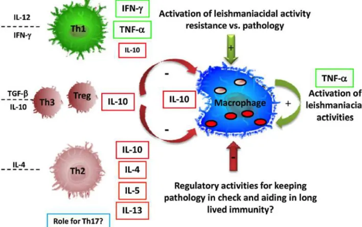
Figure 6: Models of immune responses in the infection caused by L. major (Gollob et al., 2008). CD4+ T lymphocytes balance the activation of macrophages for Leishmania control. Classically, CD4+ T cell subtypes Th1 and Th2 act controlling the anti-leishmanial activity in host macrophages. IL-12 and IFN-γ
stimulate the differentiation of the immune response to Th1 subtype, which activate anti-leishmanial mechanisms in macrophages increasing nitric oxide (NO) production by iNOS. IL-4, which differentiates
The murine model of leishmaniasis has been widely used to better understand the life cycle, course of infection and parasite-host relationship. This model has the advantage of permitting a highly controlled study, potentially clarifying important immunological aspects of the disease (Pereira and Alves, 2008).
The course of infection caused by L. amazonensis changes remarkably in different isogenic strains of mice. BALB/c animals are highly susceptible presenting progressive lesion growth (Calabrese and da Costa, 1992). C57BL/10 also present with non-healing lesions and persistent parasite loads (Afonso and Scott, 1993). C3H, C57BL/6, BDA and CBA mice are less susceptible to infection, but they do not heal and do not decrease the parasite loads (Afonso and Scott, 1993; Barral et al., 1983; Cortes et al., 2010; Jones et al., 2002; Neal and Bray, 1983; Soong et al., 1997). Thus, contrary to the established model of Th1/Th2 dichotomy in infections caused by L. major, there is not a resistant model to L. amazonensis infection. The characterization of the type of immune response generated in this infection still presents controversial data in the literature (Pereira and Alves, 2008). The capacity to mount a Th1 response is still considered as resistance factor, but the susceptible phenotype is remarkably variable. Many factors such as a Th2 dominant response (Lemos de Souza et al., 2000), the absence of a Th1 response (Afonso and Scott, 1993), or a mixed Th1/Th2 response (Ji et al., 2002; Sacks and Noben-Trauth, 2002) influences the increased susceptibility to L. amazonensis infection.
IFN-γ, cytokine associated with a Th1 response, is downregulated in draining lymph nodes of C57BL/6 mice infected with L. amazonensis, when compared to infection caused by L. braziliensis (Maioli et al., 2004). This could suggest a regulator mechanism promoted by L. amazonensis differing from other Leishmania species, related with the capacity of this specific parasite to successfully infect many mouse strains. Moreover, mice lacking IFN-γ present similar lesions compared to wild type animals at the initial stages of infection, suggesting an irrelevant role of this cytokine in the beginning of the infection. However, 2 months post infection, IFN-γ deficient mice loose control of lesion growth, increase the production of IL-4 and decrease the production of IL-12 and TNF-α. These data suggest that production of IFN-γ is crucial for the establishment of Th1 response in the disease (Pinheiro and Rossi-Bergmann, 2007).
The expression of IL-12, another important inflammatory cytokine during the Th1 response, has been shown to induce a significant reduction in parasitism during L. amazonensis infection. IL-12 is downregulated in infected C3H mice, and its expression is associated with the presence of the pathogen and it is independent of IL-4 expression (Jones et al., 2000). In addition to the reduction of IL-12 expression, those animals were also irresponsive to exogenous IL-12. This characteristic implies a deficiency in mRNA expression of IL-12b2 receptor, which is prevented somehow by the parasite (Jones et al., 2000).
The role of IL-4 during infection with L. amazonensis is very controversial. It was demonstrated that IL-4 deficient BALB/c mice express high levels of IFN-γ and anti-Leishmania
IgG2a antibodies 2 weeks post infections when compared to wild type. However, following infection with high dose of parasites (5 x 10⁶ promastigotes/footpads), the lesions progress equally in IL-4-/- or WT BALB/c mice with no differences in parasite loads. In contrast, IL-4-/- develops smaller lesions and lower parasite loads when inoculated with 105, 104 or 103 parasites/footpads (Guimaraes et al., 2006). On the other hand, some studies suggest that IL-4 is not a predominant factor of susceptibility in the infection, since its production decreases 3 weeks post infection. Moreover, IL-4 deficient mice or C57BL/6 and BALB/c mice treated with anti-IL-4 are not capable of healing lesions (Afonso and Scott, 1993; Ji et al., 2003; Jones et al., 2000).
The regulatory cytokine IL-10 also has an obscure role in the infection caused by L. amazonenis. Despite its importance as a limiting factor of the Th1 response during the acute phase of the disease, this cytokine does not have a similar role in the chronic phase: both WT and IL-10 deficient C57BL/6 mice exhibit a decreased Th1 response in the chronic phase. Moreover, the IL-10-/- mice reduce 1-2 logs of parasite burden during the infection with parasite persistence and similar lesion development. Therefore, IL-10 has a partial influence in the l. amazonensis infection (Jones et al., 2002).
Experiments with IL-10 deficient BALB/c mice showed that these animals, although unable to control the lesion progression, upregulate the production of IFN-γ, nitric oxide and mRNA levels of IL-12p40 and IL-12Rb2 when compared to wild type animals (Padigel et al., 2003). However, after anti-IL-4 treatment, the IL-10 deficient mice can control the infection and have few parasites in the lesion site. This shows that IL-10 together with IL-4 modulate the susceptibility impairing the development of a protective Th1 immune response.
system and promote disease. As with other Leishmania species causing cutaneous disease, L. amazonensis presents with three clinical manifestations varying according to the genetic background of the patient and the efficiency of the T-cell response: localized cutaneous leishmaniasis (LCL), anergic diffuse cutaneous leishmaniasis (ADCL), and borderline disseminated cutaneous leishmaniasis (BDCL) (Silveira et al., 2009).
The LCL is the most common form the leishmaniasis (about 95% of the cases) caused by L. amazonensis. This form is characterized by one or more ulcerative lesion(s) with good response to traditional antimonial therapy (Silveira et al., 2009). More than 50% of the patients do not present delayed type hypersensitivity (DTH) and lymphocyte proliferation reactivity (Silveira et al., 1998; Silveira et al., 1991). Moreover, LCL patients produce higher expression of IL-4 mRNA, without a decrease in the expression of IFN-γ mRNA. Indeed, the augmented expression of IL-4 mRNA suggests a possible mechanism of downregulation of IFN-γ activity, which could explain the lack of DTH in these patients (Silveira et al., 2004).
In contrast to LCL, patients with ADCL present with more than one non-ulcerative cutaneous lesions containing heavily parasitized macrophages (Soong et al., 2012). All ADCL patients present as DTH negative. This suggests a strong inhibition of T-cell response, which could contribute with disease dissemination (Barral et al., 1995; Petersen et al., 1982). Moreover, there are low numbers of CD4+ and CD8+ T cells in the lesions as well as very low IFN-γ mRNA expression and very high IL-4 mRNA expression, which characterize this form of disease as a well defined Th2 immune response inducer. Because of this well-defined Th2 response in ADCL, frequent recrudescence of the disease is observed after antimonial therapy (Bomfim et al., 1996; Silveira et al., 2004).
3.3.4. Evasion of immune response by the parasite
Because of the lack of the necessary machinery to invade actively the host, the Leishmania
are confined mostly to professional phagocytes. Although these parasites achieve invasion of many phagocytic cellular types, there is no evidence that the parasite can replicate in cells other than macrophages. These cells have primary defenses, including the activation of oxidative metabolism, by NADPH oxidase, iNOS, and synthesis and liberation of metabolites of arachidonic acid, which are induced by attachment and phagocytosis of microbial agents (Peters and Sacks, 2006). The microorganisms are endocyted through the plasma membrane of the phagocytic cells forming a vesicle in the cytoplasm called the phagosome. However, the phagosome does not have the machinery to digest and destroy the phagocyted intruder. So, there is a fusion of phagosome with lysosomes, a vesicle containing proteases, acid pH and ROS, capable to destroy and digest the microorganism.
The promastigote forms of Leishmania are covered by lipophosphoglycan (LPG) and a family of glycolipids named glycoinositolphospholipids (GIPs) (Naderer et al., 2004). LPG has a critical role in the infection of macrophages, protecting the promastigotes from reactive oxygen species (ROS) generated during the infection (Spath et al., 2003). Some studies showed that LPG could inhibit the fusion of the phagosome with the lysosome (Tuon et al., 2008), act as a ROS scavenger or inhibit the recruitment of NADPH oxidase to the phagosome membrane preventing damage to the parasite (Lodge et al., 2006). Another protein present on the surface of promastigotes, the gp63 protease, can inhibit the action of phagolysosome enzymes, favoring the persistence of the parasite inside the cell (Sorensen et al., 1994).
Amastigotes do not express LPG and gp63, however, they also have evasion mechanisms to avoid the host immune response. The membrane of amastigotes has high numbers of phosphatidylserine on the external portion, which permits an entrance mechanism into macrophages that is similar to that of apoptotic cells. This strategy avoids microbicidal processes and inflammatory responses (Wanderley et al., 2006). Furthermore, the opsonization of amastigotes by IgG antibodies from the host promotes the uptake of amastigotes via FC receptors, which induces the release of IL-10 (Kane and Mosser, 2001).
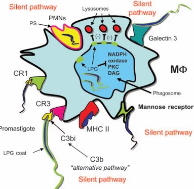
Figure 7 illustrates the major ways that Leishmania enter macrophages without triggering inflammatory and microbicidal mechanisms.
Figure 7: Many ways of inhibition of microbicidal activities used by Leishmania spp. to immune response evasion. Adapted (Peters and Sacks, 2006). Leishmania evade the microbicidal activities of macrophages in many ways. The phagocytosis of Leishmania by specific CR1 and CR3, manose receptors and phosphatidylserine receptors promotes, among other responses, release of IL-10. The uptake by these receptors and IL-10 production induce downregulation of oxidative defenses by macrophages. Inside of macrophages, the Leishmania also inhibit the fusion with lysosomes with phagosomes via LPG and eventually gp63.
3.4. NADPH oxidases, production of ROS and inflammation
reactions where the molecular oxygen (O2) is the final acceptor of electrons. However, the majority of the studies about the NADPH oxidases are concentrated on B lymphocytes. They catalyze superoxide anion (O2l
-) production by reducing oxygen using NADPH as an electron donor in the following reaction: NADPH + 2O2 à NADP+ + 2O2•- + H+ (Babior, 1999).
The superoxide generated by these enzymes is used as the initial molecule for the production of many ROS, including, free radicals and oxygen singlets, hydrogen peroxide, hydroxyl radicals and oxidized halogens. These oxidants generated are used by phagocytes to destroy invader microorganisms, but can also cause much tissue damage to the host. The damage caused by ROS is due to the spontaneous chemical reaction between ROS and cell components. There are oxidations by ROS of important proteins or even DNA, which promotes cell collapse and death. Because of this, the action of NADPH oxidase is highly regulated e activated just under certain stimuli (Babior, 1999).
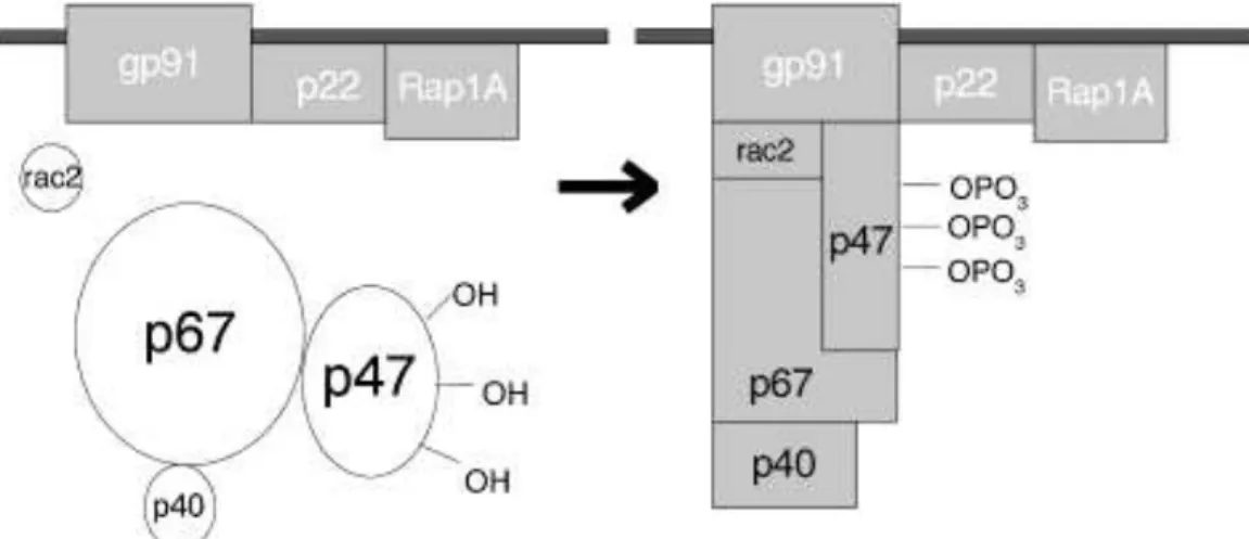
The structure of the NADPH-dependent oxidase of phagocytes (also called Nox2 or gp91phox) is composed by two transmembrane subunits (gp91phox and p22phox), three cytosolic components (p67phox, p47phox and p40phox) and a small G protein (rac1 or rac2). The rac proteins are kept inactivated by interaction with an inhibitor of guanine nucleotide dissociation, which impairs the exchanges of guanine nucleotides (GDP by GTP) by racs. The activation of NADPH oxidase is associated to attachment of cytoplasmatic components to transmembrane subunits (Babior, 2004).
The superoxide anion is the product of the multimeric NADPH oxidase enzyme complex. The NADPH complex contains the membrane-bound cytochrome b558, which two polypeptide chains (gp91phox and p22phox) and two non-identical heme groups (associated to gp91phox) are part of the membrane-linked complex (DeLeo et al., 2000; Nauseef, 2004). The gp91 subunit is expressed as a polypeptide of 58-kDa and after glycosylation in the endoplasmic reticulum becomes a 65-kDa subunit (Yu et al., 1999). After this subunit leaves the endoplasmic reticulum, it traffic trough the
trans-Golgi network, where it is additionally glycosylated, acquiring also the heme group, which increases the mass of the molecule to the final 91-kDa (DeLeo et al., 2000; Nauseef, 2004; Yu et al., 1999). The process of maturation of gp91phox increases the affinity for the p22, the other membrane-linked subunit and both are anchored to the membrane. The other subunits of NADPH oxidase, p40, p47, p67, and Rac2 are located in the cell cytoplasm and associate with the membrane-bound components upon activation (Nauseef, 2004). The assembly of the subunits to active the enzyme on the target membrane is crucial for the local release of optimal amounts of superoxide, since this molecule is unable to transpose cellular membranes.
In the presence of appropriated stimuli such as phagocytosis triggered by pathogen-associated molecular patterns (PAMPs) binding to toll receptors, there is activation of p38 MAPK via MyD88. It was demonstrated that Myd88-/- peritoneal macrophages are inefficient in eliminating Gram-negative bacteria such as Escherichia coli or Salmonella typhimurium. This deficiency was correlated with decreased production of superoxide anion mediated by NADPH oxidase. Moreover, these Myd88-/- peritoneal macrophages have defective NADPH oxidase assembly due to impaired p38 MAPK activation and consequent p47phox phosphorylation (Laroux et al., 2005). Therefore, after p38 MAPK, p47phox is phosphorylated, which exposes the binding site to p22phox assembly (Laroux et al., 2005). The assembly of the three subunits (p22phox, p47phox and gp9phox) permits the recruitment of p67phox (Leto et al., 1994), which after interaction with rac, exposes the binding site to gp91phox, activating the superoxide production (Mizrahi et al., 2006). The subunit p40phox seems to be an obligatory binding partner of p67phox, but its role in the activation of NADPH oxidase is still unclear. However, animal models deficient to subunit p40phox (Ellson et al., 2006) and granulomatous disease patients presenting mutations in the PX domain of p40phox (Matute et al., 2009) established the importance of this subunit in the activity of the NADPH oxidase. The phosphorylation of the PX domain of p40phox is an important signal to activation of NADPH oxidase (Chessa et al., 2010).
granulomatous disease (CGD) (Pollock et al., 1995). Patients with CGD have early recurrent infections that can lead to death as early as childhood. Although they show normal chemotaxis, degranulation and phagocytosis, the patients show deficiency in destruction of phagocyted microorganisms because of the lack of metabolites generated by superoxide anion. In addition to the symptoms found in humans, mice with CGD also show higher numbers of neutrophils after peritonitis caused by chemical components (Pollock et al., 1995) and an increase in polymorph nuclear cells, chemokines and inflammatory cytokines as well the global lung inflammation caused by experimental infection with Pneumococcal pneumonia (Marriott et al., 2008).
At the moment, there are seven isoforms of NADPH oxidase identified in mammals, the isoforms Nox-1 to 5, and Duox-1 and 2. Others variations in subunits of the complex, the p47phox isoform (Noxo1) and p67phox isoform (Noxa1) also were identified in mammalian cells (Banfi et al., 2003; Banfi et al., 2001; Cheng et al., 2001; De Deken et al., 2000; Dupuy et al., 1999; Geiszt et al., 2000; Suh et al., 1999). However, except for gp91phox, the others isoforms do not seem to be involved in phagocytes defense.
Superoxide may associate with other ions to generate even more toxic products such as O3, HOCl, HOBr, ONOO-, NO2•, CO3•-, H2O2, •OH, etc. (Halliwell, 2006). However, because of its negative charge, superoxide is unable to transpose biological membranes. Among the cited molecules, hydrogen peroxide (H2O2) stands out as being formed by action of superoxide dismutase (SOD) on O2•- and H2O. SOD is an antioxidant enzyme present in many organisms and is a hallmark of antioxidant defense in many systems. It catalyzes the formation of H2O2 by combination of O2•- and 2H+ ions. The SOD transforms the highly reactive superoxide anion in a lesser reactive hydrogen peroxide, which protect partially the proteins of the organism from oxidation. There are three isoforms of SOD presented in mammals: SOD1, localized in cytoplasm and dependent of Cu/Zn metals; SOD2, presented in mitochondrial matrix and dependent of Mn metal; and SOD3, a secreted isoform also dependent of Cu/Zn (Fukai and Ushio-Fukai, 2011). Mice deficient in SOD2, have uncontrolled oxidative stress and die a few days after the birth (Li et al., 1995), which illustrates the importance of SOD to protect the organism of superoxide action.