Arq Bras Cardiol volume 73, (nº 3), 1999
Mesquita et al. Propranolol and QT dispersion in congestive heart failure
2 9 5
Hospital Universitário Antônio Pedro - Niterói
Mailing address: Evandro Tinoco Mesquita - Av. Comandante Ari Parreiras, 311/ 1102 - 24230-320 - Niterói, RJ, Brazil.
Received on 1/26/99 Accepted on 4/24/99
Objective – Studies have shown that therapy with
beta-blockers reduces mortality in patients with heart fai-lure. However, there are no studies describing the effects of propranolol on the QT dispersion in this population. The objective of this study was to assess the electrophysiologi-cal profile, mainly QT dispersion, of patients with heart fai-lure regularly using propranolol.
Methods – Fifteen patients with heart failure and
using propranolol were assessed over a period of 12 months. Twelve-lead electrocardiograms (ECG) were re-corded prior to the onset of beta-blocker therapy and after 3 months of drug use.
Results – A significant reduction in heart rate, in QT
dispersion and in QTc dispersion was observed, as was also an increase in the PR interval and in the QT interval, after the use of propranolol in an average dosage of 100 mg/day.
Conclusion – Reduction in QT dispersion in patients
with heart failure using propranolol may explain the re-duction in the risk of sudden cardiac death with beta-blo-cker therapy, in this specific group of patients.
Key words: congestive heart failure, electrocardiography, beta-blocker
Arq Bras Cardiol, volume 73 (nº 3), 295-298, 1999
Evandro Tinoco Mesquita, Flávia Cristina Carvalho de Deus, Cassia Regina Guedes, Eduardo Reis Maia, César Gerson Pereira Subieta, Humberto Villacorta, Patrice Alessandra dos Santos, Helena
Cramer, Valeria Battistella Amado dos Santos, Luis José Martins Romeo
Niterói, RJ - Brazil
Effects of Propranolol on the QT Dispersion in Congestive
Heart Failure
Original Article
Currently, heart failure (HF) of all the cardiovascular diseases is a major public health problem. In addition to the significant impact on quality of life, HF drastically reduces the patient’s survival and accounts for a substantial per-centage of deaths caused by cardiovascular diseases 1.
Death in patients with HF may result from progressive failure of the heart pump, or in more than half of the patients it may be sudden, probably resulting from arrhythmias 1.
A measurement that may be very useful in this context and that has been identified as a marker of electrical myocar-dial instability is the variation in the duration of the QT in-terval between the leads, called QT dispersion.
QT dispersion is a new death predictor of HF, and it reflects variations in the repolarization in different regions of the myocardium. This results from reentrant mechanisms due to the existence of areas of slow conduction, which constitute important physiological substrate for the arrhythmias. It has already been demonstrated that heterogeneity of ventricular repolarization is associated with a higher vulnerability to arrhythmias and occurrence of sudden death 2.
Beta-blockers have been described as capable of redu-cing cardiovascular mortality after episodes of acute myocardial infarction (AMI) 3, particularly reducing the in-cidence of fatal arrhythmias. The behavior of the QT disper-sion in patients with HF using beta-blockers, however, has not yet been elucidated. This study analyzes prospectively the variations of the QT dispersion in the surface electrocar-diogram (ECG) of patients with HF, before and after the use of propranolol, to assess the electrophysiologic effect of the drug on an important marker of the risk of sudden death.
Methods
2 9 6
Mesquita et al.
Propranolol and QT dispersion in congestive heart failure
Arq Bras Cardiol volume 73, (nº 3), 1999
Antônio Pedro. The exclusion criteria for the use of propra-nolol were the following: atrial fibrillation, sinus bradycardia lower than 60 bpm, PR interval >0.25s, bronchial asthma, intermittent claudication, and decompensated diabetes mellitus. All patients signed a written consent to take part in the study, which was approved by the Committee on Ethics in Research of the Universidade Federal Fluminense.
After a clinical stabilization period of 3 months, during which the patients used digoxin (0.25mg/day), enalapril (mean dose of 20mg/day), and sometimes diuretics, 12-lead ECGs were recorded at a rate of 25mm/s. Treatment with propranolol was then started at an initial dose of 30mg/day. This dose was gradually increased (weak progression of 30% to 50% of the dose, according to clinical response) until the maximum dose of 120 mg/day or the heart rate of 60 bpm, or both were achieved. Patients were hemodynamica-lly stable during the entire period of assessment, 80% of whom were in functional class I and 20% in functional class II. The patients did not use any other cardioactive drugs and were instructed to seek the outpatient care unit in case of malaise, fatigue, edema, paroxysmal nocturnal dyspnea, asthma rales, and symptoms of hypotension or bradycardia (dizziness, somnolence, syncope). New ECGs were recor-ded after a mean period of 3 months of using propranolol.
ECG tracings were blindly analyzed by 2 independent investigators, and the following mean values of the varia-bles were calculated: heart rate (HR), PR interval, QT inter-val duration, corrected QT (QTc), QT dispersion, and QTc dispersion, in 4 successive complexes for each lead. The QT interval was measured starting from the onset of the QRS complex until the end of the T wave, which is the return of the T wave to the baseline. When the U wave was present, the QT interval ended at the middle point between the T and U waves, which was obtained at the intersection of a tan-gent line to the repolarization line with the isoelectric line. QTc was obtained using Bazett’s formula 5 (QTc = QT/√RR). QT dispersion, defined as the difference between maximum and minimum QT, was calculated based on the QT intervals obtained in the 12 leads. The same was done for the QTc dispersion, which was corrected using the RR interval.
The results obtained were statistically analyzed, and the mean values of each variable were compared in the pre-and post-beta-blocker period, using a paired t test pre-and a sig-nificant value of p <0.05.
Results
The study group consisted initially of 27 patients con-secutively referred to the Heart Failure Outpatient Care Unit. Twelve out of these 27 patients were excluded from the study due to the following reasons: 2 had thyroid dysfunc-tion (hypo- and hyperthyroidism), 4 had atrial fibrilladysfunc-tion on the ECG, and 6 did not return to the outpatient care unit.
The 15 remaining patients underwent the protocol and were followed up from January ’97 to January ’98. This gro-up consisted of 8 men (53.3%), with a mean age of 51.9 years (age ranged from 28 to 72 years); 73.3% were white and 26.6% were black.
The etiology of the HF was ischemic or hypertensive, or both in 8 patients (53.3%), idiopathic in 4 (26.7%), and rheu-matic, chagasic or alcoholic in the 3 remaining patients (20.0%). The mean dosage of propranolol was 100mg/day, and the mean time of treatment was 3 months.
A clear change on the electrocardiographic profile of the patients with HF before and after the use of propranolol was observed.
Propranolol significantly reduced the HR and promo-ted a slight increase in the PR interval. An increase also occurred in the duration of the QRS complex and also in the duration of the QT interval. The maximum QT interval was obtained in one of the leads of the horizontal plane (V1 to V6) in 61% of the ECGs (out of the 26 ECGs analyzed). There was no significant variation in the QTc; extension of the QT interval, therefore, may have resulted only from the conco-mitant reduction of the HR, because the QTc was not alte-red (table I).
The statistically significant reduction in the QT dispersion was the main finding, reflecting an electrophy-siological improvement in these patients with HF caused by the use of propranolol.
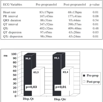
QTc dispersion calculated in 13 patients also showed a significant reduction, indicating that the variation in the QT dispersion does not depend on the HR (fig. 1) 6.
Bundle-branch blocks defined as QRS >120ms were documented in 4 (16.0%) out of the 26 ECGs of the patients with HF, who could be analyzed. Left bundle-branch block (LBBB) was present in 3 patients (12.0%) and right bundle-branch block (RBBB) in only one patient (4.0%). No relation between the values of the QT and QTc dispersions and the presence of LBBB and RBBB was observed.
Table I - Electrocardiographic variations with the use of beta-blocker
ECG Variables Pre-propranolol Post-propranolol p-value
Heart rate 83±17bpm 68±13bpm 0.01
PR interval 167±43ms 177±41ms 0.08
QRS duration 88±31ms 93±44ms 0.54
QT interval 347±52ms 390±57ms 0.01
QTc 402±32ms 409±40ms 0.48
QT dispersion 97±45ms 63±20ms 0.03
QTc dispersion 98±39ms 63±24ms 0.01
Fig. 1 - Variation of QT and QTc dispersions after the use of propranolol (Prop).
Disp. Qt Disp. Qtc
Pre-prop
Arq Bras Cardiol volume 73, (nº 3), 1999
Mesquita et al. Propranolol and QT dispersion in congestive heart failure
2 9 7
Discussion
This study is the first to analyze the effects of pro-pranolol on QT dispersion on the 12-lead ECG of patients with HF.
QT dispersion, QTc dispersion and the respective adjusted dispersions were observed to be significantly in-creased in patients with HF in this study, when compared with the normal values reported in the literature.
After 3 months of propranolol use at a mean dose of 100mg/day, an important reduction occurred in these values (fig. 2), suggesting a beneficial effect of the drug on myo-cardial electrophysiology in HF.
A number of studies have identified the QT dispersion as a marker of risk for sudden death 7. Analysis of QT dis-persion has been satisfactorily used to stratify the risks and to measure the effects of certain interventions on the ventri-cular repolarization. Baar et al 1 showed that patients with HF, who suddenly evolved to death, had values of QT dis-persion significantly higher than the values found in patien-ts who died due to progressive HF or who survived. Buja et al 8 reported that QT dispersion is greatly increased in pati-ents with diastolic HF compared with healthy patipati-ents. They also showed that QT dispersion is increased in patien-ts with severe ventricular arrhythmias or those who evolved to sudden death.
Day et al 9 concluded that the variability of the QT in-terval on the surface ECG reflects the regional myocardial electrical activity, not representing just a technical artifact. The finding of reduction in QT dispersion in patients with different diseases treated with class III antiarrhythmic drugs– sotalol and amiodarone – supports this theory. Another study confirming the reliability of the QT dispersi-on as a marker of risk for sudden death was developed by Goldner et al 10 and compares the QT dispersion with the high-resolution ECG. The study showed that QT dispersion is a sensitive indicator of spontaneous or induced ventricu-lar tachyarrhythmias with a sensitivity of 88% and specifici-ty of 57%. Higham and Campbell 11 observed high values of QT and QTc dispersion in patients in the first hours after AMI; reduction in these values occur with clinical evolution and use of thrombolytic agents.
In addition, among all risk factors analyzed by Pinsky et al 12 in the study on sudden death in patients awaiting heart transplantation, QT dispersion appeared as the best predictor of death during the waiting period. In a huge contrast, in the measurements obtained on the 12-lead ECG, variability of the QT interval was a strong predictor of mor-tality. When the patients were divided into 2 groups (QT dispersion <140ms and QT dispersion >140ms), differences of one year in mortality were observed (24% and 56%, respectively, p<0.05). Analysis of QT dispersion allowed the identification of high-risk patients only through ECG, which is a simple, low-cost, noninvasive method, available in most centers.
Pinsky et al 12 also reported that the extension of the QT interval did not prove to be a marker of risk for sudden death in patients with cardiac dysfunction. Extension of the QT interval seems not to reflect the alteration of myocardial ventricular repolarization. This alteration is indicated by QT dispersion.
Ventricular arrhythmias are common in HF, and they seem to have a multifactorial cause, including ventricular reentrant mechanisms. The reentrant circuit is formed by myocardial structural alterations, resulting in a slower and more heterogeneous ventricular repolarization 13. As the QT interval reflects the duration of the myocardial action poten-tials, the increase in the QT interval variability, i. e., the increase in QT dispersion, acts as an indicator of susceptibi-lity to possibly fatal ventricular arrhythmias 14. The results of this study support the evidence that beta-blockers have a beneficial effect on HF, possibly due to a mechanism of reduction of the arrhythmogenic potential, which is expres-sed by a decrease in the QT dispersion on the ECG.
Previous studies have shown the effects of beta-blo-ckers on the HF. Recent studies using carvedilol 15, a nonselective beta-blocker with vasodilating properties, have shown results similar to ours, with significant reducti-on in the QT and QTc dispersireducti-ons in patients with HF.
The mechanism remains unknown. It is suggested, ho-wever, that increased adrenergic tonus has a significant effect on QT dispersion 16. Patients with the long QT syndrome
Fig. 2 - Individual variations of QT and QTc dispersions (A and B, respectively) with the use of propranolol (Prop).
A. QT dispersion (ms) B. QTc dispersion (ms)
Post-Prop
2 9 8
Mesquita et al.
Propranolol and QT dispersion in congestive heart failure
Arq Bras Cardiol volume 73, (nº 3), 1999
1. Barr CS, Naas A, Freeman M, Lang CC, Struthers AD. QT Dispersion and sudden unexpected death in chronic heart failure. The Lancet 1994; 343: 327-9. 2. Merx W, Yoon MS, Han J. The role of local disparity in conduction and recovery
time on ventricular vulnerability to fibrilation. Am Heart J 1977; 94: 603-10. 3. Chadda k, Goldstein S, Curb JD, Byington R. Effect of propranolol after acute
myocardial infarction in patients with congestive heart failure. Circulation 1986; 73: 503-10.
4. Mc Kee PA, Castelli WP, Mc Namara PM, Kannel WB. The natural history of car-diac heart failure, The Framingham Study. N Engl J Med 1971; 285: 141. 5. Bazett HC. An analysis of the time-relations of electrocardiograms. Heart 1920; 7: 353. 6. Malik M, Camm AJ. Mystery of QTc Interval Dispersion. Am J Cardiol 1997; 79: 785-7. 7. Mantari M, Oikarinen L, Manninen V, Viitasalo M. QT dispersion as a risk factor for sudden cardiac death and fatal myocardial infarction in a coronary risk popu-lation. Heart 1997; 78: 268-72.
8. Buja G, Miorelli M, Turrini P, Melacini P, Nava A. Comparison of QT Dispersion in hypertrophic cardiomyopathy between patients with and without ventricular arrhythmias and sudden death. Am J Cardiol 1993; 72: 973-6.
9. Day CP, McComb JM, Campbell RWF. QT Dispersion: an indication of arrhythmia risk in patients with long QT intervals. Br Heart J 1990; 63: 342-4. 10. Goldner B, Brandspielgel HZ, Horwitz L, Jadonath R, Cohen TJ. Utility of QT
Dis-persion combined with the signal-averaged electrocardiogram in detecting patients susceptible to ventricular tachyarrhythmias. Am J Cardiol 1995; 76: 1192-3.
References
11. Higham PD, Campbell RWF. QT dispersion. Br Heart J 1994; 71: 508-10. 12. Pinsky DJ, Sciacca RR, Steinberg JS. QT Dispersion as a marker of risk in patients
awaiting heart transplantation. J Am Coll Cardiol 1997; 29: 1576-84. 13. Perkiomaki JS, Koistinen MJ, Yli-Mayry S, Huikuri HV. Dispersion of QT
interval in patients with and without susceptibility to ventricular tachyarrhyth-mias after previous myocardial infarction. J Am Coll Cardiol 1995; 26: 174-9. 14. Davey PP, Bateman J, Mulligan IP, Forfar C, Barlow C, Hart G. QT interval
disper-sion in chronic heart failure and left ventricular hypertrophy: relation to autono-mic nervous and Holter tape abnormalities. Br Heart J 1994; 71: 268-73. 15. Gill AE Jr, Gilbert EM, Lowes BD, Abraham WT, Briston MR, Reiter MJ.
Carve-dilol Reduces QT Dispersion in Patients with Congestive Heart Failure. In: Anais da “70th Scientific Session Orange” (Supplement to Circulation). Orlan-do: Country Convention Center, 1997: I-577.
16. Fisher ML, Plotnick GD, Peters RW, Carliner NH. Beta-blockers in congestive cardiomyopathy – conceptual advance or contraindication? Am J Med 1986; 80-(suppl 2B): 59-65.
17. Mesquita ET, Maia ER, Guedes CR, et al - Effects of propranolol on ventricular repolarization in patients with congestive heart failure. J Am Coll Cardiol 1998; 31(suppl C): 428C.
18. Maia ER, Subieta CGP, Tavares CMF, et al. Effects of propranolol on the parame-ters of systolic function in congestive heart failure. J Am Coll Cardiol 1998; 31 (suppl C): 428C.
treated with beta-blockers or with sympathetic denervation showed reduction in QT dispersion. This finding is particu-larly appealing when the neurohumoral hypothesis of HF, which suggests that the progression of the disease results from the chronic action of the adrenergic system 16, is consi-dered. Further investigations are still required; the excessive neuronal activation in HF may, however, influence the QT dispersion, promoting a physiopathological interaction with the heart mortality in these conditions.
Even though carvedilol is the paradigm beta-blocker in HF, no comparative studies have been conducted compa-ring the less costly first-generation beta-blockers with car-vedilol. In public hospitals, because of severe socioecono-mic problems, patients with HF have difficulty paying for ex-pensive treatments, especially those involving multiple
drugs. This study has shown that the effect of propranolol on cardiac function is similar to that reported in the literature on the most recent drugs 17,18.
This study has shown that a significant variation in the electrocardiographic tracings may be obtained after the use of propranolol. In addition, the ECGs were manually analyzed, and to minimize this bias, the tracings were sepa-rately assessed by two investigators, who ignored the clini-cal details of this complementary test.