Santa Casa de Misericórdia de Goiânia - Hospital Santa Helena
Mailing address: José Maria Dias de Azeredo Bastos - Rua C-249, 65/201 - 74280-140 - Goiânia, GO - E-mail: [email protected]
Objective - To evaluate the immediate results of
per-cutaneous mechanical mitral commissurotomy.
Methods - Thirty patients underwent percutaneous
mechanical mitral commissurotomy performed with a Cribier's metallic valvulotome from 8/11/99 to 2/4/00. Mean age was 30.7 years, and 73.3% were women. With re-gards to functional class, 63.3% were class III, and 36.7% were class IV. The echocardiographic score had a mean value of 7.5± 1.8.
Results - The mitral valve area increased from
0.97±0.15cm2 to 2.16±0.50cm2 (p>0.0001). The mean diastolic gradient decreased from 17.9±5.0mmHg to 3.2±1.4mmHg. The mean left atrial pressure decreased from 23.6±5.4mmHg to 8.6±3.1mmHg, (p>0.0001). Sys-tolic pressure in the pulmonary artery decreased from 52.7±18.3mmHg to 32.2±7.4mmHg. Twenty-nine cases were successful. One patient developed severe mitral regurgitation. Interatrial septal defect was observed and one patient. One patient had cardiac tamponade due to left ventricular perforation. No deaths occurred.
Conclusion - This method has proven to be safe and
efficient in the treatment of rheumatic mitral stenosis. The potential advantage is that it can be used multiple times after sterilization, which decreases procedural costs significantly.
Key words: commissurotomy, mitral valve, and metallic
valvulotome
Arq Bras Cardiol, volume 77 (nº 2), 126-31, 2001
José Maria Dias de Azeredo Bastos, César A. Esteves, David Araújo, Luciene Ap. M. A. Bastos, Magno Eistein, Geraldo Paulino Santana, Gilson José de Oliveira, Luiz Brasil, Antônio Calzada,
Delzirene Botelho Calzada, José Pereira, Rômulo Sales, Nivaldo Gomes de Olivera
Goiânia, GO - Brazil
Percutaneous Mechanical Mitral Commissurotomy Performed
With a Cribier’s Metallic Valvulotome. Initial Results
The therapeutic approach to mitral stenosis has evolved considerably since 1984, after the first report of percutaneous valvuloplasty by Inoue et al 1, and it has
become an accepted alternative to surgical commis-surotomy, leading to comparable immediate- and long-term results 2-4.
Currently, 2 techniques of percutaneous dilatation are applicable, the double-balloon technique and the Inoue technique, which is by far the most often used worldwide. However, the cost of the procedure with these techniques is extremely high, due to the balloon catheter used, which, in a way, is a limitation for countries with restricted financial resources. Consequently, most centers reuse these balloon catheters several times, although they are intended to be disposable catheters. Imperfect sterilization introduces potential hazards and decreased performance.
Recently, Alain Cribier 5 developed a metallic device,
replacing the latex balloon, for percutaneous dilatation of the mitral valve.
The advantage of this device, because it is a metallic material, is that it enables appropriate sterilization, thus ma-king it possible to be reused several times without any loss of performance, and significantly reducing the cost of per-forming the procedures. Other advantages are the improved efficacy and tolerance of the technique, which is aimed at acting on mitral valves fused as a result of rheumatic fever. The objective of the present study is to stress the safe-ty of the method and present the immediate results of this new technique.
Methods
detected by transesophageal echocardiography were excluded.
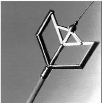
The valvulotome (Medicorp Inc.) consists of a me-tallic dilator screwed onto the distal end of a catheter. The entire system is made of the following components: metallic dilator – made of stainless steel, when closed, is a cylinder 5-cm long and 5-mm wide, with a slightly tapered tip. The distal half of the dilator consists of two hemicylindrical bars 20 mm in length that can be opened out in parallel up to a maximum extent of 40 mm by a lever-arm system. The dilator has an internal tube that allows passage of a traction wire and also the recording of distal pressure. This detachable device is screwed onto the distal end of a catheter (figs. 1 and 2); catheter – is 13 F, 4.3mm in diameter and 170cm in length; its proximal end has a connector for recording distal pressure, and it also allows connection of the activating pliers; metallic guidewire – a stainless steel guidewire that is 0.035 in in diameter and 270cm in length. A metallic bead 2 mm in diameter is soldered at the junction of the stiff core and the 10 cm long floppy distal end. Once positioned in the left ventricle, the metallic dilator is pushed over the wire across the mitral valve. When positioned, this guidewire will become a traction system that allows the dilator to be opened; for that, the metallic bead is positioned in contact with the distal end of the dilator, and the guidewire is securely locked with a threaded fastener located in the activating pliers. Squeezing the arms of the pliers will cause a backward traction of the guidewire and the metallic bead, which is transmitted to the distal end of the dilator, thus, forcing the bars to spread apart parallel to the metallic dilator; activating pliers – attached to the distal end of the catheter. Manual pressure exerted on the arms of the pliers allows the metallic dilator to open as previously described, and the release of the pressure in the guidewire closes the
dilator. The activating pliers are made of a caliper that enables the degrees of opening of the bars to be altered from outside with the help of a cursor (the programmable degrees of opening are 35, 37 and 40mm) and a safety lock that prevents the complete closure of the dilator after the release of the pressure exerted on the pliers (it holds the dilator open at 20mm). To obtain a complete closure of the metallic dilator, after withdrawal from the mitral valve, this safety lock must be activated manually. This security system was designed to avoid any accidental extraction of valvular tissue, and a thread fastener is present that was designed to block the guidewire into the commissurotomy at the time of opening. After dilation, the metallic dilator can be unscre-wed from the catheter and sterilized by autoclave for reuse. The activating pliers and guidewire can be sterilized again. Only the catheter is meant for single use 5.
The percutaneous mechanical mitral commissurotomy procedure is performed while the patient is under anesthesia and mild sedation. The entry site is the right femoral vein, which has to be punctured approximately 2cm below the inguinal ligament.
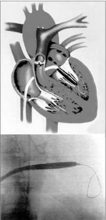
An 8 F Mullins catheter is used to makes a transseptal puncture. It is recommended that the septal puncture be made approximately 2cm below the usual site used in the Inoue technique, to facilitate the passage of the metallic dilator across the valve. Afterwards, after septal perforation, an initial dose of heparin 2,500 UI is administered; an additional dose of 2,500 UI will be administered after dilatation of the atrial septum and confirmation of the absence of pericardial effusion. Both the needle and dilator are removed, leaving the Mullins sheath in the left atrium. Left atrial pressure and left atrioventricular gradient are then measured. A floating balloon catheter (Critikon USCI) is advanced through the sheath and used to cross the mitral
Fig. 2 - Cribier’s metallic dilator open is made of 2-hemicylindrical bars 20mm in length that can be opened out in parallel.
valve. The distal end of the balloon catheter is positioned at the apex of the left ventricle, and the sheath is advanced over it, beyond the mitral valve orifice. The balloon catheter is then removed, and the guidewire of the device is advan-ced through the sheath in the left ventricle, with the metallic bead positioned in the midventricle, clearly beyond the mitral valve.
The Mullins sheath is removed, and a 14 F polye-thylene dilator is advanced over the wire to enlarge the atrial septum puncture site.
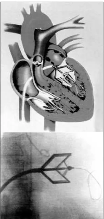
The identical maneuver is then completed by addi-tional dilation with an 18 F dilator, which is also used to enlarge the femoral vein puncture site (fig. 3). The commis-surotomy is then advanced over the wire, and its distal end is placed across the mitral valve. At that time, the guidewire is pulled back until the bead is firmly held against the tip of the valvulotome, and then securely fastened by screwing the thread fastener of the pliers (fig. 4). The dilation can then be performed by squeezing the arms of the activating pliers. The desired extent of the bar opening is obtained by use of the caliper. At least 2 openings of the dilating bars are performed (fig. 5). After dilation, the device is pulled back into the left atrium, with the guidewire kept in the left ventricle. The transvalvular gradient is assessed; left atrial pressure is measured with the pressure line of the device. Whenever available, 2-dimensional (2D) echocardiography is performed to assess the quality of commissural splitting (fig. 6) and to obtain a preliminary assessment of the mitral valve area. If necessary, an additional opening at of a larger size can be made. After valvotomy, a left ventricular angiogram is performed to assess the degree of any subsequent mitral regurgitation.
M-mode and 2-D echocardiography are used to confirm the severity of mitral stenosis and valve mor-phology. An echocardiographic score in 16 grades is used to assess valve thickness, leaflet mobility, valvular
calcification, and subvalvular disease, each of these being graded from 1 to 4 according to the severity.
The mitral valve area is determined by echocardiogra-phy with planimetry and using the Doppler pressure half-ti-me half-ti-method, and planihalf-ti-metry is the reference half-ti-method used for assessment of the results.
Left and right heart pressures and the mean transmitral pressure gradient are measured through catheterization, be-fore and after percutaneous mechanical mitral commissuro-tomy. A left ventricular angiogram in the 30° right anterior oblique view is performed, using the same amount and delivery rate of contrast to assess left ventricular function and the presence and severity of any mitral regurgitation.
Fig. 3 - Dilation of the atrial septum puncture site with an 18 F dilator.
A coronary arteriography was performed before the procedure in patients with documented ischemia, in men over 40, and women over 50 years of age. The final echocar-diographic results were recorded 24h after the procedure. Success was defined when mitral valve area was >1.5cm2
after 24h, in the absence of Seller's grade >2 mitral regurgi-tation.
Data concerning patient’s age, functional class (NY-HA), and history of previous mitral commissurotomy were observed.
Continuous variables are expressed as mean ± standard deviation. Variation in continuous variables from baseline to day 1 after completion of percutaneous mechanical mitral commis-surotomy was assessed with the paired Student’s t test.
Results
Percutaneous mechanical mitral commissurotomy was successfully performed in 29 patients (97%), with only 1 unsuccessful case, in which failure to cross the mitral valve occurred due to cardiac tamponade, which was caused by wire perforation of the left ventricle, and resulted in the need for the patient to have emergency cardiac surgery. After surgery, no further events occurred.
The maximum extent of the metallic dilator bar opening was 40mm in 27 patients (91%) and 37mm in 2 patients. The mean number of openings was 4. Through this technique a significant increase occurred in valve area, which was 0.97±0.15cm2 before percutaneous mechanical mitral
commissurotomy, evaluated with planimetry, and it became 2.16±0.50cm2 with p<0.0001 (tab. I) after the procedure.
Bilateral splitting of the commissures was noted in all
Table I - Data before and after percutaneous mechanical mitral commissurotomy
Before PMMC After PMMC P
Pressure, mm Hg
Arterial pulmonar (systolic) 52.7±18.3 32.2±7.4 P<0.0001 Left midatrial 23.6±5.4 8.6±3.1 P<0.0001 Mean gradient 17.9±5.0 3.2±1.4 P<0.0001 Valve area, cm2 (planimetry) 0.97±0.15 2.16±0.50 P<0.0001
*mitral valve area obtained 24 h after commissurotomy; PMMC-percutaneous mechanical mitral commissurotomy.
Fig. 5 - Dilation realization through metallic bead against tapered tip of the valvotome.
patients. A nonsignificant difference was noted when the valve area was correlated with the echocardiographic score, although the population was small. The mean duration of the procedure from the time the septal puncture was completed to the withdrawal of the catheters was 45min.
In addition to the above-mentioned case of hemoperi-cardium with tamponade, which resulted from left ventricle perforation, another 2 cases of deep venous thrombus occurred, and 1 of these patients evolved to mild associated pericardial effusion; both were clinically treated, without events.
One patient evolved from mild to moderate mitral re-gurgitation; however, after evident improvement in symp-toms, he continued with clinical treatment and follow-up.
On transthoracic color flow Doppler, echocardiogram interatrial septal defect was detected, which was trivial after the procedure.
No other complications occurred, and the patients were discharged an average of 3 days after the procedure. In the study series, no deaths occurred.
Discussion
Our results show that percutaneous mechanical mitral commissurotomy can be used successfully and safely instead of surgery.
Analysis performed 24h after percutaneous mecha-nical mitral commissurotomy showed that the mean valve area was 2.16cm2 ± 0.50cm2, a 122% increase from baseline.
In the literature, several reports confirm that the final valve area was a significant predictor of favorable long-term out-come. Concerning the immediate results of balloon mitral valvulotomy (BMV) 6,7, postvalvulotomy valve areas
obtai-ned are slightly less with the Inoue technique than with the double-balloon technique, as Vahanian et al 8 demonstrated
in a study where mitral valve area reached an average of 1.84cm2 with the Inoue technique and 1.93cm2 with the
double-balloon technique.
The very limited incidence of complications in this first series of patients is noteworthy. Only 1 patient had cardiac tamponade due to left ventricle perforation, and required emergency cardiac surgery. This well-known, life-threatening complication of BMV has an incidence of 1% to 9% reported in the literature 9-11 and can result from
inadver-tent atrial perforation during transseptal catheterization or from perforation of the left ventricle by guidewires in the double-balloon technique. Despite the necessity of placing a guidewire in the left ventricle with this new technique, the risk of ventricular perforation seems to be decreased because of the high flexibility of the distal 10 cm of the wire, the stability of the dilator head during opening, and the traction with subsequent backward movement of the wire associated with the opening process.
Of the 30 patients studied, only 1 (3%) developed severe mitral regurgitation after percutaneous mechanical mitral commissurotomy. In the literature, the incidence of severe mitral regurgitation after BMV is reported to vary
between 1.4% and 7.5% 9,10,12,13 and no difference occurs
between the single- or double-balloon technique.
Finally, no significant interatrial shunting could be detected after percutaneous mechanical mitral commis-surotomy with transthoracic echocardiography or transe-sophageal color flow Doppler. With BMV, the incidence of residual transatrial shunting varies between 2.5% and 87%, depending on the modality of assessment 10,11,12-14.
However, regardless of the modality of assessment used, most of the transatrial shunts closed spontaneously during follow-up 14,15.
In a randomized study, the results of closed surgical commissurotomy were not as good as those of BMV and open commissurotomy, with a mean increase in valve area to only 1.6 cm2 versus 2.2 cm2, respectively 2.
These results are in disagreement with other studies in which BMV and closed surgical commissurotomy are compared 2,3. Despite the resemblance of the instrument
used in closed surgical commissurotomy and percutaneous mechanical mitral commissurotomy, some differences do exist, such as the self-positioning of the dilator’s bars in the commissures during percutaneous mechanical mitral commissurotomy. A possibility of immediate evaluation of the hemodynamic and echocardiographic results after dilation offers the possibility of subsequent additional dilator openings of a larger size when needed.
A perfect positioning of the metallic device across the valve before opening can be achieved by several means: 1) a satisfactory position is obtained when the distal half of the dilator is located slightly ahead of the aortic orifice, which is indicated by the presence of a pigtail catheter placed against the aortic leaflets; 2) the left atrial angiogram obtained after transseptal catheterization with a fixed image in on a monitor, makes it easier to locate the free edges of the valve; 3) the transition between the left ventricular and left atrial pressure curves provides an indication of the location of the mitral valve; 4) the positioning is monitored by trans-thoracic echocardiography.
The extent of bar opening is a significant factor in the immediate percutaneous mechanical mitral commissuro-tomy results.
In adult patients of average stature, we usually open the metallic device to 40mm at first. However, in patients wi-th a body surface area <1.5m2, we start with 37mm. In
chil-dren, the initial opening is 35mm; it is worth mentioning that in all cases, the opening can be gradually increased accor-ding to the results.
In clinical practice, this device had as its mechanism of action a direct stretching associated with separation of the commissures, assessed by echocardiography, which is very important for the performance of the procedure.
1. Inoue K, Owaki T, Nakamura T, Kitamura F, Miyamoto N. Clinical application of trasvenous mitral commissurotomy by a new ballon catheter. J Thorac Cardiovasc Surg 1984; 87: 394-402.
2. Arora R, Nair M, Kalra GS, Nigan M, Khalilullah M. Immediate and long-term results of balloon and surgical closed mitral valvotomy: a randomized compa-rative study. Am Heart J 1993; 125: 1091-4.
3. Rees VP, Raju BS, Wynne J, et al. Percutaneous balloon valvuloplasty composed with open surgical commissurotomy for mitral stenosis. N.Engl J Med. 1994; 331: 961-7. 4. Palacios IF. What is the gold standard to measure mitral valve area posmitral
balloon valvuloplasty? Cathet Cardiovasc Diagn. 1993; 33: 315-6. 5. Cribier A. Percutaneous mechanical mitral commissurotomy with a newly
desig-ned metalic valvulotome. Circulation 1999; 99: 793-9.
6. Arora R, Kalra GS, Murty GSR, et al. Percutaneous transatrial mitral commissuro-tomy; immediate and intermediate results. J Am Coll Cardiol 1994; 23: 1327-32. 7. Iung B, Cormier B, Ducimetière P, et al. Immediate results of percutaneous mitral commissurotomy: a predictive model on a serie of 1514 patients. Circulation 1996; 94: 2123-30.
8. Vahanian A, Cormier B, Iung B. Percutaneous transvenous mitral commissuro-tomy using the Inoue balloon: international experience. Cathet Cardiovasc Diagn 1994; 2: 8-15.
References
9. Chen CR, Cheng TO. Percutaneous balloon mitral valvuloplasty by the Inoue technique: a multicenter study of 4832 patients in China. Am Heart J 1995; 129: 1197-203.
10. A report from the National Heart, Lung, and Blood Institute balloon valvuloplasty Registry. Complications and mortality of percutaneous balloon mitral commissurotomy. Circulation. 1992; 85: 2014-24.
11. Hermann HC, Kleaveland P, Hill JA, et al. The M-heart percutaneous balloon mitral valvuloplasty registry: initial results and early follow-up. J Am Coll Cardiol 1990; 15: 1221-6.
12. Vahanian A, Michel PL, Cormier B, et al. Immediate and mid-term results of percutaneous mitral commissurotomy. Eur Heart J 1991; 12(suppl B): 84-9. 13. Padial LR, Freitas N, Sagie A, et al. Echocardiography can predict which
patients will develop severe mitral regurgitation after percutaneous mitral valvulotomy. J Am Coll Cardiol 1996; 27: 1225-31.
14. Yoshida K, Yoshikawa J, Akasaka T, et al. Assessment of left-to-right atrial shunting after percutaneous mitral valvuloplasty by transesophageal color Doppler flow-mapping. Circulation 1989; 80: 1521-6.