C
a s eR
e p o Rt2 5 7 Arq Bras Oftalmol. 2017;80(4):257-9 http://dx.doi.org/10.5935/0004-2749.20170062
Optical coherence tomography angiography artifactual choroidal neovascularization
in optic disc pit maculopathy
Artefato de neovascularização de coroide na angiograia por tomograia de coerência óptica
em pit de disco óptico
Marina roizenblatt1, leo Muller1,Claudio z. lobos1,2, ViniCius s. saraiVa1, oCtaViano Magalhães Jr.1, andre Maia1
Submitted for publication: January 10, 2017 Accepted for publication: March 7, 2017
1 Department of Ophthalmology, Universidade Federal de São Paulo, São Paulo, SP, Brazil. 2 Facultad de Ciencias, Pontificia Universidad Católica de Valparaíso, Valparaíso, Chile.
Funding: No specific financial support was available for this study.
Disclosure of potential conflicts of interest: None of the authors have any potential conflict of interest to disclose.
Corresponding author: Marina Roizenblatt. Universidade Federal de São Paulo. Departamento de Oftalmologia - Secretaria Administrativa. Rua Botucatu, 821, 1o andar - São Paulo, SP - 04023-062 Brazil - E-mail: [email protected]
ABSTRACT
This case report describes a 19-year-old Caucasian man presented with decreased visual acuity in the right eye for 3 months. Dilated funds exam revealed optic disk pit associated with serous macular detachment. Optical coherence tomography identified communication between the optic disk pit and the macular serous de-tachment, and optical coherence tomography angiography displayed a subfoveal area suggestive of subfoveal choroidal neovascularization. However, there was no evidence of leakage in the fluorescein angiogram and no evidence of choroidal neovascularization in optical coherence tomography in the area corresponding to the suspicious subfoveal choroidal neovascularization. The patient underwent 23-gauge pars plana vitrectomy in the right eye. Six weeks after surgery, multimodal imaging was repeated and there was near-complete resorption of the subretinal fluid. Optical coherence tomography angiography signal superimposed on optical coherence tomography B-scan also demonstrated normal choriocapillaris signal throughout the macula. In conclusion, optical coherence tomography angiogra-phy may produce artifacts in optic disk pit maculopathy that simulate choroidal neovascularization.
Keywords: Optic disk; Tomography, optical coherence; Fluorescein angiography; Vitrectomy; Retinal detachment; Artifacts
RESUMO
O presente estudo relatou o caso de um homem caucasiano de 19 anos com diminui-ção da acuidade visual no olho direito há 3 meses. Na fundoscopia havia um pit de papila associado ao descolamento seroso macular. A tomografia de coerência óptica identificou uma comunicação entre o pit e o descolamento seroso e a angiografia por tomografia de coerência óptica demonstrou uma área subfoveal sugestiva de membrana neovascular sub-retiniana. No entanto, não houve evidência de vaza-mento na angiofluoresceínografia com contraste e nem de membrana neovascular sub-retiniana na tomografia de coerência óptica na área suspeita. O paciente foi submetido a vitrectomia pars plana 23-gauge no olho direito. Seis semanas após a cirurgia, os exames foram repetidos e houve reabsorção quase completa do líquido sub-retiniano. O sinal da angiografia por tomografia de coerência óptica sobre-posto à tomografia de coerência óptica B-scan era normal na região da mácula. Em conclusão, a angiografia por tomografia de coerência óptica pode produzir artefatos em maculopatia secundária a pit de papila que simulam uma membrana neovascular sub-retiniana.
Descritores: Disco óptico; Tomografia de coerência óptica; Angiofluoresceínografia; Vitrectomia; Descolamento retiniano;Artefatos
INTRODUCTION
Optical coherence tomography angiography (OCTA) is a novel technology that generates volumetric angiography images with applicability for the diagnosis and follow-up of a wide range of retinal diseases. However, OCTA image artifacts can alter vascular appea-rance, leading to false clinical interpretations(1). Optic disc pit (ODP) is
a rare clinical entity characterized by a congenital cavity of the optic nerve head(2). The disease may be complicated by serous macular
detachment, causing progressive visual loss. ODP may be rarely asso-ciated with peripapillary choroidal neovascularization (CNV)(3). We
report a case of ODP maculopathy in which preoperative OCTA revea-led artifactual subfoveal CNV and postoperative OCTA was normal.
CASE REPORT
A 19-year-old caucasian, otherwise healthy man presented with decreased visual acuity in the right eye for 3 months. Best-corrected visual acuity (BCVA) was 20/60 in the right eye and 20/20 in the left
Op t i c a lc O h e r e n c et O m O g r a p h ya n g i O g r a p h ya rt i f a c t ua lc h O r O i d a l n e O va s c u l a r i z at i O ni nO p t i cd i s cp i tm a c u l O pat h y
2 5 8 Arq Bras Oftalmol. 2017;80(4):257-9
CNV= choroidal neovascularization; OCT= optical coherence tomography; OCTA= optical coherence tomography.
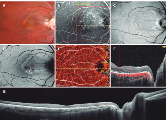
Figure 1. Preoperative multimodal imaging of the right eye: optic disc pit maculopathy in a 19-year-old man. A) Color image shows an optic disk pit associated with serous macular detachment. B) Fluorescein angiography. C) En-face OCT (supericial retina). D) Fundus autoluorescence. E) OCTA: image suggesting CNV (white arrow). F) OCT B-scan indicating the presence of communication between the optic nerve and serous retinal detachment. G) OCT showing subretinal luid (yellow arrow).
A B C
F E
D
G
CNV= choroidal neovascularization; OCT= optical coherence tomography; OCTA= optical coherence tomography.
Figure 2. Postoperative multimodal imaging of the right eye: resolution of optic disc pit maculopathy in a 19-year-old man. A) Color image. B) Fluorescein angiography. C) En-face OCT (supericial retina). D) Fundus autoluorescence. E) OCTA: disappearance of suspicious CNV image. F and G) OCT B-scan indicating near-complete absorption of subretinal luid.
A B C
F E
D
Ro i z e n b l at t M, e ta l.
2 5 9 Arq Bras Oftalmol. 2017;80(4):257-9
The patient underwent 23-gauge pars plana vitrectomy in the right eye. Triamcinolone-assisted posterior hyaloid detachment, as well as fluid-air exchange, was performed. No laser or peeling was performed during the surgery and air was used as the vitreous subs-titute. Six weeks after surgery, multimodal imaging was repeated (Figure 2). There was subtotal resorption of the subretinal fluid. BCVA improved to 20/20 in the right eye. OCTA demonstrated normal cho-riocapillaris signal throughout the macula (Figure 2 E). OCTA signal superimposed on OCT B-scan also demonstrated normal chorioca-pillaris signal throughout the macula (Figure 2 F).
DISCUSSION
OCTA images can produce both positive and negative artifacts, which are important to recognize when interpreting clinical images. For instance, reflection artifacts may project superficial vessels into deeper layers and shadowing or eye movement artifacts may create areas devoid of vessels(1).
We presented a case of a young patient diagnosed with ODP maculopathy whose preoperative OCTA suggested subfoveal CNV. Note that OCTA displayed a ring of relatively decreased choriocapilla-ris signal surrounding a small, subfoveal area of relatively increased choriocapillaris signal (Figure 1 E). The same phenomenon was
pre-sent in the OCTA signal superimposed on the OCT B-scan (Figure 1 F). However, fluorescein angiogram and OCT did not demonstrate any signs of CNV in the same area, which suggests an artifactual lesion.
The patient was scheduled for PPV, which caused near-complete resolution of subretinal fluid 6 weeks postoperatively. Postoperative OCTA revealed normal choriocapillaris, confirming the artifactual na -ture of the suspected subfoveal CNV. Possible mechanisms for the abovementioned artifactual CNV include light scattering by the elevated retina and/or presence of subretinal fluid.
In conclusion, optical coherence tomography angiography may produce artifacts in optic disc pit maculopathy that simulate choroi-dal neovascularization. Multimochoroi-dal imaging is important to accura-tely interpret unusual optical coherence tomography angiography findings.
REFERENCES
1. Spaide RF, Fujimoto JG, Waheed NK. Image Artifacts in Optical Coherence Tomogra-phy AngiograTomogra-phy. Retina. 2015;35(11):2163-80.
2. Georgalas I, Ladas I, Georgopouls G, Petrou P. Optic disc pit: a review. Arch Clin Exp Ophthalmol. 2011;249(8):1113-22.
3. Borodic GE, Gragoudas ES, Edward WO, Brockhurst RJ. Peripapillary subretinal neovas-cularization and serous macular detachment. Association with congenital optic nerve pits. Arch Ophthalmol. 1984;102(2):229-31.