INTRODUCTION
Pterygium is a chronic ocular surface disease characterized by invasion of the cornea by fibrovascular, wing-shaped conjunctival tissue that leads to impairment of visual acuity and cosmetic appea-rance. Risk of pterygium is increased by inflammation, viral infection, age, and genetic factors. Epidemiological studies have implicated ultraviolet light exposure as having a significant causative association with pterygia(1-3). Interleukin (IL)-6 and IL-8 are released by pterygium epithelial cells after ultraviolet irradiation(4), and accumulation of these proinflammatory cytokines in tears may induce chronic inflam-mation, fibrovascular proliferation, and pterygium formation.
Vitamin D is a circulating steroid hormone with antiangiogenic, anti-inflammatory, and antifibrotic activity(5-7). Several ocular condi-tions including age-related macular degeneration, myopia, and aller-gic conjunctivitis have been associated with vitamin D, but there is limited information on vitamin D level in patients with pterygium(8-10). The primary study hypothesis was based on a possible relationship between pterygium and vitamin-D level. Pterygium is caused by chronic inflammation and fibrovascular ingrowth after ultraviolet
ex-posure(4). Because vitamin D has antiangiogenic, anti-inflammatory, and antifibrotic activity, an inverse relationship between vitamin D level and pterygium would be expected(5,6). Second, pterygium is one of the most common UV-induced ocular diseases, and outdoor activity has been reported to be significantly associated with ptery-gium formation(11). Because the primary natural source of vitamin D is in synthesis in the skin secondary to sun exposure, vitamin D levels may be increased in patients with pterygium, especially outdoor workers. In this study, we investigated blood vitamin D level in patients with pterygium.
METHODS
STUDY DESIGN AND POPULATION
This prospective case-control study was conducted in the De-partment of Ophthalmology at the Gaziantep University School of Medicine. All participants gave written informed consent. The study was approved by the ethics committee of Gaziantep University School of Medicine, and was carried out following the principles of the Declaration of Helsinki.
Vitamin D level in patients with pterygium
Níveis de vitamina D em pacientes com pterígio
Necip Kara1, Seda ceri1
Submitted for publication: December 8, 2016 Accepted for publication: February 2, 2017
1 Department of Ophthalmology, Gaziantep University School of Medicine, Gaziantep, Turkey.
Funding: No specific financial support was available for this study.
Disclosure of potential conflicts of interest: None of the authors have any potential conflict of interest to disclose.
Corresponding author: Necip Kara. Department of Ophthalmology. Gaziantep University School of Medicine - Gaziantep - Turkey - E-mail: [email protected]
Approved by the following research ethics committee: Gaziantep University School of Medicine (#56/2015).
ABSTRACT
Purpose: To evaluate blood vitamin D level in patients with pterygium.
Methods: This prospective study, compared 58 eyes of 58 healthy individuals (control group) with 63 eyes of 63 patients with pterygium (study group). Sub-jects were stratified by time spent indoors or outdoors. Participants were given comprehensive ophthalmic examinations; blood 25-hydroxyvitamin D (nmol/L) was assayed.
Results: Vitamin D level was significantly higher in men with pterygium than without it (p=0.020), but the difference was not significant in women (p=0.86). In the pterygium group, vitamin D level was significantly increased in participants with outdoor activity (p=0.010). In the control group, vitamin D levels did not differ significantly with indoor and outdoor activity (p=0.126).
Conclusion: Vitamin D level in participants with pterygium was significantly increased only in men and in those with more outdoor activity.
Keywords: Vitamin D/metabolism; Pterygium/metabolism; Ultraviolet rays; Sunlight
RESUMO
Objetivo: Avaliar os níveis sanguíneos de vitamina D em pacientes com pterígio.
Métodos: Neste estudo prospectivo e comparativo, foram incluídos 58 olhos de 58 indivíduos saudáveis (grupo controle) e 63 olhos de 63 pacientes com pterígio (grupo de estudo). Os indivíduos também foram categorizados quanto ao tempo foi gasto dentro de casa ou ao ar livre. Todos os participantes foram submetidos a exames oftálmicos e avaliações de nível sanguíneo 25-hidroxivitamina D (nmol/L).
Resultados: O nível de vitamina D foi significativamente maior em pacientes mas-culinos com pterígio do que aqueles sem pterígio (p=0,020). Não houve diferença significativa entre mulheres do grupo de estudo e controle (p=0,86). No grupo pterígio, o nível de vitamina D foi significativamente maior no subgrupo com atividade ao ar livre (p=0,010). No grupo controle, o nível de vitamina D não foi significativamente diferente entre a atividade interna e ao ar livre (p=0,126).
Conclusão: Os achados deste estudo revelaram que o nível de vitamina D nos casos de pterígio foi significativamente maior apenas nos homens e nos casos com maior atividade ao ar livre.
The study group included patients with pterygium, and the control group consisted of age- and sex-matched healthy volunteers without pterygium. All subjects were of the same race and had lived in the same region for at least 5 years. The participants were stratified by the amount of time spent indoors or outdoors by asking questions about their job and routine daily activities including walking, playing outdoor sports, and others. The outdoor group included those who spent >75% of the daytime hours in outdoor activities. The indoor group spent >75% of the daytime hours in indoor activities.
Participants with ocular and systemic diseases that may be asso-ciated with altered vitamin D levels, including myopia, age-related macular degeneration, diabetic retinopathy, glaucoma, diabetes, Crohn’s disease, Alzheimer disease, and others were excluded. To avoid a possible confounding influence on activity we also excluded adult subjects with a body mass index above 30 kg/m2.
EVALUATION
Clinical evaluation included visual acuity using an Snellen chart, intraocular pressure measurement, slit-lamp biomicroscopy, and a fundus examination. The pterygium diagnosis was made by a single ophthalmologist (NK). Pterygium size was measured using a slit lamp, a horizontal slit beam of light from the limbus to the head of the pterygium and a vertical beam to the highest vertical extent of pterygium tissue on the cornea.
VITAMIN D MEASUREMENT
Blood samples were collected from both study and control par-ticipants during the same autumn season and assayed at the Ga-ziantep University School of Medicine clinical laboratory. Serum 25-hydroxyvitamin D (25OHD, nmol/L) is the primary indicator of vitamin D status and is the form that was assayed.
STATISTICAL ANALYSIS
Statistical analysis was performed with SPSS (version 16.0, Chica-go, IL). The normality of all data distributions was confirmed with the Shapiro-Wilk test. Between-group differences in continuous variables were compared by the independent-sample t-test. The chi-squared test was used to compare differences in categorical variables. P-values <0.05 were considered statistically significant.
RESULTS
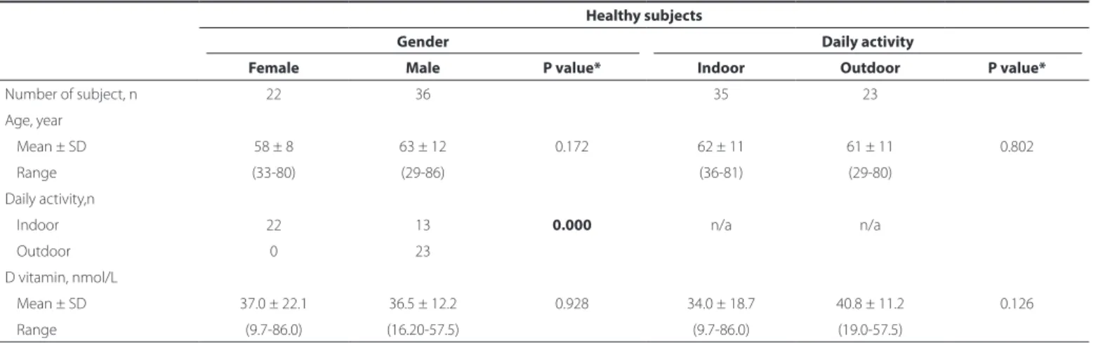
The demographic and clinical characteristics of the study and control groups are shown in table 1. There were no significant between-group differences in age and sex ratio. None of the partici-pants routinely used sunglasses or hats regularly. Overall mean vita-min D levels (nmol/L) in patients with pterygium and controls were not significantly different (p=0.731, Table 1). In women, vitamin D levels in the study and control groups were not significantly different (P=0.86). In men, the vitamin D level was significantly higher in those with pterygium than in those without it (P=0.020, Table 2). In the pterygium group, vitamin D levels were significantly increased in men (P=0.000) and in the outdoor activity group (P=0.010, Table 3). In the control group, vitamin D levels in men and women (P=0.928) and the indoor and outdoor activity groups (P=0.126) were not signi-ficantly different (Table 4). Vitamin D level in the pterygium group was not significantly correlated with either age or horizontal and vertical pterygium length (P>0.05, Table 5).
DISCUSSION
Vitamin D research has become increasingly popular in ophthal-mology Dadaci et al. found decreased plasma vitamin D levels in patients with seasonal allergic conjunctivitis(9). In a population-based study, Yoo et al. reported that the lowest serum vitamin D quintile
had a significantly increased odds ratio of open angle glaucoma(12). Galor et al. reported that increased vitamin D was associated with decreased dry eye syndrome symptoms(13). There is also evidence that low-level outdoor sunlight exposure may be involved in myopia pathogenesis(14). Grotting et al. found that decreased vitamin D was associated with increased risk of anterior uveitis(15), and Jee et al. reported that increased serum vitamin D was associated with de-crea sed risk of age-related cataracts in humans(16).
In this study, vitamin D levels were significantly higher in male, but not female, patients with pterygium than in controls. In patients with pterygium, vitamin D levels were significantly higher in men than in women, possibly because men had more outdoor activity than women, which resulted in more UV exposure. Vitamin D level was also significantly higher in pterygium group participants with more outdoor activity than in those with more indoor activity. That is in line with a recent study by Jee et al.(17) who found a positive association between blood vitamin D level and pterygium even after adjusting for sun exposure. They also reported that the relative odds of pterygium were significantly increased in third, fourth, and fifth vitamin D quintiles versus the lowest quintile.
The pathogenesis of pterygium is poorly understood, but his-topathological evidence suggests that genetic factors, cytokines, growth factors, antiapoptosis activity, extracellular matrix modeling, immunological responses, and viral activity may be involved(18). It has been proposed that elevated vitamin D levels may increase cellular calcium, which in turn would activate S100 protein(19). S100 is a calcium-activated signaling protein that interacts with other proteins to modulate diverse biological processes(20). An increase in S100 protein has been seen in pterygium tissue compared with normal conjunctiva(19). Although increase in vitamin D level has been associated with more time spent outdoors, Mutti et al. did not find
Table 1. Demographic and clinical characteristics of patients with pterygium and healthy controls
Pterygium Control P value*
Number of subject,n 63 58 Age (year),
Mean ± SD 58 ± 12 61 ± 11 0.143 Range (21-85) (29-86)
Gender, n
Female 33 22 0.079
Male 30 36
Daily activity, n
Indoor 38 35 0.573 Outdoor 25 23
Pterygium length, mm Horizontal,
Mean ± SD 02.9 ± 01.2
Range (1-6) n/a Vertical,
Mean ± SD 04.7 ± 01.6 Range (1-8) D vitamin, nmol/L
Mean ± SD 37.5 ± 16.8 36.3 ± 18.9 0.731 Range (8.6-92.0) (9.7-86.0)
a such a relationship(21). Similarly, in this study, vitamin D level in healthy controls did not differ significantly with the extent of indoor or outdoor activity. Patients with pterygium with more outdoor time had higher vitamin D levels than patients who spent most of the day indoors. These findings are consistent with an association of vitamin D level and pterygium formation.
An alternative explanation is that pterygium formation may occur as a direct effect of focal UV exposure of conjunctival tissue inde-pendent of vitamin D level. Conjunctival damage from UV exposure may activate a complex series of oxidative reactions and cell-death pathways(22). UV exposure activates epidermal growth factors, expres-sion of proinflammatory cytokines, and matrix metalloproteinases
Table 2. Demographic and clinical characteristics of men and women with and without pterygium
Female Male
Pterygium Control P* Pterygium Control P*
Number of subject, n 33 22 30 36 Age (year),
Mean ± SD 59 ± 12 58 ± 8 0.869 57 ± 13 63 ± 12 0.063 Range (21-81) (33-80) (35-85) (29-86)
Daily activity, n
Indoor 32 22 1.0 6 13 0.291
Outdoor 1 0 24 23
Pterygium length, mm n/a
Horizontal n/a
Mean ± SD 02.8 ± 01.3 02.9 ± 01.1
Range (1-5) (1-6)
Vertical 04.4 ± 01.8 05.0 ± 01.3
Mean ± SD (1-8) (2-8)
Range
D vitamin, nmol/L 0.869
Mean ± SD 30.7 ± 12.9 36.1 ± 21.2 45.0 ± 10.1 36.5 ± 12.0 0.020
Range (8.6-58.5) (9.7-86.0) (10.17-92.0 (16.2-57.5)
N= number; SD= standard deviation; mm= millimeter, n/a= non-applicable; *= independent-t test.
Table 3. Demographic and clinical characteristics of pterygium patients stratiied by sex and daily activity
Pterygium patients
Gender Daily activity
Female Male P Value* Indoor Outdoor P value*
Number of subject, n 33 30 38 25 Age, year
Mean ± SD 59 ± 12 57 ± 13 0.471 59 ± 10 57 ± 15 0.520 Range (21-81) (35-85) (36-81) (21-85)
Daily activity,n
Indoor 32 6 0.000 n/a n/a
Outdoor 1 24
Pterygium length, mm Horizontal
Mean ± SD 02.8 ± 01.3 05.0 ± 01.3 0.704 02.9 ± 01.3 02.8 ± 01.1 0.790 Range (1-5) (2-8) (1-5) (1-6)
Vertical
Mean ± SD 04.4 ± 01.8 02.9 ± 01.1 0.203 04.5 ± 01.7 05.0 ± 01.3 0.307 Range (1-8) (1-6) (1-8) (2-8)
Vitamin D, nmol/L 0.000
Mean ± SD 30.7 ± 12.9 45.0 ± 10.1 33.2 ± 14.4 44.0 ± 17.1 0.010
Range (8.6-58.5) (10.17-92.0) (8.68-63.7) (10.17-92.0)
Table 4. Clinical characteristics of healthy control participants stratiied by sex and daily activity
Healthy subjects
Gender Daily activity
Female Male P value* Indoor Outdoor P value*
Number of subject, n 22 36 35 23 Age, year
Mean ± SD 58 ± 8 63 ± 12 0.172 62 ± 11 61 ± 11 0.802 Range (33-80) (29-86) (36-81) (29-80)
Daily activity,n
Indoor 22 13 0.000 n/a n/a
Outdoor 0 23
D vitamin, nmol/L
Mean ± SD 37.0 ± 22.1 36.5 ± 12.2 0.928 34.0 ± 18.7 40.8 ± 11.2 0.126 Range (9.7-86.0) (16.20-57.5) (9.7-86.0) (19.0-57.5)
N= number; SD= standard deviation; mm= millimeter; n/a= non-applicable; *= independent-t test.
Table 5. Correlation of sex, indoor and outdoor activity in pterygium patients
All Female Male Indoor Outdoor
Age
Pearson corr -0.143 -0.017 -0.190 -0.156 -0.096 P value* -0.264 -0.924 -0.315 -0.348 -0.647 Horizontal
Pearson corr -0.015 -0.025 -0.039 -0.074 -0.169 P value* -0.907 -0.896 -0.838 -0.671 -0.420 Vertical
Pearson corr -0.284 -0.229 -0.271 -0.289 -0.224 P value* -0.058 -0.224 -0.148 -0.093 -0.282 Pearson corr= pearson correlation coefficient; *= pearson correlation test.
in pterygium cells. Focal UV radiation may also destroy limbal stem cells, which may then result in conjunctival invasion of the cornea and pterygium formation(23). A previous case-control study reported a significant association of outdoor work and pterygium formation, possibly because sunlight exposure increased the risk compared with indoor workers(11). The same investigators also confirmed an increased risk of pterygium formation in individuals who spent more time in the sun, especially with cumulative exposure over 5-10 years. Epidemiological studies have also reported an association between pterygium and increased ultraviolet radiation exposure in lower lati-tude regions(24). Patients with Pterygium thus tend to live in regions with high UV indices ant to work outdoors.
Jee et al.(17) reported an association of vitamin D and pterygium formation in both men and women, but we found it in men only. There is evidence of a significant gender effect on vitamin D status(25). Low vitamin D levels appear to be common in postmenopausal women(26), and Jee et al. found a negative correlation of vitamin D level and development of nonproliferative or proliferative diabetic retinopathy in men but not in women.(27) In observational studies of patients with type 2 diabetes mellitus, both Scragg et al. and Suzuki et al. concluded that mean vitamin D concentrations were significantly higher in men than in women(28,29). Gender-spe cific association of vitamin D receptor polymorphism has an effect on susceptibility to diabetes(30), but the relation has not been studied in pterygium. It is not clear why vitamin D is associated with pterygium in men but
not in women, but the relationship may depend on outdoor activity. Further studies are needed to identify the factors responsible for this difference and to explain the nature of the sex-related biological mechanisms.
One of the study limitations was not estimating the food and supplement intake of the patients. The primary source of vitamin D is production in the skin secondary to sun exposure, but small amounts are obtained from foods such as fatty fish and milk. Another was not comparing vitamin D levels in women with primarily indoor and outdoor activity. This was because most women were homemakers who spent most of the day indoors. We did not perform histopa-thological evaluations, which would have confirmed conjunctival squamous metaplasia or primary acquired melanosis in pterygia samples. Also, because we did not conduct a genetic analysis we do not know whether any patients may have had a genetic predis-position for pterygium. In summary, in this series of patients with pterygium, vitamin D level was significantly increased in men and in participants with more outdoor activity.
REFERENCES
1. Detorakis ET, Drakonaki EE, Spandidos DA. Molecular genetic alterations and viral presence in ophthalmic pterygium. Int J Mol Med. 2000;6(1):35-41.
2. Moran DJ, Hollows FC. Pterygium and ultraviolet radiation: a positive correlation. Br J Ophthalmol. 1984;68(5):343-6.
3. Mackenzie FD, Hirst LW, Battistutta D, Green A. Risk analysis in the development of pterygia. Ophthalmology. 1992;99(7):1056-61.
4. Di Girolamo N, Kumar RK, Coroneo MT, Wakefield D. UVB-mediated induction of in-terleukin-6 and -8 in pterygia and cultured human pterygium epithelial cells. Investig Ophthalmol Vis Sci. 2002;43(11):3430-7.
5. Albert DM, Scheef EA, Wang S, Mehraein F, Darjatmoko SR, Sorenson CM, et al. Cal-citriol is a potent inhibitor of retinal neovascularization. Invest Ophthalmol Vis Sci. 2007;48(5):2327-34.
6. Yilmaz SS, Hizli D, Yilmaz E, Eryilmaz OG, Hizli F, Halta H. Effect of vitamin D on pos-toperative adhesion formation in a rat uterine horn adhesion model. J Reprod Med. 2013;58(11-12):511-6.
7. Krishnan AV, Feldman D. Mechanisms of the anti-cancer and anti-inflammatory actions of vitamin D. Annu Rev Pharmacol Toxicol. 2011;51:311-36.
8. Itty S, Day S, Lyles KW, Stinnett SS, Vajzovic LM, Mruthyunjaya P. Vitamin D deficiency in neovascular versus nonneovascular age-related macular degeneration. Retina. 2014;34(9):1779-86.
9. Dadaci Z, Borazan M, Kiyici A, Oncel Acir N. Plasma vitamin D and serum total immu-noglobulin E levels in patients with seasonal allergic conjunctivitis. Acta Ophthalmol. 2014;92(6):e443-6.
10. Yazar S, Hewitt AW, Black LJ, McKnight CM, Mountain JA, Sherwin JC, et al. Myopia is associated with lower vitamin D status in young adults. Invest Ophthalmol Vis Sci. 2014;55(7):4552-9.
12. Yoo TK, Oh E, Hong S. Is vitamin D status associated with open-angle glaucoma? A cross-sectional study from South Korea. Public Health Nutr. 2014;17(4):833-43. 13. Galor A, Gardener H, Pouyeh B, Feuer W, Florez H. Effect of a mediterranean dietary
pattern and vitamin D levels on dry eye syndrome. Cornea. 2014;33(5):437-41. 14. Sherwin JC, Reacher MH, Keogh RH, Khawaja AP, Mackey DA, Foster PJ. The
associa-tion between time spent outdoors and myopia in children and adolescents: a systematic review and meta-analysis. Ophthalmology. 2012;119(10):2141-51. 15. Grotting LA, Davoudi S, Palenzuela D, Papaliodis GN, Sobrin L. Association of low
vitamin D Levels With noninfectious anterior uveitis. JAMA Ophthalmol. 2016 Dec 22. 16. Jee D, Kim EC. Association between serum 25-hydroxyvitamin D levels and
age-re-lated cataracts. J Cataract Refract Surg. 2015;41(8):1705-15.
17. Jee D, Kim EC, Cho E, Arroyo JG. Positive association between blood 25 hydroxyvi-tamin d levels and pterygium after control for sunlight exposure. PLoS One. 2016; 11(6):e0157501.
18. Di Girolamo N, Wakefield D, Coroneo MT. UVB-mediated induction of cytokines and growth factors in pterygium epithelial cells involves cell surface receptors and intracellular signaling. Invest Ophthalmol Vis Sci. 2006;47(6):2430-7.
19. Riau AK, Wong TT, Beuerman RW, Tong L. Calcium-binding S100 protein expression in pterygium. Molecular Vis. 2009;15:335-42.
20. Tu CL, Chang W, Bikle DD. The extracellular calcium-sensing receptor is required for calcium-induced differentiation in human keratinocytes. J Biol Chem 2001;276: 41079-85.
21. Mutti DO, Marks AR. Blood levels of vitamin D in teens and young adults with myopia. Optom Vis Sci. 2011;88(3):377-82.
22. Buron N, Micheau O, Cathelin E, Lafontaine PO, Creuzot-Garcher C, Solary E. Diffe-rential mechanisms of conjunctival cell death induction by ultraviolet irradiation and benzalkonium chloride. Inv Ophthalmol Vis Sci. 2006;47(10):4221-30.
23. Chui J, Coroneo MT, Tat LT, Crouch R, Wakefield D, Di Girolamo N. Ophthalmic ptery-gium: a stem cell disorder with premalignant features. Am J Pathol. 2011;178(2):817-27. 24. Taylor HR. Aetiology of climatic droplet keratopathy and pterygium. Br J Ophthalmol.
1980;64(3):154-63.
25. Verdoia M, Schaffer A, Barbieri L, Di Giovine G, Marino P, Suryapranata H, et al. Impact of gender difference on vitamin D status and its relationship with the extent of coronary artery disease. Nutr Metab Cardiovasc Dis. 2015;25(5):464-70.
26. Masoni AM, Menoyo I, Bocanera R, Pezzotto SM, Morosano ME. Hypovitaminosis D and Associated Risk Factors in Postmenopausal Women. Health. 2014;6(11):1180-90. 27. Jee D, Han Kd, Kim EC. Inverse association between high blood 25-hydroxyvitamin
D levels and diabetic retinopathy in a representative Korean population. PLoS One. 2014;9(12):e115199.
28. Scragg R, Sowers M, Bell C, Third National Health and Nutrition Examination Survey. Serum 25-hydroxyvitamin D, diabetes, and ethnicity in the Third National Health and Nutrition Examination Survey. Diabetes Care. 2004;27(12):2813-8.
29. Suzuki A, Kotake M, Ono Y, Kato T, Oda N, Hayakawa N, et al. Hypovitaminosis D in type 2 diabetes mellitus: Association with microvascular complications and type of treatment. Endocr J. 2006;53(4):503-10.
30. Gyorffy B, Va´sa´ rhelyi B, Krikovszky D, Mada´csy L, Tordai A, Tulassay T, et al. Gender-specific association of vitamin D receptor polymorphism combinations with type 1 diabetes mellitus. Eur J Endocrinol. 2002;147(6):803-8.