Cop
yright
© ABE&M t
odos os dir
eit
os r
eser
vados
.
Y chromosome aberration
in a patient with
cloacal-bladder exstrophy-epispadias
complex: an unusual inding
Aberração cromossômica do Y em uma paciente com extroia de bexiga e de cloaca e epispadia: um achado raro
Mirian Yumie Nishi1, Thais Cotrim Martins1, Elaine Maria Frade Costa1,
Berenice Bilharinho Mendonca1, Amilcar Martins Giron2, Sorahia Domenice1
SUMMARY
Chromosome aberrations or genetic syndromes associated with cloacal-bladder exstrophy complex have rarely been reported. The aim of this report is to describe a 14 year-old female Brazilian patient with a complex urogenital malformation, short stature, lack of secondary se-xual characteristics and Y chromosome aberration. A girl with cloacal bladder exstrophy com-plex was referred for evaluation of short stature and absence of secondary sexual characteris-tics. Pre-pubertal levels of gonadotropins and sex steroids were observed at the beginning of monitoring, but follow-up showed a progressive increase in testosterone levels. The patient underwent gonadectomy and testicular tissue was identiied without dysgenetic characteris-tics. She had a 46,X,inv(Y)(p11.1q11.2) karyotype, normal SRY sequence, and no Y deletions. The pericentric inversion of Y chromosome apparently did not contribute to the development of the complex urogenital malformation in this patient. Currently, no teratogenic agent, environmen-tal factor, or defective genes have been recognized as etiologic factors for this type of urogenienvironmen-tal malformation. Arq Bras Endocrinol Metab. 2013;57(2):148-52
SUMÁRIO
Aberrações cromossômicas ou síndromes genéticas associadas ao complexo extroia de be-xiga e de cloaca e epispadia são raramente relatadas. O objetivo é descrever uma paciente brasileira com 14 anos que apresenta uma malformação urogenital complexa, baixa estatura, ausência de características sexuais secundárias e alteração do cromossomo Y. Uma menina com extroia de bexiga e de cloaca e epispadia foi encaminhada para avaliação de baixa estatu-ra e ausência de desenvolvimento de caestatu-racterísticas sexuais secundárias. Níveis pré-pubeestatu-rais de gonadotroinas e esteroides sexuais foram observados no início da avaliação, mas durante o seguimento notou-se um aumento progressivo dos níveis de testosterona. Ela foi submetida à gonadectomia e identiicou-se a presença de testículos sem características disgenéticas. O cariótipo era 46,X,inv(Y)(p11.1q11.2), com sequência normal do SRY e ausência de deleções do Y. A inversão pericêntrica do cromossomo Y, aparentemente, não contribuiu para o desenvolvi-mento da malformação urogenital complexa nessa paciente. Atualmente, nenhum agente te-ratogênico, fator ambiental ou mutações gênicas foram reconhecidos como fatores etiológicos para essa malformação urogenital. Arq Bras Endocrinol Metab. 2013;57(2):148-52
1 Unidade de Endocrinologia do
Desenvolvimento, Laboratório de Hormônios e Genética Molecular LIM-42, Hospital das Clínicas, Faculdade de Medicina, Universidade de São Paulo (HC-FMUSP), Sao Paulo, SP, Brazil
2 Divisão de Urologia,
HC-FMUSP, Sao Paulo, SP, Brazil
Correspondence to:
Sorahia Domenice Laboratório de Hormônios e Genética Molecular LIM-42 Hospital das Clínicas, Faculdade de Medicina, Universidade de São Paulo Av. Dr. Eneias de Carvalho Aguiar, 155, PAMB, 2º andar, bloco 6 05403-900 – Sao Paulo, SP, Brazil [email protected]
Received on May/18/2012 Accepted on Nov/17/2012
INTRODUCTION
T
he cloacal-bladder exstrophy-epispadias complex (BEEC) is an anterior midline defect with variable expression comprising anomalies involving theCop
yright
© ABE&M t
odos os dir
eit
os r
eser
vados
.
its most severe form, cloacal exstrophy (CE) or “OEIS” complex (omphalocele, exstrophy of the bladder, im-perforate anus, and spinal defects). The incidence of BEEC varies with regard to ethnic background, sex, and phenotypic expression, but it is twice as common among males (2).
The great majority of BEEC cases are classiied as non-syndromic, and the etiology of this disorder is still unknown. No single teratogenic agent or environ-mental factor that could play a role in the expression of BEEC, has been identiied. However, Reutter and cols., studying a large cohort of patients with BEEC and their families, concluded that smoking and medical radiation during the irst trimester of pregnancy might be associated with a more severe BEEC phenotype (2). Chromosomal aberrations or genetic syndromes associ-ated with BEEC have only rarely been reported.
CASE REPORT
The present study was approved by the Ethics Com-mittee of the Hospital das Clinicas, University of Sao Paulo Medical School. Written consent was obtained from the patient’s mother.
An infant was born from a nonconsanguineous couple by normal vaginal delivery. Multiple anomalies were present at birth, including omphalocele, cloacal exstrophy, imperforate anus, ambiguous genitalia with prominent rugated labioscrotal folds, and no apparent genital tubercle. Abdominal sonography showed left-kidney agenesia. The family history was unremarkable, with three healthy siblings (one female and two males), and no other similarly affected individuals. The mother had short stature without sexual dysfunction, was 26 years of age and the father 29 years old at the time of the child’s birth. At the age of three days, the pa-tient was submitted to corrective surgery. A colostomy was created, and the cloacal exstrophy and abdominal wall were closed. At four years of age, she underwent surgery to close the colostomy. In the same year, the (exstrophic) bladder was closed and enlarged with the ileum and colon bowel. The bladder neck was closed and the construction of a urinary continent stoma was performed with the cecal appendices (Mitrofanoff prin-ciple). External female genitalia were created. Latter, other surgeries were performed to correct an enterocu-taneous istula and to remove bladder lithiasis.
At 14 years of age she was referred to an endocri-nologist to evaluate her short stature and absence of
development of secondary sexual characteristics. Physi-cal examination revealed (Table 1): height 135 cm, (SDS -4.3 female; SDS -3.7 male), height age of 11.5 years and bone age of 12 years, and low weight 38.8 kg (SDS -2.9 female; SDS -2.3 male), low-set hairline, shield-like chest with widely separated nipples, acne in the dorsal area, hair in the nasolabial region, and deep voice. Neuropsychomotor and intellectual develop-ment were normal for the patient’s age.
Genital examination identiied labia major with ex-cess of skin and posterior fusion, exex-cess of skin in the clitoris region without palpable tissue, a single perineal opening, and Tanner B1P1.
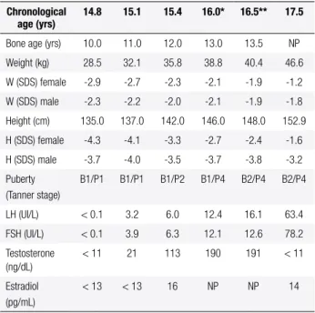
Initial hormonal data showed prepubertal levels of gonadotropins and sex steroids, but in the follow-up, testosterone levels gradually increased (Table 1).
Table 1. Clinical and laboratory follow-up of the patient with BEEC and 46,X,inv(Y)(p11.1q11.2) karyotype
Chronological age (yrs)
14.8 15.1 15.4 16.0* 16.5** 17.5
Bone age (yrs) 10.0 11.0 12.0 13.0 13.5 NP
Weight (kg) 28.5 32.1 35.8 38.8 40.4 46.6
W (SDS) female -2.9 -2.7 -2.3 -2.1 -1.9 -1.2
W (SDS) male -2.3 -2.2 -2.0 -2.1 -1.9 -1.8
Height (cm) 135.0 137.0 142.0 146.0 148.0 152.9
H (SDS) female -4.3 -4.1 -3.3 -2.7 -2.4 -1.6
H (SDS) male -3.7 -4.0 -3.5 -3.7 -3.8 -3.2
Puberty (Tanner stage)
B1/P1 B1/P1 B1/P2 B1/P4 B2/P4 B2/P4
LH (UI/L) < 0.1 3.2 6.0 12.4 16.1 63.4
FSH (UI/L) < 0.1 3.9 6.3 12.1 12.6 78.2
Testosterone (ng/dL)
< 11 21 113 190 191 < 11
Estradiol (pg/mL)
< 13 < 13 16 NP NP 14
B: breast; F: female; H: height; M: male; NP: not performed; P: pubic hair; SDS: standard deviation score; W: weight.
* Data obtained before the irst surgery; ** Data obtained before the second surgery. Normal hormone levels: female pre-pubertal range – LH: ≤ 0.6 IU/L; FSH: ≤ 3.2 IU/L; testosterone: < 14 ng/dL; estradiol: < 21 pg/mL; pubertal range – LH: 1.1-6.3 IU/L; FSH: 1.4 – 5.7 IU/L; testosterone: ≤ 98 ng/dL; estradiol: 22 – 232 pg/mL; male pre-pubertal range- testosterone: < 19 ng/dL; pubertal range – testosterone: ≤ 669 ng/dL; estradiol: ≤ 35 pg/mL.
identi-Cop
yright
© ABE&M t
odos os dir
eit
os r
eser
vados
.
ied. After the surgery, testosterone levels remained elevated (198 ng/dL), conirming the presence of the other testis. The patient underwent surgery in which the testis was identiied near the left kidney and re-moved. Histological analysis showed normal testicu-lar tissue. Psychological evaluation was performed to elucidate her gender orientation, which turned out to be female.
Cytogenetic analysis of lymphocyte and gonadal tissue showed a 46,X,inv(Y)(p11.1q11.2) karyotype (Figure 1). FISH analysis conirmed the presence of the SRY gene (Yp11.31), DYZ3 (centromere) and DYZ1 (Yq12) regions of the Y chromosome, and clari-ied the mechanism that generated the Y chromosome inversion (Figure 1). Eight loci of the Y chromosome: PAR1, SRY, TSPY, AMELY (Yp), DYZ3 (centromere), DYS280, DYS1, and DYZ1 (Yq) were ampliied by PCR using genomic DNA extracted from blood, in-dicating that these regions were preserved. The SRY gene sequence was normal. The mother’s karyotype was 45,X[10]/46,XX[113].
DISCUSSION
Advanced parental age (3), increased parity even after adjusting for age (4) and in vitro fertilization (5) have been reported as risk factors to BEEC development. Spontaneous errors of development such as somatic mutation or complex gene-environment interactions may be responsible for BEEC.
A role of genetic factors in the pathogenesis of BEEC has also been suggested. This hypothesis was based on observations of rare familial cases, high but incomplete concordance in monozygotic twins, and a single report of increased recurrence risk for BEEC in the offspring of an affected parent (6-8).
Cytogenetic and molecular analyses have revealed chromosomal anomalies in a few patients with BEEC. Numerical chromosomal aberrations (47,XXX; 47,XXY; 47,XYY; 45,X/46,XX) were observed. In some of these cases Down syndrome was observed (9). Aneuploidy of sex chromosomes in some of these cases might point to loci involved in the formation of BEEC (1). The obser-vation of different sexes in two subsequent sponta neous
1 2 3 4 5
6 7 8 9 10 11 12
13 14 15 16 17 18
19 20 21 22 x Y
Figure 1. (A) Karyotype BTGW: 46,X,inv(Y)(p11.1q11.2); (B) Metaphase chromosome spread submitted by FISH technique, indicating the X chromosome, and the locations of the SRY gene and of the DXZ1 locus on the Y chromosome (arrows); (C) Schematic representation of the mechanism that generates pericentric inversion of the Y chromosome.
A
C
Cop
yright
© ABE&M t
odos os dir
eit
os r
eser
vados
.
abortions, however, did not support this hypothesis (8). Apparently, none of these chromosomal abnormalities could be conirmed as the cause of this disorder.
Structural aberrations involving the chromosome 9 at region q32-ter have been identiied in six BEEC cases (1). The SF-1 (Steroidogenic Factor 1; 9q33.3) and SET genes (Suppressor of variegation, Enhancer of zeste and Thrithorax; 9q34.11) have been investi-gated in patients with BEEC (10). However, no muta-tions were detected in these studies. Nonetheless, other genes located in this region might be involved in the etiology of BEEC.
Our patient had 46,X,inv(Y)(p11.1q11.2) karyo-type, and apparently neither large deletions in Y chro-mosome nor SRY gene mutations were identiied. Pos-sibly, the inverted Y chromosome was inherited from the father, but the father’s blood sample was not available.
Pericentric inversion of the Y chromosome occurs in approximately 1:1,000 males in the general population, and it is considered a chromosome heteromorphism that does not inluence male phenotype (11). However, in the literature, there are some cases of pericentric in-version of the Y chromosome leading to XY female with gonadal dysgenesis and no SRY mutations. Gimelli and cols. reported a young woman with gonadal dysgen-esis, a gonadoblastoma and a dysgerminoma in both gonads, normal external genitalia and a 46,X,inv(Y) (p11.31q12) karyotype (12). This inversion led to a si-lencing of SRY due to the position-effect variegation, which caused the gonadal dysgenesis (12). Mitsuhashi and cols. also described a XY female with normal ex-ternal genitalia, 46,X,inv(Y)(p11.2q11.2) karyotype, and gonadal dysgenesis. This inversion did not lead to position-effect variegation, but the streak gonads pre-sented abnormally prolonged SRY expression. Thus, the authors believed that the regulation of the SRY gene was impaired, causing gonadal dysgenesis (13). Different from the previously described patients, in the patient described here, histological study identiied a testis without characteristics of a dysgenetic gonad, and no abnormalities in the encoding region of SRY or 5’-UTR were detected.
Interestingly, our patient’s mother had a 45,X lin-eage in the karyotype consistent with Turner syndrome. The age of the patient’s mother was considered in the cytogenetic analysis, because of the loss of sex chromo-somes with increasing age (14). She had some Turner stigmata, including webbed neck, short stature, recur-rent otitis with hearing loss, and facial dysmorphia, but
she had four spontaneous pregnancies. Despite the fact that women with Turner syndrome present a high risk of having malformed offspring (15), the other three siblings were phenotypically normal. Also, there are no cases in the literature describing women with Turner syndrome who had been pregnant with offspring with BEEC.At the moment, no defective gene has been rec-ognized as the cause of BEEC development, neither has any teratogenic agent or environmental factor. The Y chromosome aberration identiied in this patient with a complex urogenital malformation is an unusual ind-ing, and apparently did not contribute to the develop-ment of the disease. The mother’s chromosomal condi-tion was not related to BEEC, either.
Acknowledgment: this study was supported by a grant from Con-selho Nacional de Desenvolvimento Cientíico e Tecnológico (CNPq) 305743/2011-2 to B.B.M.
Disclosure: no potential conlict of interest relevant to this article was reported.
REFERENCES
1. Ebert AK, Reutter H, Ludwig M, Rosch WH. The exstrophy-epispa-dias complex. Orphanet J Rare Dis. 2009;4:23.
2. Reutter H, Boyadjiev SA, Gambhir L, Ebert AK, Rosch WH, Stein R, et al. Phenotype severity in the bladder exstrophy-epispadias complex: analysis of genetic and nongenetic contributing fac-tors in 441 families from North America and Europe. J Pediatr. 2001;159(5):825-31. e1.
3. Boyadjiev SA, Dodson JL, Radford CL, Ashrai GH, Beaty TH, Mathews RI, et al. Clinical and molecular characterization of the bladder exstrophy-epispadias complex: analysis of 232 families. BJU Int. 2004;94(9):1337-43.
4. Byron-Scott R, Haan E, Chan A, Bower C, Scott H, Clark K. A population-based study of abdominal wall defects in South Australia and Western Australia. Paediatr Perinat Epidemiol. 1998;12(2):136-51.
5. Wood HM, Babineau D, Gearhart JP. In vitro fertilization and the cloacal/bladder exstrophy-epispadias complex: a continuing as-sociation. J Pediatr Urol. 2007;3(4):305-10.
6. Messelink EJ, Aronson DC, Knuist M, Heij HA, Vos A. Four cases of bladder exstrophy in two families. J Med Genet. 1994;31(6):490-2. 7. Reutter H, Shapiro E, Gruen JR. Seven new cases of familial iso-lated bladder exstrophy and epispadias complex (BEEC) and re-view of the literature. Am J Med Genet A. 2003;120A(2):215-21. 8. Smith NM, Chambers HM, Furness ME, Haan EA. The OEIS
com-plex (omphalocele-exstrophy-imperforate anus-spinal defects): recurrence in sibs. J Med Genet. 1992;29(10):730-2.
9. Ludwig M, Ching B, Reutter H, Boyadjiev SA. Bladder exstro-phy-epispadias complex. Birth Defects Res A Clin Mol Teratol. 2009;85(6):509-22.
Cop
yright
© ABE&M t
odos os dir
eit
os r
eser
vados
.
11. Toth A, Gaal M, Laszlo J. Familial pericentric inversion of the Y chromosome. Ann Genet. 1984;27(1):60-1.
12. Gimelli G, Giorda R, Beri S, Gimelli S, Zuffardi O. A 46,X,inv(Y) young woman with gonadal dysgenesis and gonadoblastoma: cytogenetics, molecular, and methylation studies. Am J Med Genet A. 2006;140(1):40-5.
13. Mitsuhashi T, Warita K, Sugawara T, Tabuchi Y, Takasaki I, Kondo T, et al. Epigenetic abnormality of SRY gene in the adult XY female
with pericentric inversion of the Y chromosome. Congenit Anom (Kyoto). 2010;50(2):85-94.
14. Russell LM, Strike P, Browne CE, Jacobs PA. X chromosome loss and ageing. Cytogenet Genome Res. 2007;116(3):181-5.