Coronary Tomography for Predicting Adverse Events in Patients with
Suspected Coronary Disease
Márcio Vinicius Lins Barros
1,2,Daniel Rocha Rabelo
1, Maria do Carmo Pereira Nunes
1,3,Maria Helena
Albernaz Siqueira
1Hospital Mater Dei1; Faculdade de Saúde e Ecologia Humana (FASEH)2, Vespasiano; Faculdade de Medicina , Universidade Federal de Minas Gerais3, MG – Brazil
Mailing Address: Márcio Vinícius Lins Barros • Rua Carangola, 57, Apto 1.201, Santo Antonio. Postal Code 30330-240, Belo Horizonte, MG - Brazil E-mail: [email protected]
Manuscript received December 4, 2011; manuscript revised December 6,
Abstract
Background: Although studies have demonstrated a high diagnostic accuracy of multi-detector Coronary CT (MDCT) in the detection of Coronary Artery Disease (CAD), data on the prognostic value of this method are limited.
Objective: Determine the value of MDCT in predicting adverse clinical outcomes in patients with suspected CAD. Methods: We prospectively evaluated 355 consecutive patients (mean age 58 ± 12 years, 252 males) with suspected CAD between January 2008 and June 2010. CAD at MDCT was defined as the presence of atheromatous plaque observed in any coronary artery. The adverse clinical outcomes were defined as death, myocardial infarction, unstable angina or myocardial revascularization.
Results: During a mean follow-up of 15 months, there were 55 cardiac events. In the multivariate analysis using the Cox regression model, NYHA functional class, diabetes, smoking and atherosclerosis at the MDCT were predictors of adverse clinical outcome, and the presence of plaque at the MDCT was strongly associated with adverse clinical outcomes, regardless of established risk factors for CAD (hazard ratio 5.29; 95% confidence interval: 2.4 -11.8, p <0.001). Conclusion: The presence of atherosclerosis demonstrated by MDCT in patients with suspected CAD showed independent and incremental value when compared to conventional risk factors in the prediction of adverse clinical outcomes, and may prove useful in risk stratification of these patients. (Arq Bras Cardiol 2012;99(6):1142-1148) Keywords: Coronary Disease; Computed Tomography; Prognosis.
Introduction
Coronary Artery Disease (CAD) is an important cause of mortality and morbidity worldwide, with significant socioeconomic impact 1. Multidetector coronary computed
tomography (MDCT) is an important method in the evaluation of coronary arteries with high accuracy in the diagnosis of CAD2-5. However, although previous studies have demonstrated
its role in adverse event prediction 6-12, its validation has not been
fully determined. The aim of this study was to evaluate the value of MDCT in predicting adverse clinical outcomes in patients with suspected CAD.
Methods
From January 2008 to December 2010, consecutive patients with suspected CAD who underwent MDCT were enrolled in a cohort with prospective data collection. The patients were referred due to different indications, including symptom assessment, signs of heart disease (abnormal resting
ECG or positive stress test), or of asymptomatic patients with two or more risk factors for CAD. Exclusion criteria were known CAD, acute coronary syndrome, cardiac arrhythmia, allergy to iodinated contrast media and kidney failure. Free and informed consent was obtained from all patients. On admission, a collection of standardized data on the presence of cardiac risk factors was completed by each individual. Systemic arterial hypertension was defined as a documented history of high blood pressure or treatment with antihypertensive medications. The presence of diabetes mellitus was defined by previous diagnosis of diabetes and / or use of insulin or oral hypoglycemic agents. Dyslipidemia was defined as a history of dyslipidemia or current treatment with lipid-lowering drugs. We considered smoking as the current habit of smoking or smoking cessation within three months of the examination.
The New York Heart Association (NYHA) classification was used to define functional class, and family history of coronary artery disease was defined as the presence of CAD in first-degree relatives younger than 55 (men) or 65 (women) years.
Original Article
Barros et al. Coronary Tomography for predicting cardiac events
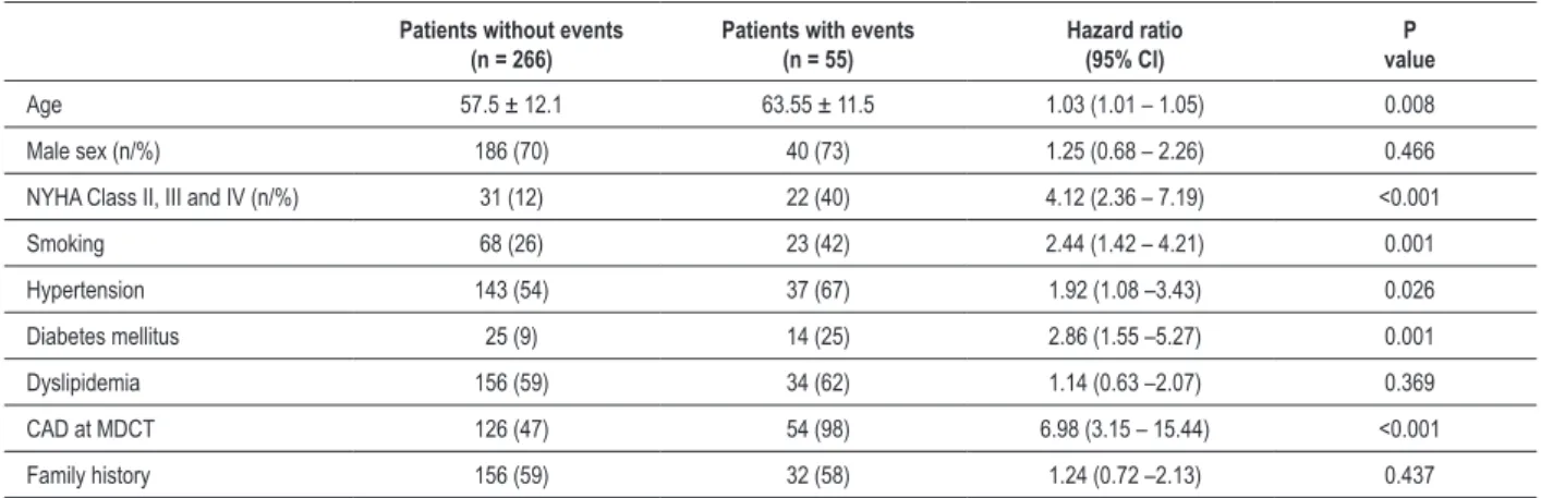
Table 1 – Basal characteristics of the study population according to the occurrence of adverse clinical outcomes
Patients without events (n = 266)
Patients with events (n = 55)
Hazard ratio (95% CI)
P value
Age 57.5 ± 12.1 63.55 ± 11.5 1.03 (1.01 – 1.05) 0.008
Male sex (n/%) 186 (70) 40 (73) 1.25 (0.68 – 2.26) 0.466
NYHA Class II, III and IV (n/%) 31 (12) 22 (40) 4.12 (2.36 – 7.19) <0.001
Smoking 68 (26) 23 (42) 2.44 (1.42 – 4.21) 0.001
Hypertension 143 (54) 37 (67) 1.92 (1.08 –3.43) 0.026
Diabetes mellitus 25 (9) 14 (25) 2.86 (1.55 –5.27) 0.001
Dyslipidemia 156 (59) 34 (62) 1.14 (0.63 –2.07) 0.369
CAD at MDCT 126 (47) 54 (98) 6.98 (3.15 – 15.44) <0.001
Family history 156 (59) 32 (58) 1.24 (0.72 –2.13) 0.437
NYHA: New York Heart Association; CAD: coronary artery disease; MDCT: multi-detector coronary tomography
image acquisition. Patients with HR > 70 bpm and with no contraindications received 15 mg of intravenous metoprolol. Images were reconstructed with slices of 0.3 mm thickness at 0.4-mm intervals, with retrospective gating. All data were analyzed by physicians experienced in the method, using multiplanar reformatting and 3-D reconstruction using volume rendering technique.
The coronary arteries were divided into 17 segments 13. Segments were evaluated for the presence of any
atherosclerotic plaque, defined as structures ≥ 1 mm 2 within
and / or adjacent to the coronary artery lumen. Thereafter, it was determined whether the lesion was obstructive or not, using a threshold of 50% of luminal narrowing. The percentage of coronary artery luminal obstruction was based on a comparison of luminal obstruction in relation to lumen diameter immediately proximal to the plate. Patients without coronary artery calcium or coronary plaques at the MDCT were considered normal, and abnormal MDCT results were defined as the presence of any coronary plaque.
Patient adverse clinical outcomes were obtained through telephone interviews and were classified as the occurrence of: 1) cardiac death, 2) myocardial infarction, 3) unstable angina associated with hospitalization, or 4) revascularization.
Statistical Analysis
The demographic and clinical characteristics of the study population were expressed as numbers and percentages for categorical variables and as mean and standard deviation for continuous variables, compared with the chi-square test for categorical variables and Student’s t test for continuous variables. To satisfy the assumption that the events are independent, the recurrence of cardiac events in one participant was not included in the analysis.
Cox regression model was used to assess the value of clinical variables and the presence of plaque on MDCT in predicting cardiac events. Initially, the univariate analysis of clinical characteristics and variables by MDCT was performed to identify potential predictors. Hazard ratios were calculated
with a 95% confidence interval as an estimate of risk associated with a particular variable. Subsequently, the multivariate analysis was performed including all variables selected in the univariate analysis. The incremental value of each variable in the model was tested using the likelihood ratio, yielding the chi-square and comparing it to the previous model. Statistical analyzes were performed using the SPSS software (version 18.0, SPSS Inc., Chicago, Illinois) and p <0.05 was considered statistically significant.
Results
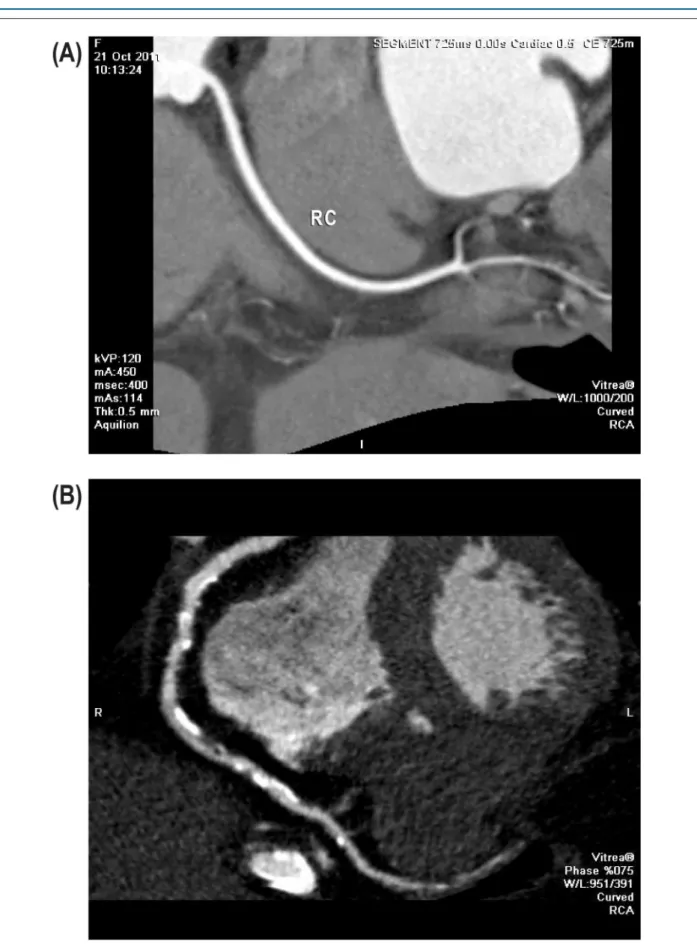
During the study period, 405 patients were initially selected; 21 were excluded for indication of post-CABG surgery and 29 for post-angioplasty evaluation. In total, 355 consecutive patients were included in the study, 71.8% men, with a mean age of 58.4 ± 12.4 years. The general characteristics of the patients are summarized in Table 1. The indications for MDCT assessment were as follows: evaluation of chest pain in 131 patients (32.4%), positive stress test (133 patients [33%]); asymptomatic patients with two or more risk factors (110 patients [27.2%]), and other causes in 30 patients (7.4%). The presence of coronary plaques (Figure 1a) was observed in 55.8% of patients with 28.5% in the right coronary artery, 23.7% in the circumflex artery and 40.6% in the left anterior descending artery. Coronary arteries with lesions > 50% (Figure 1b) were found in 7.7% in the right coronary artery, 5.4% in the circumflex artery and 9.4% in the left anterior descending artery. Mean follow-up was 15 months (range 3-43 months), carried out in 321 patients (90.4%). To determine whether loss at follow-up would influence the results, we performed a comparative analysis between the groups with and without follow-up, demonstrating no difference regarding age, sex, smoking status, hypertension, family history, dyslipidemia, NYHA FC and presence of atherosclerosis at CT. Only the presence of diabetes showed differences between the two groups (p = 0.03).
Original Article
Barros et al. Coronary Tomography for predicting cardiac events
Table 2 – Multivariate analysis by Cox regression model for predicting adverse clinical outcomes
Variables Hazard ratio 95% Conidence Interval p value
NYHA Functional Class 2.034 1.507 – 2.745 < 0.001
Diabetes mellitus 2.360 1.203 – 4.630 0.012
Smoking 2.108 1.186 – 3.745 0.011
CAD at MDCT 5.219 2.337 – 11.656 < 0.001
NYHA: New York Heart Association; CAD: coronary artery disease; MDCT: multi-detector coronary tomography
died of acute myocardial infarction, non-fatal myocardial infarction occurred in five patients; unstable angina requiring hospitalization occurred in 15 patients; 32 patients underwent coronary revascularization, with percutaneous coronary intervention being performed in 27 patients, while the remaining five patients underwent CABG. The decision for revascularization was based on worsening of angina and/ or ischemia at noninvasive testing. Univariate analysis of clinical characteristics and at MDCT to predict adverse clinical outcomes is shown in Table 1. Variables that were significant in the univariate analysis were included in the multivariate analysis. Independent predictors of cardiac events obtained in the multivariate analysis are shown in Table 2.
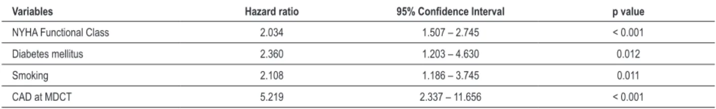
Presence of coronary plaques on MDCT, symptoms of dyspnea by NYHA functional class, smoking and diabetes mellitus were independent predictors of adverse clinical outcomes. Figure 2 shows the different curves related to event-free survival of patients with and without plaques at MDCT.
To determine whether the presence of hemodynamically non-significant coronary atherosclerosis (<50%) could be associated with the occurrence of adverse cardiovascular events we conducted a sub-analysis by withdrawing from the study the patients with significant atherosclerotic lesions. In this new model, the presence of atherosclerosis demonstrated by MDCT in patients with non-significant obstructive lesions remained significant as a predictor of adverse clinical outcomes (hazard ratio 3.21, 95% CI: 1.5 -7.1, p = 0.004).
Discussion
The reference standard test for the diagnosis of CAD is the conventional coronary angiography (CA). Although the incidence of morbidity and mortality is low, the CA can cause severe complications, with total risk of all major complications of 1.7%, mortality of 0.11% and a vascular complication rate of 0.43% 14. Multidetector computed tomography for coronary
assessment rapidly emerged as an accurate diagnostic tool for CAD, with high sensitivity (ranging from 94% to 99%), specificity (range 95% to 97%), positive predictive value (range 76% to 97%) and negative predictive value (ranging from 97% to 100%)
2-4, and the introduction of the 64- detector MDCT allowed a
substantial improvement in method accuracy 15-17. The high
negative predictive value strongly suggests that MDCT could be useful to exclude CAD diagnosis.
In our study, after an average follow-up of 15 months, only one patient with no evidence of coronary atherosclerosis at
MDCT showed adverse clinical outcome, demonstrating a negative predictive value of 98.2% for predicting adverse cardiac events. Gilard et al. 7 showed a low incidence of
cardiac events after normal MDCT in patients with suspected CAD in a study of 141 patients. Pundziute et al. 8 showed
100% absence of events in patients with no abnormalities at MDCT, highlighting an excellent negative predictive value of a normal MDCT. Hadamitzky et al. 9 demonstrated a significant
impact in the prediction of cardiac events for a mean period of 18 months of follow-up.
Chow et al. 18, in a large international multicenter registry,
showed that significant CAD estimated by MDCT had incremental value over left ventricular ejection fraction and clinical variables 18. These results confirm that MDCT is useful as
a diagnostic tool in patients with suspected CAD and may, when the result is normal, strongly suggest ruling out the diagnosis. This finding is of major clinical relevance, as these patients may indeed be safely reassured without additional testing.
Other noninvasive tests also provide useful prognostic information for risk stratification. Myocardial scintigraphy showed an annual mortality or infarction rate below 1% a year 19 when the study is normal. Stress echocardiography has
an excellent negative predictive value for the occurrence of adverse cardiac events 20-22. In this study, we support the idea
that the presence of any plaque, regardless of the stenosis severity, can predict adverse clinical outcomes. The MDCT directly identifies coronary plaques, while conventional CA images determine the contour of the coronary vessel lumen, providing no information on the vessel wall and plaque volume and can therefore, underestimate the atherosclerotic lesion and possible vulnerable plaques that may lead to acute coronary syndrome 23.
Previous studies have shown that non-obstructive lesions can contribute to coronary events 24. Because less obstructive
plaques are more frequent than severely obstructive ones, as shown here, coronary occlusion and myocardial infarction may in fact be more often related to non-obstructive lesions25,26.
Figure 2 – Comparison of event-free survival between patients with atherosclerotic plaque at MDCT and patients without plaque
larger cohorts (with longer follow-up) are clearly needed to confirm these results. Chow et al., in a large multicenter international registry, showed that the severity of CAD estimated by MDCT had incremental value over left ventricular ejection fraction and clinical variables 18.
MDCT results have the potential to influence the decision of revascularization indication and can change the outcome in this study. However, we considered as adverse clinical outcome only those that occurred after three months of follow-up (mean 15
CABG was based on clinical decision, according to established guidelines. Nevertheless, the results of MDCT may be a confounding factor, being associated with the revascularization outcome.
Conclusion
Original Article
Barros et al. Coronary Tomography for predicting cardiac events
1. World Health Organization. Cardiovascular diseases. Fact sheet No. 317. Geneva; 2007 [Cited in 2012 jan 10].Available from: http://www.who.int/ mediacentre/factsheets/fs317/en/index.html
2. Schuijf JD, Pundziute G, Jukema JW, Lamb HJ, va der Hoeven BL, de Roos A, et al. Diagnostic accuracy of 64-slice multislice computed tomography in the noninvasive evaluation of significant coronary artery disease. Am J Cardiol. 2006;98(2):145-8.
3. Hamon M, Giuseppe GB, Malagutti P, Agostini P, Morello R, Valgimigli M, et al. Diagnostic performance of multislice spiral computed tomography of coronary arteries as compared with conventional invasive coronary angiography: a meta-analysis. J Am Coll Cardiol. 2006;48(9):1896-910.
4. Mowatt G, Cook JA, Hillis GS, Walker S, Fraser C, Jia X, et al. 64-Slice computed tomography angiography in the diagnosis and assessment of coronary artery disease: systematic review and meta-analysis. Heart. 2008;94(11):1386-93.
5. Rochitte CE, Pinto IM, Fernandes JL, Azevedo Filho CF, Jatene AD, Carvalho AS, et al.; Sociedade Brasileira de Cardiologia. I Diretriz de ressonância e tomografia cardiovascular da Sociedade Brasileira de Cardiologia - sumário executivo. Arq Bras Cardiol. 2006;87(3):e48-59.
6. Min JK, Shaw LJ, Devereux RB, Okin PM, Weinsaft JW, Russo DJ, et al. Prognostic value of multidetector coronary computed tomographic angiography for prediction of all-cause mortality. J Am Coll Cardiol. 2007;50(12):1161-70.
7. Gilard M, Le Gal G, Cornily JC, Vinsonneau U, Joret C, Pennec PY, et al. Midterm prognosis of patients with suspected coronary artery disease and normal multislice computed tomographic findings: a prospective management outcome study. Arch Intern Med. 2007;167(15):1686-9.
8. Pundziute G, Schuijf JD, Jukema JW, Boersma E, de Roos A, van der Wall EE, et al. Prognostic value of multislice computed tomography coronary angiography in patients with known or suspected coronary artery disease. J Am Coll Cardiol. 2007;49(1):62-70.
9. Hadamitzky M, Freissmuth B, Meyer T, Hein F, Kastrati A, Martinoff S, et al. Prognostic value of coronary computed tomographic angiography for prediction of cardiac events in patients with suspected coronary artery disease. JACC Cardiovasc Imaging. 2009;2(4):404-11.
10. Russo V, Zavalloni A, Bacchi Reggiani ML, Buttazzi K, Gostoli V, Bartolini S, et al. Incremental prognostic value of coronary CT angiography in patients with suspected coronary artery disease. Circ Cardiovasc Imaging. 2010;3(4):351-9.
11. van Werkhoven JM, Schuijf JD, Gaemperli O, Jukema JW, Boersma E, Wijns W, et al. Prognostic value of multislice computed tomography and gated single-photon emission computed tomography in patients with suspected coronary artery disease. J Am Coll Cardiol. 2009;53(7):623-32.
12. Carrigan TP, Nair D, Schoenhagen P, Curtin RJ, Popovic ZB, Halliburton S, et al. Prognostic utility of 64-slice computed tomography in patients with suspected but no documented coronary artery disease. Eur Heart J. 2009;30(3):362-71.
13. Austen WG, Edwards JE, Frye RL, Gensini GG, Gott VL, Griffith LS, et al. A reporting system on patients evaluated for coronary artery disease. Report of the Ad Hoc Committee for Grading of Coronary Artery Disease, Council on Cardiovascular Surgery, American Heart Association. Circulation. 1975;51(4 Suppl):5-40.
14. Scanlon PJ, Faxon DP, Audet AM, Carabello B, Dehmer GJ, Eagle KA,et al. ACC/ AHA guidelines for coronary angiography: a report of the American College of Cardiology/American Heart Association Task Force on practice guidelines (Committee on Coronary Angiography): Developed in collaboration with the Society for Cardiac Angiography and Interventions. J Am Coll Cardiol. 1999;33(6):1756-824.
15. Budoff MJ, Dowe D, Jollis JG, Gitter M, Sutherland J, Halamert E, et al. Diagnostic performance of 64-multidetector row coronary computed tomographic angiography for evaluation of coronary artery stenosis in individuals without known coronary artery disease. Results from the prospective multicenter ACCURACY (Assessment by Coronary Computed Tomographic Angiography of Individuals Undergoing Invasive Coronary Angiography) Trial. J Am Coll Cardiol. 2008;52(21):1724-32.
16. Miller JM, Rochitte CE, Dewey M, Arbab-Zadeh A, Niimura H, Gotlieb I, et al. Diagnostic performance of coronary angiography by 64-Row CT. N Engl J Med. 2008;359(22):2324-36.
17. Meijboom WB, Meijs MVL, Schuijf JD, Cramer MJ, Mollet NR, van Mieghem CA, et al. Diagnostic accuracy of 64-slice computed tomography coronary angiography: a prospective, multicenter, multivendor study. J Am Coll Cardiol. 2008;52(25):2135-44.
18. Chow BJ, Small G, Yam Y, Chen L, Achenbach S, Al-Mallah M, et al.; CONFIRM Investigators. Incremental prognostic value of cardiac computed tomography in coronary artery disease using CONFIRM: COroNary computed tomography angiography evaluation for clinical outcomes: an InteRnational Multicenter registry. Circ Cardiovasc Imaging. 2011;4(5):463-72.
19. Gibbons RJ, Balady GJ, Bricker JT, Chaitman BR, Fletcher GF, Froelicher VF, et al.; American College of Cardiology/American Heart Association Task Force on Practice Guidelines. Committee to Update the 1997 Exercise Testing Guidelines. ACC/AHA 2002 guideline update for exercise testing: summary article. A report of the American College of Cardiology/American Heart Association Task Force on Practice Guidelines (Committee to Update the 1997 Exercise Testing Guidelines). J Am Coll Cardiol. 2002;40(8):1531-40.
20. Sozzi FB, Elhendy A, Roelandt JR, van Domburg RT, Schinkel AF, Vourvouri EC, et al. Long-term prognosis after normal dobutamine stress echocardiography. Am J Cardiol. 2003;92(11):1267-70.
21. Krivokapich J, Child JS, Walter DO, Garfinkel A. Prognostic value of dobutamine stress echocardiography in predicting cardiac events in patients with known or suspected coronary artery disease. J Am Coll Cardiol. 1999;33(3):708-16.
22. Pingitore A, Picano E, Varga A, Gigli G, Cortigiani L, Previtali M, et al. Prognostic value of pharmacological stress echocardiography in patients with known or suspected coronary artery disease: a prospective, large-scale, multicenter, head-to-head comparison between dipyridamole and dobutamine test. Echo-Persantine International Cooperative (EPIC) and Echo-Dobutamine International Cooperative (EDIC) Study Groups. J Am Coll Cardiol. 1999;34(6):1769-77.
23. Aldrovandi A, Cademartiri F, Menozzi A, Ugo F, Lina D, Maffei E, et al. Evaluation of coronary atherosclerosis by multislice computed tomography in patients with acute myocardial infarction and without significant coronary artery stenosis: a comparative study with quantitative coronary angiography. Circ Cardiovasc Imaging. 2008;1(3):205-11.
References
clinicaloutcomes. MDCT may prove useful in the risk stratification of these patients.
Potential Conflict of Interest
No potential conflict of interest relevant to this article was reported.
Sources of Funding
There were no external funding sources for this study.
Study Association
24. Libby P, Theroux P. Pathophysiology of coronary artery disease. Circulation. 2005;111(25):3481-8.
25. Mann JM, Davies MJ. Vulnerable plaque: relation to degree of stenosis in human coronary arteries. Circulation. 1996;94(5):928-31.