Quantitative determinations and imaging in different structures of
buried human bones from the XVIII-XIXth centuries by energy
dispersive X-ray
fluorescence – Postmortem evaluation
D. Guimarães
a,b,n, A.A. Dias
c, M. Carvalho
c, M.L. Carvalho
c, J.P. Santos
c, F.R. Henriques
d,
F. Curate
e,f, S. Pessanha
ca
Laboratory of Inorganic and Nuclear Chemistry, Wadsworth Center, New York State Department of Health, P.O. Box 509, Albany, NY 12201-0509, USA
b
Department of Environmental Health Sciences, School of Public Health, the University at Albany, P.O. Box 509, Albany, NY 12201-0509, USA
c
LIBPhys-UNL, Laboratório de Instrumentação, Engenharia Biomédica e Física da Radiação and Departamento de Física da Faculdade de Ciências e Tec-nologia, Universidade Nova de Lisboa, 2829-516 Caparica, Portugal
d
Museums Division and Local History, City Hall Almada, Portugal
e
Research Centre for Anthropology and Health, University of Coimbra, Portugal
f
Interdisciplinary Center for Archaeology and Evolution of Human Behavior, University of Algarve, Portugal
a r t i c l e i n f o
Article history:
Received 22 February 2016 Received in revised form 13 April 2016
Accepted 13 April 2016 Available online 26 April 2016 Keywords:
Human bone
Elemental concentration, Micro-X Ray fluorescence
Triaxial EDXRF
a b s t r a c t
In this work, a non-commercial triaxial geometry energy dispersive X-ray Fluorescence (EDXRF) setup and a benchtopm-XRF system were used to identify postmortem contamination in buried bones. For two of the individuals, unusually high concentrations of Cu and Pb, but also Zn (in one individual) were observed. The pigments of the burial shroud coverings have been identified as the source of con-tamination.
Accurate and precise quantitative results were obtained by nondestructive process using fundamental parameters method taking into account the matrix absorption effects.
A total of 30 bones from 13 individuals, buried between the mid-XVIIIth to early XIXth centuries, were analyzed to study the elemental composition and elemental distribution. The bones were collected from a church in Almada (Portugal), called Ermida do Espírito Santo, located near the Tagus River and at the sea neighbourhood.
The triaxial geometry setup was used to quantify Ca, Fe, Cu, Zn, Br, Sr and Pb of powder pressed bone pellets (n¼9 for each bone). Cluster analysis was performed considering the elemental concentrations for the different bones. There was a clear association between some bones regarding Fe, Cu, Zn, Br and Pb content but not a categorization between cortical and trabecular bones. The elemental distribution of Cu, Zn and Pb were assessed by the benchtop
μ
-analysis, the M4 Tornado, based on a polycapillary system which provides multi-elemental 2D maps. The results showed that contamination was mostly on the surface of the bone confirming that it was related to the burial shroud covering the individuals.& 2016 Elsevier B.V. All rights reserved.
1. Introduction
Trace elements are chemical elements that accumulate in a particular sample or environment in concentrations inferior to 100mg/g[1]. These elements can be essential or non essential to the living organism. Determining the elemental constitution of mineralized human remains, such as bones, can give information about ante-mortem and post-mortem exposure[2,3].
Human bones are dynamic organs that remodel throughout life
and can be differentiated based on their structure. At a macro-scopic level we can distinguish between cortical bone: compact, constituting the external bone surface, conferring their strength, rigidity and shape; and trabecular bone: spongy, making up the interior of bones, very porous, more vascular and metabolically active than compact bones. It is estimated that, in adults, about of 5–10% of the skeleton is replaced per year, with a higher bone turnover for trabecular compared to cortical [4,5]. Consequently, depending on in vivo uptake of food and water and environmental exposure, new minerals are continuously being integrated into newly formed bone[6]. Analyzing the elemental constitution of ancient bones can lead to important conclusions on populations’ socioeconomic status, occupational practices and even dietary habits[7–9]. Zinc, Sr, and Br in bone are well established as dietary Contents lists available atScienceDirect
journal homepage:www.elsevier.com/locate/talanta
Talanta
http://dx.doi.org/10.1016/j.talanta.2016.04.028
0039-9140/& 2016 Elsevier B.V. All rights reserved.
nCorresponding author at: Laboratory of Inorganic and Nuclear Chemistry,
Wadsworth Center, New York State Department of Health, P.O. Box 509, Albany, NY 12201-0509, USA; [email protected]
Although bone is a good biomarker, its sparse morphological composition makes it prune to post-mortem contamination, also known as diagenesis, particularly in the spongy bone, in part due to its higher surface area[13,18]. The burial soil (elemental com-position and pH) and the weather and environmental conditions (rain, ground water, erosion) play a crucial role in diagenesis, af-fecting the elemental concentration of the buried bones[8].
Energy Dispersive X-ray Fluorescence (EDXRF) technique is a widespread fully matured instrumental analytical method. It plays an important role in biological samples analysis providing non-de-structive multi-elemental analysis, for all the elements with atomic number higher than 13 (or even less in vacuum), in a wide range of concentrations[19]. Due to the minor sample preparation EDXRF has the advantage of being a fast technique at relatively low costs. Since the 1960s XRF has been implemented in archaeological studies[20]
being widely used to determine the concentration of trace elements in human bone remains [8,11,21–23]and even to distinguish be-tween human bone and dental remains from other material [24]. EDXRF has also been used to map the spatial distribution of several elements in bone, once it is advantageous over other destructive techniques as laser ablation - inductively coupled plasma - mass spectrometry (LA-ICP-MS)[22,25,26].
In this work the elemental composition of human bone remains (XVIIIth-XIXth century) is measured by energy dispersive X-ray fluorescence (EDXRF). Two complementary setups were used to measure the bones: EDXRF with triaxial geometry andm-EDXRF with conventional geometry. Thefirst technique allows improved detec-tion limits and the second allows micro beam analysis enabling mapping and spatial distribution of elements within the bone.
The analyzed bones belonged to a group of skeletons recovered from the chapel Ermida do Espirito Santo (EES) in Almada (Por-tugal), that was built in the XV century[27]. After the big Lisbon earthquake in 1755 that was followed by a tsunami, the chapel started being used as the parish seat, becoming a burial space up to 1833. At this time burials in churches became forbidden by royal decree, which forced inhumations to be in open space cemeteries. The recent restructuring of the area, promoted by the Municipality, allowed full archaeological intervention of the abandoned chapel. Following the work carried out between 2010 and 2011, human remains of 83 individuals have been exhumed[28], of which 13 will be the subject of the present work. Everything indicates that they remained untouched after death, because they were all or-iented on the same way, Southwest-Northeast, according to the ritual of the Catholic Church [29]. The scrapings of the burial shrouds covering two individuals were also analyzed.
2. Materials and methods 2.1. Samples
2.1.1. Collection and cleaning
A total of 30 bones from 13 individuals, dating from XVIIIth-XIXth centuries, were recovered from inside a chapel (EES). This
wrapping the bodies. These samples (from individual #32 and #37) were collected and dusted with a brush.
2.1.2. Preparation for Tri-axial EDXRF analysis
For each bone, a few grams were taken with a polyester tool (Retsch GmbH, Germany). Due to the poor preservation state of some bones, the use of a mortar and pestle was enough to reduce them to powder. For the more intact bones the samples were powdered in a polyester mill (Retsch GmbH, Germany). In both cases the resulting bone powder was pressed into pellets 15 mm in diameter, by a 10 t manual hydraulic press from Specac. There was no further chemical treatment or additive to the samples.
For each bone n¼9 pellets were made to minimize effects of inhomogeneity. Each pellet, with a thickness of about 1 mm, was glued (heptane mixture) on a Mylarfilm, fixed into a slide mount and placed on the sample holder (50 mm x 50 mm) in front of the X-ray beam for the element determination.
The remains of two burial shrouds were also analyzed by this instrument but no further treatment was needed, a small piece was placed in front of the beam and the X-ray spectrum was obtained.
Table 1
List of the individuals and bones collected from the chapel Ermida do Espírito Santo and analyzed in this work.
Individual Bone anatomy Bone structure
#1 Fibula Compact Ribs Trabecular #4 Tibia Compact Ribs Trabecular #6 Humerus Compact Ribs Trabecular Scapula Compact #7 Humerus Trabecular Ribs Trabecular #8 Femur Compact Ribs Trabecular #10 Fibula Trabecular Tibia Compact Humerus Compact #11 Femur Compact Ribs Compact #14 Humerus Compact Ribs Trabecular #15 Ulna Trabecular Ribs Trabecular Radius Trabecular #17 Ulna Trabecular Ribs Trabecular Radius Compact #32 Radius Compact Ribs Trabecular #37 Tibia Compact Fibula Trabecular Femur Trabecular #83 Ulna Compact Ribs Trabecular
2.1.3. Preparation form-EDXRF analysis. For the bones of interest transversal sections, around 1 mm thick, were obtained using a microtome equipped with a diamond saw (Buehler Isomet 1000, USA). The resulting sample slice would then be placed inside the XRF chamber on a table attached to a XY translatable stage and directly exposed to the X-ray microbeam to perform the elemental mappings. For each bone of interest one map was acquired.
3. Instrumentation 3.1. Tri-axial geometry EDXRF
A self-constructed spectrometer was used to analyze all the samples. It consists of a commercial X-ray tube with a W anode (Philips PW 2184) operated at 50 kV and 20 mA. A Mo secondary target was placed at a 90° angle with the sample. With this tri-axial arrangement the bremsstrahlung radiation from the X-ray tube anode excites the characteristic lines of the secondary target (17.44 keV for Kα, 19.60 keV for Kβ), which will irradiate the sample. The use of a triaxial geometry, a silver filter and two collimators provide a decrease in the scattering and background radiation[19].
The spot size is ellipse shaped with a major axis of about 2 cm and a minor axis of about 1.5 cm. This system uses a nitrogcooled Si(Li) detector by Oxford Instruments (England) with en-ergy resolution of 135 eV at 5.9 keV. The acquisition live time per measurement is 1000 s. The concentrations were calculated by our
personal software XRFAES (X-ray Fluorescence Automatic Evalua-tion System) and the quantification method is the Fundamental Parameters method[19,30]. In this software, the sample mass is obtained through the information in the coherent and incoherent scattered radiation with a normalizing condition, which means that all concentrations within the sample should add up to unity. The self-attenuation factor is obtained iteratively by constructing a three element virtual matrix that represents the sample. In the present study the chosen matrix was Ca, C and O for the bone samples and C, H, O for the burial shrouds.
3.2. Conventional geometrym-EDXRF
A commercial instrument, the M4 TORNADO (Bruker, Germany) was used for elemental mapping on the cross sections of the more interesting bones. This instrument consists on a low power X-ray tube with a Rh anode and was operated at 50 kV and 300 uA. Placed after the X-ray tube, a poly-capillary lens focus the beam to a spot size that can go down to 25mm for Mo-K
α
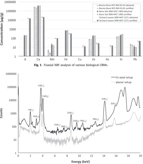
. The use offilters (100mm Al, 50 mm Ti and 25 mm Cu filters) contributes to almost eliminate radiation below 9 keV. This, together with the use of the poly-capillary allows a quasi-monochromatic beam between 9 and 15 keV to irradiate the sample. This system uses a thermoelec-trically cooled Silicon-Drift-Detector with energy resolution of 142 eV at 5.9 keV. The measurements were done under 20 mbar vacuum conditions (to improve detection limits), with a scanning spatial resolution of 25mm, with an acquisition live time per measurement of 3.76 s for each point. In general the maps tookFig. 1. Triaxial XRF analysis of various biological CRMs.
about 3 h to ensure high resolution 2D maps for each element.
4. Statistical analysis
In this study a multivariate statistical analysis of the data was performed. In particular, the Hierarchical Cluster Analysis (HCA) was used as statistical techniques on a data set of 30 bones and 5 variables (the concentration values for Fe, Cu, Zn, Br and Pb).
The HCA method is a statistical treatment designed to reveal groupings (or clusters) within a data set that could be not evident by the reading of data table. The purpose of this analysis is to join together objects into successively larger clusters, by using some measure of similarity or dissimilarities.
Distance is the basic criterion for any clustering and probably the most straightforward way of computing the distances among objects in a multidimensional space is to compute Euclidean dis-tances. Objects that are near each other should belong to the same cluster, and objects that are far from each other should belong to different clusters. The dendrogram is the graphical summary of the cluster analysis. The cases (or the variables) are listed along the horizontal axis, while the distance among the clusters is showed along the vertical axis.
The agglomerative hierarchical cluster analysis dendrogram was obtained using Microsofts Excel 2011/XLSTAT©-(Version
2015.5.01.23264). The distance between the clusters was defined using the single linkage method with center and reduce to avoid scaling effects. This statistical analysis allows classifying the dif-ferent bones into groups, or clusters, being the association be-tween different bones strong within cluster and weak bebe-tween clusters.
NIST 1571. The results are shown inFig. 1. Detection limits are also improved for the tri-axial system, and can differ up to one order of magnitude for lighter elements (seeFig. 2)[8].
5.2. Bone analysis
5.2.1. Elemental concentrations by Tri-axial EDXRF
Using the tri-axial spectrometer all the 30 bones shown in
Table 1(belonging to 13 different individuals) were analyzed in-cluding trabecular and cortical regions for each individual, when possible. Ca, Zn, Fe, Cu, Br, Pb and Sr mean elemental concentra-tions were obtained and the results are presented inTable 2and
Fig. 3. The error affecting the measurements represents the stan-dard deviation for each sample (n¼9 pellets). The results were separated in a table and afigure because when analyzing the data it was possible to identify immediately two distinct behaviors: elements that maintained a similar concentration across the dif-ferent bones and elements that did not.
Belonging to the former group, Ca and Sr (Table 2), have a moderately constant concentration independently of being a spongy bone or a compact bone, from the same or from a different individual. While Ca is a major essential element of the bone composition, Sr is a non-essential trace element that competes to replace Ca [32,33] and has been linked to food consumption habits.
According toTable 2, Ca is the most stable element with re-lative standard deviations (RSD) of 7% followed by Sr with a RSD of 16%, across the 30 bones. The values obtained range from 27 to 38% dry wt for Ca and from 130 to 270mg/g for Sr. Although the concentration range for Sr is broad, this is only due to 3 out of 30 bones: compactfibula of individual #1 (130 mg/g), compact hu-merus of individual #10 (130mg/g) and compact radius of in-dividual #32 (270mg/g). Other than that, all other values are within 25% of the mean value, confirming the constant behavior of this element across all bones. The concentrations obtained for Ca and Sr are comparable to the values found in human bones de-termined by other authors[8,13,34,35]. Yet, it is important to state that establishing a precise comparison with other studies is ex-tremely difficult because there may be diversity on the type of bones analyzed; cortical vs trabecular; different sample prepara-tion methods (concentraprepara-tions presented as dry, wet, ash weight, or sometimes not specified); and particular health conditions specific to each individual.
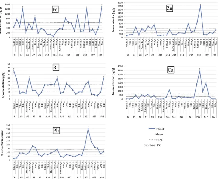
The second group features the elements Fe, Zn, Br, Cu, and Pb, and the results are presented in Fig. 3. These elements do not present homogeneous concentration levels across all bones and reported values are often found outside 30% of the average value (dashed line inFig. 3). Additionally, the minimum %RSD across all the 30 bones is 58% (for Br) but it can go up to 158% (for Cu), showing clearly the variability of Fe, Zn, Br, Cu, and Pb on this set of bones.
From these 5 elements, only for Fe there is a clear distinction between cortical and trabecular bones. For each individual (with the exception of individual #14,Fig. 3), it is noticed enrichment in
#7 Humerus 3273 190720 Ribs 3573 210720 #8 Femur 3473 170710 Ribs 3072 170710 #10 Fibula 3473 240720 Tibia 3373 250720 Humerus 2772 130710 #11 Femur 3072 200720 Ribs 3473 230720 #14 Humerus 3373 220720 Ribs 3573 210720 #15 Ulna 3773 220720 Ribs 3473 220720 Radius 3373 220720 #17 Ulna 3773 180710 Ribs 3873 210720 Radius 3673 200720 #32 Radius 3673 270720 Ribs 3373 250720 #37 Tibia 3473 200720 Fibula 3773 230720 Femur 3373 230720 #83 Ulna 3273 170710 Ribs 3573 200720
the spongy bones comparing to the compact bones, in particular for the ribs that can go up to a 4-fold increase in individuals #17 and #32. Because the spongy tissue is porous and with larger voids it is more susceptible to Fe accumulation, probably due to ex-change mechanism between archaeological bone and its sur-roundings. This is not unexpected and has been reported in other
works[13,36]. In fact when comparing the range of average con-centrations obtained for Fe in this work, with what is reported for contemporary citizens, less than 200mg/g[35,37,38], the former are (with few exceptions) higher, supporting the likelihood of contamination post-mortem.
Zinc in bone, known as a dietary marker, is usually described as
Fig. 3. Results obtained by triaxial XRF (n¼9) for Fe, Zn, Br, Cu and Pb in 30 buried human bones from 13 different individuals; “_c” indicates compact bone and “_s” spongy bone.
not influenced by diagenesis informing about bone ante-mortem conditions [10,13]. However, in the present work, Zinc does not show a steady level (Fig. 3), as stated in otherfindings[8,13]. The results obtained across the 30 bones range from 90 to 1800mg/g and have a %RSD of 94, what clearly shows the variability of Zn concentrations in these samples. With an average value of 371mg/ g, the results obtained are higher than what is reported by other studies done in contemporary bones [2,35,37,38]. In particular, individual #32, with Zn concentration of 1800mg/g in the spongy part of the ribs, warrants further investigation. It is important to notice that although not frequently linked to post-mortem con-tamination Elliot and Grime[39], attribute Zn concentration values found in bone to contamination from burial ground.
Fig. 3shows enhanced levels of Br in these samples ranging from 5 to 40mg/g. There is a lack of studies reporting Br in bone, because it is not always present in significant quantities in the calcified bone mineral. Some studies reporting values of Br sound
in human bone, link it to environmental influence, as marine surroundings, and therefore seafood consumption habits, or nuts
[11,14]. As mentioned before, the chapel is located near the River Tagus as well as less than 10 km away from the Atlantic Ocean. Fishery has been the major source of sustenance on this part of the country, what corroborates with thefinding of high Br concentrations.
Copper and Pb measurements (Fig. 3) also show enhanced le-vels of these elements across all the 30 bones. With concentrations ranging from 13 to 3400mg/g for Cu and 20 to 400mg/g for Pb, the results are in general higher than the concentrations found in other studies, independently of the sample being ancient bones
[8,11,13,36]or contemporary bones[2,35,37]. For example Darrah et al.[37]report bone concentrations of Cu and Pb found in 58 Rochester residents as ranging from 0 to 31.8mg/g for Cu and o1 to 9.5mg/g for Pb. The fact that all these bones, from 13 different individuals, have high levels of Cu and Pb suggest, at same extent,
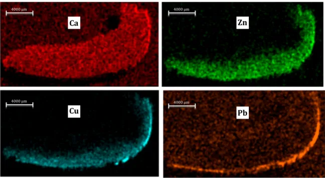
Fig. 5. Calcium, zinc, copper and lead mapping obtained using them-EDXRF system for a humerus transversal cross section of individual #32.
contamination due Cu and Pb present in burial soil. However it might not explain all the results. In fact, for individuals #32 and #37 Pb and Cu concentrations are drastically increased over the other 11 individuals. To better explain these results elemental mapping of the bones of these two individuals is warranted. 5.2.2. Statistical analysis
To support the elemental concentration data discussion and to ensure that the large number of analyzed bones is not covering any similarities impossible to detect without statistical analysis, an agglomerative hierarchical cluster analysis was performed. The dendrogram shown inFig. 4was constructed using the elemental concentration of the elements that are variable across the 30 bones: Fe, Zn, Br, Cu, and Pb. According to this dendrogram the 30 bones are grouped in three separate clusters, with different sizes. Thefirst group comprehends all the bones from individuals #32, #37 and #11. As seen before individuals #32 and #37 were already pointed out as specific cases that needed further investigation, but not individual #11. Unfortunately the bones from the latter in-dividual were no longer available for further investigation. The second group is the smaller and includes all the bones from in-dividual #8 and the compact bones of inin-dividual #17. According to
Fig. 3, it is possible to determine that all these bones have con-centrations lower than the average across all bones for the ele-ments Fe, Br, Cu and Pb. The ribs of individual #17 were probably out of the cluster due to its high Fe content of 920mg/g. The third group is the biggest and contains all the other bones, with the strongest associations belonging to bones of the same individual as can be seen betweenfibula and ribs of individual #1 and ulna and radius of individual #15. Eventually the second and the third group will merge into an unique cluster at higher distances, with the samples of thefirst cluster standing out of the rest of the bones. The way the samples were grouped don’t show a clear distinction between cortical and trabecular bone.
5.2.3. Elemental distribution bym-EDXRF
InFigs. 5and6are presented the elemental distribution for Ca,
Zn, Cu and Pb in a cross section of humerus from individual #32 and in a tibia from individual #37, respectively.
While Ca distribution is uniform in both of the bones, Zn and Cu are clearly accumulating in the outer surface. It is evident that the diffusion of these two elements is from the outside to the inner structure of the compact bone, whilst the spongy part re-mained non-affected. This result suggests that there was con-tamination post-mortem that might not have been due to ex-changes with soil, or it would be seen also in the surface of the endosteal edge. Interestingly both these individuals were wrapped in burial shroud that could be the source of contamination. A fragment of each one of these shrouds was further analyzed.
When comparing the two bones it is noticed that in individual #37 (Fig. 6) Zn and Cu penetrate much deeper than in individual #32 (Fig. 5). Unfortunately it is not possible to use the mappings to determine which bone had higher levels of these elements be-cause the intensity scale is different for each map.
5.3. Burial shroud analysis
Burial shroud fragments, covering the remains of individuals #32 and #37, were analyzed by the tri-axial instrument and the elements present were identified. According toFig. 7, it is evident that the shrouds had high levels of Fe, Cu, Zn and Pb. The con-centrations measured were 3.2% Fe, 0.4% Cu, 215
μ
g/g Zn, 180μ
g/g Pb for the shroud of individual #32 and 0.5% Fe, 0.3% Cu, 50μ
g/g Zn and 120μ
g/g for individual #37. In fact contamination of hu-man bone remains by the use of shrouds is not unheard of. Ar-chaeological evidence for the use of burial shrouds fastened using copper alloys pins is abundant[40]. In some instances the human bones would even be found with green staining due to the dete-rioration of the pin with time and consequent contamination of the bone. Whether the elements measured in the shroud were from the shroud fabric itself or from the use of copper alloy pins is not possible to determine, because no pin was found at the burial site. Nonetheless the identification of the shroud as the cause of contamination for the bones of individual #32 and possiblythese bones were higher than found by other authors and that was attributed to probably soil contamination and diet (marine sur-roundings), respectively. Concerning the results for Zn, Pb and Cu, they were in average higher than reported in other studies, but two individuals stood out due to their unexpected high con-centrations. The imaging of these individuals’ bones using a m-EDXRF instrument based on a polycapillary system showed that these elements were accumulating mainly in the periosteal edge of the bone and that the contamination was clearly post-mortem. In fact both of the individuals were covered in burial shrouds with high concentrations of these elements, accrediting it as the most probable source of contamination.
This work shows how the use of non-destructive X-ray fluor-escence techniques can befit for purpose in the study of biologi-cal/archaeological samples. Coupling tri-axial EDXRF with XRF imaging also shows its advantages in respect to assessment about post-mortem vs pre-mortem contamination.
Acknowledgements
Authors would like to thank Almada Municipality for allowing this investigation.
S. Pessanha acknowledges Portuguese Fundação para a Ciência e Tecnologia for the postdoc grant SFRH/BPD/94234/2013.
References
[1] Control of preanalytical variation in trace element determinations: Approved Guideline., National Committee for Clinical Laboratory Standards, PA, Wayne, 1997.
[2]M.L. Carvalho, J. Brito, M.A. Barreiros, Study of trace element concentrations in human tissues by EDXRF spectrometry, X-Ray Spectrom. 27 (1998) 198–204. [3]J. Rebôcho, M.L. Carvalho, A.F. Marques, F.R. Ferreira, D.R. Chettle, Lead post-mortem intake in human bones of ancient populations by 109Cd-based X-ray fluorescence and EDXRF, Talanta 70 (2006) 957–961.
[4]U. Kini, B.N. Nandeesh, Physiology of bone formation, remodeling and meta-bolism, in: I. Fogelman, G. Gnanasegaran, H. VanderWall (Eds.), Radionuclide and Hybrid Bone Imaging, Springer-Verlag, Berlin Heidelberg, 2012, pp. 29–57. [5]I. Fernández-Tresguerres Hernández-Gil, M.A. Alobera Gracia, Md Canto
Pin-garrón, L. Blanco Jerez, Basesfisiológicas de la regeneración ósea II: El proceso de remodelado, Med. Oral. Patol. Oral. Y. Cir. Bucal 11 (2006) 151–157. [6]J. Burton, T.D. Price, The use and abuse of trace elements for paleodietary
research, in: S. Ambrose, M.A. Katzenberg (Eds.), Biogeochemical Approaches to Paleodietary Analysis, Springer, US, 2002, pp. 159–171.
[7]L.E. Wittmers, A.C. Aufderheide, J.G. Pounds, K.W. Jones, J.L. Angel, Problems in determination of skeletal lead burden in archaeological samples: an example from the First African Baptist Church population, Am. J. Phys. Anthropol. 136 (2008) 379–386.
[8]M.L. Carvalho, A.F. Marques, Diagenesis evaluation in middle ages human bones using EDXRF, X-Ray Spectrom. 37 (2008) 32–36.
[9] A.A. Dias, M. Carvalho, M.L. Carvalho, S. Pessanha, Quantitative evaluation of ante-mortem lead in human remains of the 18th century by triaxial geometry and bench top micro X-rayfluorescence spectrometry, J. Anal. Atomic Spec-trom. 30 (2015) 2488–2495http://dx.doi.org/10.1039/C5JA00340G10.1039/ C5JA00340G.
[10]I. János, L. Szathmáry, E. Nádas, A. Béni, Z. Dinya, E. Máthé, Evaluation of elemental status of ancient human bone samples from Northeastern Hungary dated to the 10th century AD by XRF, Nucl. Instrum. Methods Phys. Res. Sect. B: Beam Interact. Mater. At. 269 (2011) 2593–2599.
Copper deficit as a potential pathogenic factor of reduced bone mineral den-sity and severe tooth wear, Osteoporos. Int. 25 (2014) 447–454.
[17]A.C. Todd, S. Carroll, C. Geraghty, F.A. Khan, E.L. Moshier, S. Tang, P.J. Parsons, L-shell x-rayfluorescence measurements of lead in bone: accuracy and preci-sion, Phys. Med. Biol. 47 (2002) 1399–1419.
[18]A. Perrone, J.E. Finlayson, E.J. Bartelink, K.D. Dalton, -Application of portable x-rayfluorescence (XRF) for sorting commingled human remains (Chapter 7), in: B.J.A.E. Byrd (Ed.), Commingled Human Remains, Academic Press, San Diego, 2014, pp. 145–165.
[19]R. Van Grieken, A. Markowicz, Handbook of X-Ray Spectrometry: Methods and Techniques, Marcel Dekker,, New York, 1992.
[20]M.S. Shackley, An Introduction to X-Ray Fluorescece (XRF) Analysis in Ar-chaeology, X-Ray Fluorescence Spectrometry (XRF) in Geoarcheology, Springer,, New York 2011, pp. 7–44.
[21]B. Pemmer, A. Roschger, A. Wastl, J.G. Hofstaetter, P. Wobrauschek, R. Simon, H.W. Thaler, P. Roschger, K. Klaushofer, C. Streli, Spatial distribution of the trace elements zinc, strontium and lead in human bone tissue, Bone 57 (2013) 184–193.
[22]N. Zoeger, C. Streli, P. Wobrauschek, C. Jokubonis, G. Pepponi, P. Roschger, J. Hofstaetter, A. Berzlanovich, D. Wegrzynek, E. Chinea-Cano, A. Markowicz, R. Simon, G. Falkenberg, Determination of the elemental distribution in hu-man joint bones by SR micro XRF, X-Ray Spectrom. 37 (2008) 3–11. [23]G. Piga, A. Santos-Cubedo, S. Moya Solà, A. Brunetti, A. Malgosa, S. Enzo, An
X-ray diffraction (XRD) and X-ray Fluorescence (XRF) investigation in human and animal fossil bones from Holocene to Middle Triassic, J. Archaeol. Sci. 36 (2009) 1857–1868.
[24]A.M. Christensen, M.A. Smith, R.M. Thomas, Validation of X-rayfluorescence spectrometry for determining osseous or dental origin of unknown material, J. Forensic Sci. 57 (2012) 47–51.
[25]D.J. Bellis, D. Li, Z. Chen, W.M. Gibson, P.J. Parsons, Measurement of the mi-crodistribution of strontium and lead in bone via benchtop monochromatic microbeam X-rayfluorescence with a low power source, J. Anal. . Spectrom. 24 (2009) 622–626.
[26]P. Roschger, I. Manjubala, N. Zoeger, F. Meirer, R. Simon, C. Li, N. Fratzl-Zelman, B.M. Misof, E.P. Paschalis, C. Streli, P. Fratzl, K. Klaushofer, Bone material quality in transiliac bone biopsies of postmenopausal osteoporotic women after 3 years of strontium ranelate treatment, J. Bone Mineral. Res. 25 (2010) 891–900.
[27] T. Antonio, F. Henriques, A Ermida do Espirito Santo de Almada, Camara Municipal de Almada Divisao de Museus e Patrimonio Cultural, 2012, pp. 150– 154.
[28] F. Curate, T. Antonio, S. rosa, F. Robles, Fracturas bilaterales de tibia y perone en un individuo femenino de la "Ermida de Espirito Santo. (Almada, Portugal), in: A. Malgosa, A. Isidro, P. Ibanez-Gimeno, G. Prats-Munoz (Eds.) Actas del XI Congreso Nacional DE Paleopatologia, 2013.
[29]F. Curate, F.R. Henriques, S. Rosa, M. Vitor M. J, A. Tavares, T. Antonio, Morta-lidade Perinatal na Ermida do Espírito Santo (Almada): Entre o afecto e a marginalização, Al-Madan, Lisboa, Portugal 2015, pp. 68–76.
[30]A. Rindby, Software for energy-dispersive X-rayfluorescence, X-Ray Spectrom. 18 (1989) 113–118.
[31]S. Pessanha, M. Carvalho, M.L. Carvalho, A. Dias, Quantitative analysis of hu-man remains from 18th–19th centuries using X-ray fluorescence techniques: the mysterious high content of mercury in hair, J. Trace Elem. Med. Biol. 33 (2016) 26–30.
[32]A.L. Rheingold, S. Hues, M.N. Cohen, Strontium and zinc content in bones as an indication of diet, J. Chem. Educ. 60 (1983) 233–234.
[33]F. Bronner, Metals in Bone: Aluminum, Boron, Cadmium, Chromium, Lantha-num, Lead, Silicon, and Strontium (Chapter 25), in: J.P.B.G.R.J. Martin (Ed.), Principles of Bone Biology, Third ed.,Academic Press, San Diego, 2008, pp. 515–531.
[34]V. Zaichick, M. Tzaphlidou, Determination of calcium, phosphorus, and the calcium/phosphorus ratio in cortical bone from the human femoral neck by neutron activation analysis, Appl. Radiat. Isot. 56 (2002) 781–786.
[35]H.S. Mahanti, R.M. Barnes, Determination of major, minor and trace elements in bone by inductively-coupled plasma emission spectrometry, Anal. Chim. Acta 151 (1983) 409–417.
[36]I. Reiche, L. Favre-Quattropani, T. Calligaro, J. Salomon, H. Bocherens, L. Charlet, M. Menu, Trace element composition of archaeological bones and post-mor-tem alteration in the burial environment, Nucl. Instrum. Methods Phys. Res. Sect. B: Beam Interact. Mater. At. 150 (1999) 656–662.
[37]T. Darrah, M.E. Campbell, J. Prustman-Pfeiffer, R. Poreda, R. Hannigan, Trace element composition of modern human bone, in: P. Censi, T. Darrah, Y. Erel (Eds.), Medical Geochemistry, Springer, Netherlands, 2013, pp. 167–191. [38]M.L. Carvalho, A.F. Marques, J. Brito, Synchrotron radiation and energy
dis-persive x-rayfluorescence applications on elemental distribution in human hair and bones, AIP Conf. Proc. 652 (2003) 522–528.
[39] T.A. Elliott, G.W. Grime, Examining the diagenetic alteration of human bone material from a range of archaeological burial sites using nuclear microscopy, Nucl. Instrum. Methods Phys. Res. Sect. B: Beam Interact. Mater. At. 77 (1993) 537–547.
[40] M. Hall, S.W. Silliman, Historical Archaeology, Blackwell Pub,, Malden, MA, 2006.