UNIVERSIDADE DE LISBOA
FACULDADE DE MEDICINA VETERINÁRIA
CONTRIBUTION TO THE DISCRIMINANT POWER OF SOME OF THE VARIABLES
INVOLVED IN THE STAGING OF SEVERE EQUINE ASTHMA SYNDROME
JOANA DE SOUSA AZEVEDO SIMÕES
Orientador(es): Professora Doutora Paula Alexandra Botelho Garcia de Andrade Pimenta Tilley
Professor Doutor José Paulo Pacheco Sales Luís
Tese especialmente elaborada para a obtenção do grau de Doutor em Ciências Veterinárias na especialidade de Clínica
i
UNIVERSIDADE DE LISBOA
FACULDADE DE MEDICINA VETERINÁRIA
CONTRIBUTION TO THE DISCRIMINANT POWER OF SOME OF THE VARIABLES
INVOLVED IN THE STAGING OF SEVERE EQUINE ASTHMA SYNDROME
JOANA DE SOUSA AZEVEDO SIMÕES
Orientador(es): Professora Doutora Paula Alexandra Botelho Garcia de Andrade Pimenta Tilley
Professor Doutor José Paulo Pacheco Sales Luís
Tese especialmente elaborada para a obtenção do grau de Doutor em Ciências Veterinárias na especialidade de Clínica
Júri
Presidente: Professor Doutor Luís Filipe Lopes da Costa Vogais:
- Professora Doutora Ana Colette Pereira de Castro Osório Maurício - Professora Doutora Susana Oliveira Serrano Monteiro
- Professor Doutor Mário Pedro Gonçalves Cotovio
- Professora Doutora Paula Alexandra Botelho Garcia de Andrade Pimenta Tilley - Professora Doutora Maria Rita Martins Garcia da Fonseca Pequito
ii
Anexo 3 – DECLARAÇÃO RELATIVA ÀS CONDIÇÕES DE REPRODUÇÃO DA TESE OU DISSERTAÇÃO
Nome: Joana de Sousa Azevedo Simões
Título da Tese ou Dissertação: Contribution to the discriminant power of some of the variables involved in the staging of severe equine asthma syndrome
Ano de conclusão (indicar o da data da realização das provas públicas): 2020 Designação do curso de
Mestrado ou de
Doutoramento: Doutoramento em Ciências Veterinárias Área científica em que melhor se enquadra (assinale uma):
Clínica Produção Animal e Segurança Alimentar Morfologia e Função Sanidade Animal
Declaro sob compromisso de honra que a tese ou dissertação agora entregue corresponde à que foi aprovada pelo júri constituído pela Faculdade de Medicina Veterinária da ULISBOA.
Declaro que concedo à Faculdade de Medicina Veterinária e aos seus agentes uma licença não-exclusiva para arquivar e tornar acessível, nomeadamente através do seu repositório institucional, nas condições abaixo indicadas, a minha tese ou dissertação, no todo ou em parte, em suporte digital.
Declaro que autorizo a Faculdade de Medicina Veterinária a arquivar mais de uma cópia da tese ou dissertação e a, sem alterar o seu conteúdo, converter o documento entregue, para qualquer formato de ficheiro, meio ou suporte, para efeitos de preservação e acesso.
Retenho todos os direitos de autor relativos à tese ou dissertação, e o direito de a usar em trabalhos futuros (como artigos ou livros).
Concordo que a minha tese ou dissertação seja colocada no repositório da Faculdade de Medicina Veterinária com o seguinte estatuto (assinale um):
1. Disponibilização imediata do conjunto do trabalho para acesso mundial;
2. Disponibilização do conjunto do trabalho para acesso exclusivo na Faculdade de Medicina Veterinária durante o período de 6 meses, 12 meses, sendo que após o tempo assinalado autorizo o acesso mundial*; * Indique o motivo do embargo (OBRIGATÓRIO)
Nos exemplares das dissertações de mestrado ou teses de doutoramento entregues para a prestação de provas na Universidade e dos quais é obrigatoriamente enviado um exemplar para depósito na Biblioteca da Faculdade de Medicina Veterinária da Universidade de Lisboa deve constar uma das seguintes declarações (incluir apenas uma das três, retirando as que não interessam):
1. É AUTORIZADA A REPRODUÇÃO INTEGRAL DESTA TESE/TRABALHO APENAS PARA EFEITOS DE INVESTIGAÇÃO, MEDIANTE DECLARAÇÃO ESCRITA DO INTERESSADO, QUE A TAL SE COMPROMETE.
Faculdade de Medicina Veterinária da Universidade de Lisboa, 14 de Fevereiro de 2020
(indicar aqui a data da realização das provas públicas) Assinatura: _________________________________________________________________________
iii
The Road goes ever on and on Down from the door where it began. Now far ahead the Road has gone, And I must follow, if I can, Pursuing it with eager feet, Until it joins some larger way Where many paths and errands meet. And whither then? I cannot say.
The Road goes ever on and on Out from the door where it began. Now far ahead the Road has gone, Let others follow it who can! Let them a journey new begin, But I at last with weary feet Will turn towards the lighted inn, My evening-rest and sleep to meet.
Still round the corner there may wait A new road or a secret gate, And though I oft have passed them by, A day will come at last when I Shall take the hidden paths that run West of the Moon, East of the Sun.
The Lord of the Rings J.R.R. Tolkien
iv
Acknowledgments
The task of writing a PhD thesis is an incredible journey. At the starting point we set out full of hope and ideals, blissfully unaware of all the bumps and forks that await us further down the road. Some of our plans, pristine on paper, need to be readjusted.
Were it not for a multitude of people, that reassured and helped me overcome the obstacles, I might have fled to an island in the South Pacific. I’m a lucky person! To all who have contributed to this work, through guidance, friendship or just by smiling at the end of a particularly long day, Thank you! I am forever grateful to you all.
Dear family and friends, you’ve been my anchors throughout hardships and my life is infinitely richer because of your presence.
Mom and dad, your unconditional love is my pillar of strength. Ana, I know I’ll always have your support in “sticking it to the man”.
Raaj and Joy, fur therapy is the best way to begin and end each day! You’ve never failed to put a smile in my face.
My dearest friends, the coffees, cinema sessions, karaoke parties, dinners and dancing are treasured memories. Without you this story might have had an alternative ending.
Catarina, there are no words that can transpire how incredibly grateful I am to have known you.
Ana and Cláudia, we are “three peas in a pod”, if I say jump, you’ll simply ask how high. Didis, you believed when I didn’t… Thank you!
Adriana, I still miss your snappy remarks and energy! I’m not entirely sure who was the ‘bad influence’…
Lajja, you inspire me to see the world through a fresh perspective and remind me that hard work is the key to success.
Mariana, you’re the best grammar fascist ever!
My wonderful teachers, with whom I’ve had the privilege of working alongside for many years, your guidance and knowledge helped shape this work.
Professor Paula Tilley, my supervisor and friend, this work would not have been possible without her dedication, passion and expertise. It will always be a pleasure to continue to learn and grow under your tutelage.
Professor José Sales Luís, my co-supervisor, an adamant enthusiast of this work whom possessed a godly patience for my solemn demeanour.
Professor Luís Madeira de Carvalho, without whom the interpretation of the coprology results would be a herculean task.
v
I would also like to thank Professor Vinzenz Gerber, whom in spite of the distance was always available for help and advice and Professor Andrew Hoffman, without whom the interpretation of the open pleth data would be nothing more than hieroglyphs.
Professor Conceição Peleteiro, Dr. Hugo Pissara, Sandra and especially Maria do Rosário, for their help in interpreting and processing the BALF cytology’s.
Bruno Fortunato, once again you came to my rescue and ensured the success of this work. João Cid, always ready to lend a helping hand and with an infinite amount of patience for dealing with the horses… and me. Carlos Morgado and Jorge Runa, whom ensured all the animals were taken care of.
Daniela Silva, a curious and adventurous student, whom agreed to spend long hours on road trips. Your precious help was the key to completing the histamine challenge protocol. I wish you all the best.
Marta Vacas de Carvalho, thank you for all your help in interpreting the statistic results of this work.
Helena Almeida I’m sorry for all the last-minute requests and for ransacking the pharmacy. Adriana and Cátia, thank you for letting me join your fortress of solitude.
Lastly, I would like to express my profound gratitude to the referral veterinarians, horse owners, Monsanto’s Forrest Police and GNR who participated in this work. But most importantly to all the horses that, within their best ability, cooperated with us. This work has the sole goal of improving the veterinarian care we are able to provide them.
I would also like to acknowledge CIISA for financing this work through the project INOV CIISA 8.
vi
Resumo
A síndrome de asma equina (SAE) grave é uma doença respiratória crónica, recorrente e altamente prevalente em animais adultos, estando associada à domesticação dos equinos. Devido à sua natureza insidiosa, o tratamento é muitas vezes frustrante, originando perdas económicas significantes.
Quando expostos a ambientes com elevadas concentrações de partículas respiráveis, tais como esporos de fungos, ácaros, endotoxinas, pólen e outras partículas antigénicas, os animais geneticamente suscetíveis desenvolvem inflamação, hiper-reactividade e obstrução das vias aéreas. As concentrações de partículas respiráveis elevadas encontram-se normalmente presentes em sistemas de estabulação de equinos tradicionais, enquanto que em sistemas em extensivo (pastagem) estas concentrações tendem a ser menores.
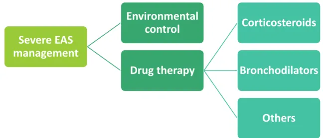
Assim, no maneio da SAE grave é essencial o controlo ambiental de modo a assegurar a melhoria da função pulmonar e em alguns casos o tratamento médico com corticosteroides e broncodilatadores para reduzir os sinais clínicos de inflamação das vias aéreas e broncospasmo.
Tendo em conta a importância clínica desta síndrome, esta dissertação tem por objetivo contribuir para o conhecimento científico da SAE grave. Assim sendo, investigámos a influencia dos testes de função pulmonar no diagnóstico e estadiamento da doença e desenvolvemos um método de estadiamento, utilizando apenas equipamento portátil, o qual poderá ser utilizado na clínica em regime de ambulatório.
Ainda, investigámos a relação entre SAE grave e a resistência a parasitas gastrointestinais, a qual até à data ainda não havia sido reportada em cavalos Puro Sangue Lusitanos. Esta associação foi, contudo, descrita em equinos de outras raças diagnosticados com SAE grave ou outras hipersensibilidades múltiplas.
Por fim, considerando que a remoção de aeroalérgenos é fundamental para a remissão da SAE grave, procurámos avaliar a complacência dos donos dos animais asmáticos a um conjunto de recomendações de maneio ambiental.
Palavras-chave: Síndrome de asma equina grave; estadiamento; função pulmonar;
vii
Abstract
Severe Equine Asthma Syndrome (EAS) is a highly prevalent, chronic and recurrent respiratory disease which appears to be related to equine domestication. Due to its insidious nature, treatment may sometimes be frustrating and severe economic loses occur.
Genetically susceptible individuals develop airway inflammation, hyperresponsiveness and obstruction when exposed to environments with high concentrations of respirable dust particles, which include mould spores, mites, endotoxins, pollen and other antigenic materials. These hazardous respirable dust concentrations are usually found in traditional equine housing systems, while lower concentrations tend to be present in outdoor systems (pasture). The management of severe EAS essentially requires environmental control to ensure improvement of lung function and in some cases medical treatment with corticosteroids and bronchodilators to ameliorate the clinical signs of airway inflammation and bronchospasm. Considering the clinical importance of this syndrome, the dissertation focuses on further contributing to scientific knowledge of severe EAS. As such, we investigated the influence of lung function tests on the diagnosis and staging of the disease and developed a staging method using only portable equipment, which has the potential of being used in equine ambulatory practice.
We also investigated the relation between severe EAS and resistance to gastrointestinal parasites, which had not been, to the author’s knowledge, previously reported in the Lusitano breed horses. This association has been reported in other equine breeds with severe EAS or with other multiple hypersensitivities (MHS).
Lastly, because allergen avoidance is fundamental for the remission of severe EAS we examined owner compliance to a set of recommended guidelines for environmental management.
Keywords: severe equine asthma, staging, lung function, gastrointestinal parasite resistance,
viii
Table of contents
Resumo vi
Abstract vii
Table of contents viii
List of figures x
List of tables xi
List of abbreviations xii
Introduction 1
Chapter 1 – Severe Equine Asthma Syndrome: A Review 2
1. Severe Equine Asthma Syndrome – literature’s terminology 3
2. Epidemiology and genetics 4
2.1. Prevalence 4 2.2. Socioeconomic impact 5 2.3. Genetics 5 2.3.1. Parasitic resistance 6 2.4. Risk factors 7 3. Aetiology 8 4. Pathophysiology 10 5. Clinical signs 13 6. Diagnosis 15
6.1. Medical history and clinical examination 15
6.2. Venous blood and serum analysis 15
6.3. Thoracic radiography 16
6.4. Respiratory endoscopy 17
6.5. Tracheal wash (TW) cytology 20
6.6. Bronchoalveolar lavage (BAL) 20
6.7. Pulmonary function tests 23
6.7.1. Arterial blood gas (ABG) analysis 23
6.7.2. Conventional lung mechanics 25
6.7.3. Flowmetric plethysmography 25
6.7.4. Forced oscillatory mechanics (FOM) 27
6.7.5. Forced expiratory maneuvers (FEM) 28
6.7.6. Evaluation of airway reactivity 28
6.7.7. Bronchodilator challenge 29
6.8. Immunological testing 30
6.9. New imaging techniques – EBUS 30
6.10. Staging methods for severe EAS 31
7. Management of severe equine asthma syndrome 31
7.1. Environmental management 32
7.1.1. Bedding 33
7.1.2. Feed 34
7.1.3. Housing and ventilation 35
7.1.4. Stable activity 36 7.1.5. Ridding arena 36 7.2. Therapeutic management 37 7.2.1. Corticosteroids 37 7.2.1.1. Systemic corticosteroids 38 7.2.1.2. Inhaled corticosteroids 40 7.2.2. Bronchodilators 41 7.2.2.1. β2-adrenoreceptor agonists 41 7.2.2.2. Anticholinergics 43 7.2.2.3. Others 43
ix
8. References 45
Chapter 2 – Objectives 69
Chapter 3 – Experimental work 72
1. Contribution of lung function tests to the staging of severe equine asthma
syndrome in the field 73
2. A study on intestinal parasitic infection in Lusitano horses with severe Equine
Asthma Syndrome 90
3. Owner compliance to environmental management protocol for severe Equine
Asthma Syndrome 102
Chapter 4 – Discussion 117
1. Contribution of lung function tests to the staging of severe equine asthma
syndrome in the field 118
2. Intestinal parasitic infection in horses with severe Equine Asthma Syndrome 121 3. Owner compliance to an environmental management protocol for severe Equine
Asthma Syndrome 123
4. References 126
Chapter 5 - Conclusion 129
Chapter 6 – Future work 132
1. Clinical staging of severe equine asthma syndrome 133 2. Link between equine asthma and parasite resistance 134
3. Disease immunology 134
x
List of figures
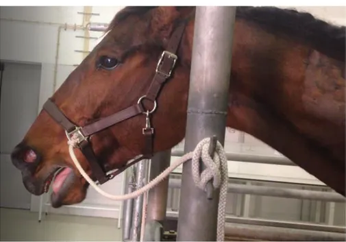
Fig. 1 – Marked nasal flare and cough in a severe EAS-affected horse 14 Fig. 2 – Lung x-ray imaging of an asthmatic horse during disease exacerbation. 17 Fig. 3 – Respiratory endoscopic examination of an asthmatic horse 19
Fig. 4 – Tracheal endoscopy of asthmatic horses. 19
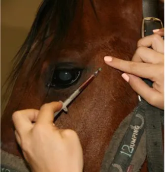
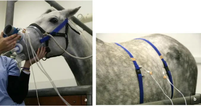
Fig. 5 – Bronchoalveolar lavage proceeding in a severe EAS-affected horse 22 Fig. 6 – Bronchoalveolar lavage cytology of a severe EAS-affected horse 22 Fig. 7 – Arterial blood collection from the transverse facial artery 24
Fig. 8 – Open Pleth System™ 27
Fig. 9 – Basic principles of severe EAS management 32
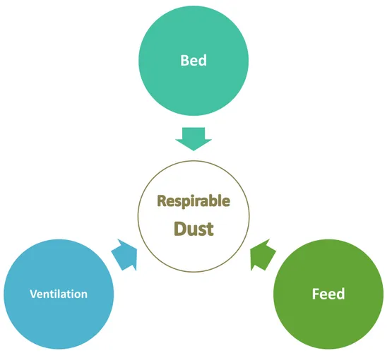
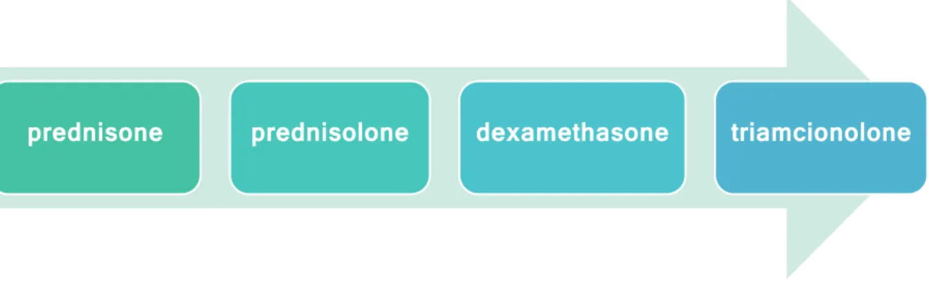
Fig. 10 – Three major aspects of environmental management 37 Fig. 11 – Systemic corticosteroids for severe EAS treatment ordered according
to their potency 39
Fig. 12 – Inhaled corticosteroids used in the treatment of severe EAS ordered
by increasing potency 41
Fig. 13 – Discriminant analysis model adjusted on SEAS staging suggested
(canonical discriminant functions). 81
Fig. 14 – Strongyle egg count per gram (EPG) in the Control group (C) and in
the severe Equine Asthma Syndrome (sEAS) group 94
Fig. 15 – Larvae count per gram (LPG) in the Control group (C) and in the severe
Equine Asthma Syndrome (sEAS) group 95
Fig. 16 – Number of horses positive for the larvae species identified per group
of animals and in the total population 96
xi
List of tables
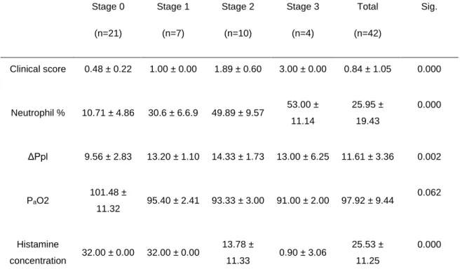
Table 1 – Descriptive statistics (mean and standard deviation) for SEAS-affected
and control horses and significance level of group mean differences 79 Table 2 – Discriminant analysis correlation matrices between groups 80 Table 3 – Horse owner questionnaire used in both interviews to evaluate
environmental management 105
Table 4 Table 4 – Environmental management guidelines given to each SEA-affected horse owner after diagnosis
106 Table 5 – Adoption of each environmental management parameter by horse
owners and health status of SEA-affected horses, before and after the implementation of the environmental guidelines
108
Table 6 – Influence of the environmental parameters adopted by horse owners on the SEA-affected horses health status
109 Table 7 – Influence of owner compliance on SEA-affected horses health status 111
xii
List of abbreviations:
ABG – Arterial blood gas ABD - Abdominal
AHR – Airway hyperreactivity BAL – Bronchoalveolar lavage BALF – Bronchoalveolar lavage fluid BE – Base excess
Bwt – Body weight
COPD – Chronic Obstructive Pulmonary Disease Cdyn – Dynamic compliance
EAS – Equine Asthma Syndrome EBUS – Endobronchial ultrasound EDTA – Ethylenediamine tetraacetic acid
EPG – Egg per gram
FEM – Forced expiratory manoeuvres FOM – Forced oscillatory mechanics HCO3 – Bicarbonate
HOARSI – Horse owner assessed respiratory signs index IAD – Inflammatory Airway Disease
IBH – Insect bite hypersensitivity IDT – Intradermal testing
IFN - Interferon IgE – Immunoglobulin E IM – Intramuscular IL-4 – Interleukin 4 IL-8 – Interleukin 8 IL-17 – Interleukin 17
IL-4R – Interleukin 4 receptor IV – Intravenous
LPG – Larvae per gram
L3 – Third stage infective larvae MHS – Multiple hypersensitivities
xiii miRNA – MicroRNA MMPs – Metalloproteinases MPO – Myeloperoxidase NANC – Nonadrenergic-noncholinergic NH3 – Ammonia
PaO2 – Arterial oxygen pressure
PaCO2 – Arterial carbon dioxide pressure
PO – Per Os
RAO – Recurrent airway obstruction RIB - Ribcage
RIP – Respiratory inductance plethysmography RL – Pulmonary resistance
RRS – Resistance
sO2 – Oxygen saturation
SPRAO – Summer Pasture-Associated Recurrent Airway Obstruction SPT – Skin prick tests
TCO2 – Carbon dioxide tension
Th1 – T-helper type 1 Th2 – T-helper type 2 Th17 – T-helper type 17 TW – Tracheal wash UK – United Kingdom W – Work of breathing
([Hist] – Histamine concentration Δflow – Delta flow (flow differential)
1
Introduction
This dissertation focuses on the study of Severe Equine Asthma Syndrome (SEAS), a highly prevalent and incurable respiratory disease, which due its chronicity and recurrence induces significant losses in the equine industry.
The main on goal of the experimental work here presented was the development of a staging method for ambulatory practice, as well as the study of certain characteristics associated with SEAS, namely the parasitic resistance and owner compliance associated with management recommendations.
For this purpose, this dissertation will be divided in six chapters. The first chapter encompasses a compilation of the current scientific knowledge about SEAS, so as to provide the reader the necessary tools to better understand the experimental clinical work later presented. As such, the disease’s nomenclature and economic impact, its genetic predisposition and risk factors, aetiology, pathophysiology, clinical presentation, diagnosis and management are addressed. The objectives of experimental work and its findings are reported in the second and third chapters. The findings are presented in the form of published or submitted scientific publications. The first publication describes a staging method for SEAS using lung function tests which can be employed in ambulatory practice, the second reports helminth parasitic resistance in SEAS-affected Lusitano horses, and the third describes the owner compliance to a set of guidelines for environmental management of asthmatic horses one year after the initial diagnosis of the disease. Lastly a general discussion and conclusion integrating the three clinical studies will be addressed, as well as future areas which must be addressed in order to further improve our knowledge of this syndrome – chapters four, five and six.
2
CHAPTER 1
3
1. Severe Equine Asthma Syndrome – literature’s terminology
Equine asthma syndrome is a fairly new designation for a disease that may be as old has the beginning equine domestication and stabling (Couëtil et al., 2016; Bullone & Lavoie, 2017). As such, an array of terms has so far been used to describe this clinical entity based on the state-of-the-art knowledge of the time.
With the refinement of research techniques, the syndrome’s terminology evolved to encompass the new findings, like clinical signs, aetiology and pathology. As such, ‘heaves’, ‘broken wind’, ‘equine emphysema’, ‘chronic bronchitis or bronchiolitis’, ‘equine chronic obstructive pulmonary disease’, ‘recurrent airway obstruction’, among others, were once used to describe the same entity, now referred to as Equine Asthma Syndrome (EAS) (Bullone & Lavoie, 2017).
The first terms used to describe the disease centuries ago were ‘broken wind’ and ‘heaves’, due to the clinical signs exhibited by stabled horses fed hay, which tended to present severe breathing difficulties (Markham, 1656).
Later, based on post-mortem findings the term ‘equine emphysema’ was proposed (Obel & Schmiterlöw, 1948). However, since lung hyperinflation observed in severely affected horses results from air trapping and not from alveoli wall disruption, this terminology was considered inadequate (Leclere, Lavoie-Lamoureux & Lavoie, 2011b).
The discovery that neutrophils were the predominant cells found in the bronchoalveolar lavage cytology of affected-horses led to the proposal of the term chronic obstructive pulmonary disease (COPD), since human COPD is also characterised by airway neutrophilia in adult patients, as well as airway inflammation and obstruction (Lavoie, 2015). Nonetheless, this term was also dropped due to significant differences between the two clinical entities, mainly due to the reversibility of lung function changes in the equine patients (Robinson, 2001).
To encompass the variable degree of airway obstruction detected in lung function evaluation and its reversibility when the horse is removed from the offending environment, the designation ‘recurrent airway obstruction’ (RAO) was proposed (Derksen, Scott, Miller, Slocombe & Robinson, 1985a).
In addition, a similar condition but with different aetiology was identified, in which horses developed clinical signs of airway inflammation and obstruction while kept at pasture, and thus unlike RAO was not related to stabling and hay feeding. This syndrome was called ‘summer pasture-associated recurrent airway obstruction’ (SPARAO) (Seahorn & Beadle, 1993; Lavoie, 2007).
In order to simplify terminology, the use of RAO was proposed for severe cases of airway obstruction with signs of increased respiratory effort at rest and ‘inflammatory airway disease’ (IAD) was reserved for milder cases of respiratory impairment and airway inflammation (Robinson, 2001). However, this new nomenclature failed to encompass other similar
4
syndromes, such as SPARAO. Thus, it was deemed necessary to further update the terminology.
Recently, researchers became aware of the similarities in phenotypical presentation, aetiology, pathology and treatment response of equine chronic non-infectious lower airway diseases and human asthma, having introduced the term ‘equine asthma’ (Leclere et al., 2011b; Bullone & Lavoie, 2015; Bond et al., 2018).
In 2016, an issued consensus statement by the American College of Veterinary Internal Medicine proposed the use of ‘equine asthma syndrome’ (EAS) when referring to the spectrum of chronic airway inflammatory diseases similar to human asthma. The panel further defined EAS presentation as varying in severity, having considered IAD as mild-moderate EAS and RAO and SPARAO as severe EAS. However, it should not be assumed that mild EAS will necessarily progress to severe EAS and these two entities should not be interpreted as a continuum (Couëtil et al., 2016; Pirie, Couëtil, Robinson & Lavoie, 2016)).
Therefore, following the panels’ recommendation, severe equine asthma syndrome is currently the correct terminology to describe horses with severe airway inflammation and obstruction, cough, mucus discharge and increased respiratory effort at rest.
This broad terminology will not only allow the inclusion of different and new phenotypes and endotypes, but also simplify future information exchange (Lavoie, 2015; Pirie et al., 2016; Bond et al., 2018).
2. Epidemiology and genetics
2.1. Prevalence
Severe EAS has a significant impact on equine health and is traditionally considered a disease of the northern hemisphere, where horses are stabled throughout the year, particularly during the Winter (Hotchkiss, Reid & Christley, 2007a, b).
Although reports vary, an estimated prevalence of 14-20% has been reported in the United Kingdom (UK) (Hotchkiss et al., 2007a, b; Ireland, Christley, McGowan, Clegg & Pinchbeck, 2015), being as high as 54% when horses are housed in traditional stables (Bracher, von Fellenberg, Winder, Gruening & Hermann, 1991). This prevalence is considered representative of the northern hemisphere.
The incidence of severe EAS is also higher in countries with a cool and wet climate, such as Switzerland and the UK, whereas it becomes virtually insignificant in countries such as Australia, where a dry a warm climate is found (Bracher et al., 1991; Dixon, Railton, McGorum, 1995; Robinson, Derksen, Olszewski & Buechner-Maxwell, 1996; Ward & Couëtil, 2005). This is most likely caused by the different equine management systems (housing and feeding) practiced in such climates, and consequent exposure to disease risk factors.
5
2.2. Socioeconomic impact
Lower airway diseases are the most common cause of poor performance in the equine athlete (Allen, Tremaine & Franklin, 2006). Since severe EAS is a recurrent disease with no known cure up to date, it will inevitably lead to economic losses associated with environmental adjustments, therapeutic management and early retirement of affected horses (Bond et al., 2018). Some animals are refractory to treatment, which may become frustrating to some owners increasing the risk of euthanasia in severely asthmatic horses (Couëtil & Ward, 2003).
2.3. Genetics
Allergic diseases occur in most mammals, including horses, and their relevance has been increasing in western industrialized countries (Weiss, 2000; Marti et al., 2008; Hopkin, 2009; Lambrecht & Hammad, 2017).
Severe EAS is one of such diseases, where susceptible individuals develop airway inflammation and obstruction after exposure to an offending environment (aeroallergen) (Bond et al., 2018). This susceptibility is most likely due to the particular genetic makeup of the affected individuals. In fact, a strong genetic predisposition for equine asthma has been identified, although the exact genetic background for this disease still remains unclear (Ramseyer et al., 2007; Gerber, Baleri, Klukowska-Rötzler, Swinburne & Dolf, 2009).
Two modes of disease inheritance – an autosomal recessive and an autosomal dominant – have recently been described in two Warmblood families and it is believed that several major genes play a crucial role in severe EAS (Gerber et al., 2008; Gerber et al., 2009). Although inheritance mode differed between these two families, the disease clinical phenotype expressed by affected individuals was the same (Laumen, Doherr & Gerber, 2010). As such, it is believed that the overall mode of severe EAS inheritance is probably complex and that different mechanisms may be involved (Graubner, Drogemuller, Fouche & Gerber, 2012). Several studies have also established an association with chromosome 13 and interleukin 4 receptor (IL-4R) (Jost et al., 2007; Gerber et al., 2009; Swinborne et al., 2009; Racine et al., 2011; Schnider, Rieder, Leeb, Gerber & Neuditschko, 2017). In one particular family, the expression of this receptor was increased during severe EAS exacerbation. However, this was not true in the other family, further confirming the genetic heterogeneity of the disease (Klukowska-Rotzler et al., 2012).
In humans, polymorphic differences in the interleukin 4 receptor α chain (IL4Rα) gene play an important role in the development of human asthma and other atopic diseases, since it induces isotopic switch to immunoglobulin E (IgE) and differentiation of T-helper type 2 (Th2) lymphocytes (Jost et al., 2007; Marti et al., 2008).
Likewise, IL4Rα plays a role in regulating susceptibility to equine asthma in some high-prevalence horse families with particular predominant Th2-type cytokine response (Lavoie et
6
al., 2001; Jost et al., 2007). Interestingly, this receptor has also been associated with defence against parasites in both humans and animals (Scales, Ierna & Lawrence, 2007).
The identification and differential expression analysis of MicroRNAs (miRNAs) present in the serum of severe EAS-affected horses, showed a downregulation of miR-128 and miR-744. These findings suggest that a Th2/Th17 immunological response may characterize severe EAS (Pacholewska, Kraft, Gerber & Jagannathan, 2017).
Asthmatic and non-asthmatic horses presented differences in E2F transcription and gene expression after a hay challenge, indicating that asthmatic horses may suffer from impaired epithelial regeneration associated to subepithelial remodelling (Tessier et al., 2018b).
Also, using RNA sequence it was detected a change in the PACRG and RTTN proteins of asthmatic horses, which have been related to ciliary function (Tessier, Côte & Bienzle, 2018a).
2.3.1. Parasitic resistance
In humans, as in horses, there have been several reports of a reverse relation between helminth parasitic infections and allergic diseases, such as asthma and atopy (Lynch et al., 1998; McKay, 2006; Hopkin, 2009; Neuhaus et al., 2010; Bründler et al., 2011; Medeiros et al., 2011;).
Studies conducted in two families of Warmblood horses reported a lower shedding of
gastrointestinal nematode parasite eggs in asthma-affected individuals, which may
suggest increased resistance to parasitic infections (Neuhaus et al., 2010; Brundler et
al., 2011; Schleuniger et al., 2011; Graubner et al., 2012). In Portugal, a similar study
indicated that asthmatic horses may present a lower shedding of helminth eggs
(Medeiros et al., 2011).
Currently, two theories exist that may explain this relationship: the hygiene hypothesis and the genetic hypothesis.
The hygiene hypothesis states that environmental factors, such as infections and gastrointestinal parasites, determine the prevalence of allergic diseases (Strachan, 1989; Von Mutius, 2007). In essence, helminth infections would be capable of inhibiting allergic disease (Hopkin, 2009).
Nowadays, due to modernization and population hygiene, chronic infectious diseases have become less prevalent, which resulted in the loss of cellular and humoral immunoregulatory pathways thus leading to a higher prevalence of allergic diseases (Lambrecht & Hammad, 2017).
Advocating this hypothesis, studies conducted in humans found that atopic children presented a lower helminth burden and higher IgE levels in comparison with non-atopic children (Lynch et al., 1998), and that the removal of helminth infection has the potential to diminish associated
7
immune-regulatory mechanisms and thus promote asthma and allergy (Capron, Dombrowicz & Capron, 2004).
On the other hand, the genetic hypothesis states that the increased resistance of asthmatic-individuals to helminth infection is determined by their genetic makeup. As such, genes which confer resistance to parasites must also be responsible for an increased risk of developing allergic diseases (Neuhaus et al., 2010; Lanz et al., 2013).
This hypothesis is further supported in horses by the fact that from the two Warmblood families with a high prevalence of severe EAS, only in one was IL4Rα linked to the disease occurrence. Therefore, in one of the families one of the mechanisms that could be responsible for parasitic resistance was absent, further supporting the genetic hypothesis (Scales et al., 2007; Neuhaus et al., 2010).
On a similar note, insect bite hypersensitivity (IBH) has also been associated with the presence of airway hyperreactivity, suggesting a probable link with equine asthma, although further investigation is required (Kehrli, Jandova, Fey, Jhan & Gerber, 2015; Lanz, Brunner, Graubner, Marti & Gerber, 2017). The airways of horses with IBH showed hyperreactivity similar to the airways of horses with severe EAS, thus concluding that IBH is associated with airway hyperreactivity and decreased PaO2, even in the absence of overt respiratory clinical signs. Therefore, horses suffering from IBH appear to have a higher risk for airway hyperreactivity and might be predisposed to develop severe EAS in the future (Lanz et al., 2017).
Also, an increased risk for IBH is to be expected in severe EAS-affected horses, and multiple hypersensitivities (MHS) is significantly associated with the absence of nematode eggs in the faeces (Kehrli et al., 2015).
2.4. Risk factors
Severe EAS is usually diagnosed in adult animals (Ireland et al., 2015). In fact, some studies have reported an association between disease occurrence and the age of the horse (≥4 years) (Couëtil & Ward, 2003; Hotchkiss et al., 2007b), while another study reported an association between increased age and the risk of developing airway inflammation (Robinson, Karmaus, Holcombe, Carr & Derksen, 2006).
Breed has also been reported as a risk factor in severe EAS, with Thoroughbreds and Warmbloods presenting a higher risk than other breeds (Couëtil & Ward, 2003; Ramseyer et al., 2007). However, this may be a consequence of the type of management on which these horses were kept, or it may indicate a genetic predisposition (Marti, Gerber, Essich, Ouleha & Lazary, 1991; Ramseyer et al., 2007).
Furthermore, residence in an urbanised environment, exposure to hay and/or straw in early stages of life, as well as history of respiratory infection may also increase the risk of developing severe EAS (Hotchkiss et al., 2007b).
8
3. Aetiology
As previously mentioned, severe EAS-affected horses present clinical signs when exposed to offending environmental allergens (Marti et al., 1991; Marti et al., 2008).
This disease has been associated with hay feeding and stabling, mostly due to the presence of high concentrations of airborne respirable dust particles, which induce airway inflammation (Ramseyer et al., 2007; Ivester & Couëtil, 2014).
Respirable dust concentration is defined as the number of particles with an aerodynamic diameter small enough (≤ 5 µm) to reach the distal airways, and it is considered a good index of the health hazard presented by airborne dust inhalation (Clarke, 1987; Derksen & Woods, 1994; Art, McGorum & Lekeux, 2002).
In equine housing systems the amount of respirable dust concentration represents about 17 to 29% of total dust found in the environment (Woods et al., 1993). Unsurprisingly, respirable dust concentrations are significantly higher in indoor housing systems, where they are 3 to 14-fold higher than those found outdoors (McGorum, Ellison & Cullen, 1998).
As such, studies have classified equine indoor stable environments as hazardous for human respiratory health, due to a high concentration of respirable dust particles, such as endotoxins and moulds, which were considerably above recommended levels (McGorum et al., 1998; Mazan et al., 2009). Thus, it is only logical that the same environment would have a negative impact on equine respiratory health, particularly in individuals genetically predisposed for developing airway inflammation.
No single aeroallergen has been, up to date, pinpointed as the sole major risk factor for severe EAS development, and researchers hypothesise a synergistic effect of several dust components, with horses often exhibiting hypersensitivity to multiple allergens (McPherson et al., 1979; Pirie, Collie, Dixon & McGorum, 2003a; Pirie, Dixon & McGorum, 2003b).
Because aeroallergens have a wide range of size, with the smallest being fungal spores (0.3 to 30 µm in diameter) and the largest pollen grains (10 to 100 µm in diameter), a greater significance has been attributed to the smaller ones which can penetrate the airway of horses (Robinson, 2001; Pirie & McGorum, 2017).
However, pollen grains although larger in size are still present in the respirable dust found indoors and outdoors, and they are thought to play a role in the disease pathogenesis, as is the case in human respiratory allergies (Burge, 2002; Ward and Couëtil, 2005).
Some of the most hazardous (pro-inflammatory) agents found in the stables include fungal spores (>50 species identified), bacterial endotoxins, forage and storage mites, microbial toxins, peptidoglycan, proteases, pollen and plant debris, as well as inorganic dust (McGorum & Pirie, 2008). Although spores such as Aspergillus fumigatus, Faenia rectivirgula and Thermoactinomyces vulgaris have been widely recognised as significant risk factors, a
9
considerable number of horses have shown an increased sensitivity to mite allergens (Clarke, 1987; Seguin et al., 2012; Niedzwiedz, Jaworski & Kubiak, 2015; Klier et al., 2018).
Other pollutants, especially high levels of ammonia, may further aggravate airway inflammation, which is why a good stable and bed hygiene and ventilation are fundamental for the wellbeing of asthmatic horses (Lawrence, Bump & McLaren, 1988; Pirie, 2014).
Urban pollution has been identified as a significant risk factor for asthmatic humans and it may also have a detrimental impact on equine respiratory health (Ward & Couëtil, 2005).
While in the most common phenotypical presentation of severe EAS disease exacerbation occurs with exposure to indoor aeroallergens, mainly mites and fungi spores, in pasture-associated severe EAS exacerbation occurs paradoxically by exposure to pollen and other outdoor allergens (Seahorn & Beadle, 1993; Seahorn, Groves, Harrington & Beadle, 1996; Swiderski et al., 2017; Ferrari et al., 2018)
Environmental conditions also appear to impact disease manifestation, mainly by conditioning the concentration of environmental aeroallergens. Minimum temperature and humidity will likely affect pollen and fungal concentrations, two of the major pro-inflammatory agents of severe EAS (Ward & Couëtil, 2005; Costa, Johnson, Baur & Beadle, 2006; Bullone, Murcia & Lavoie, 2016).
It is believed that horses require a prolonged exposure to high concentrations of respirable dust in order to develop clinical signs of severe EAS (Hotchkiss et al., 2007b).
Nonetheless, equine traditional management, which consist of stall housing, hay feeding and straw bedding, leads to an increased exposure to high concentrations of respirable dust particles and is associated with an increased incidence of severe EAS (Clarke, 1987; Derksen, 1993; Kirschvink et al., 2002). In this environment, respirable dust concentrations around the horse’s breathing zone can reach alarming values of 17.51 mg/m3, due to the dust content of
hay and straw. Thus, the use of alternative feeds and bedding materials suffice to reduce dust concentrations to 0.52 mg/m3, with feed having a greater impact (Woods et al., 1993; Clements
& Pirie, 2007a).
The dust concentration found in hay can be affected by the season (heat and humidity) and by storage conditions (type of bale and storage facilities) (Woods et al., 1993, McGorum et al., 1998).
A study by Robinson and colleagues reported that horses fed hay in round bales had an increased risk of >20% neutrophil counts in the tracheal wash (Robinson et al., 2006).
Also, hay bailed while containing a high moisture content presents an increased number of fungi and actinomycetes spores (Aspergillus fumigatus, Faenia rectivirgula and Thermoactinomyces vulgaris (Clarke, 1987; Seguin et al., 2010; Seguin et al., 2012). The inhalation of A. fumigatus and F. rectivirgula has been shown to result in neutrophilic airway inflammation in severe EAS-affected horses (McGorum, Dixon & Halliwell, 1993; Derksen et
10
al., 1988). Fungal infection can also further contribute to airway inflammation, one of the hallmark features of equine asthma (Dauvillier, ter Woort & van Erck-Westergren, 2018). Additionally, due to airway hyperreactivity, which is one of the clinical features found in severe EAS-affected horses, inhalation of dry or cold air and of particulate matter, such as plant materials, can further aggravate airway inflammation and obstruction (Davis et al., 2006; Pirie, 2014).
4. Pathophysiology
The pathological changes which characterise severe EAS have been vastly described in the literature. Airway inflammation along with neutrophil recruitment, mucus plugging, bronchospasm and airway remodelling, which includes airway wall thickening and smooth muscle hyperplasia, are some of the most common features of the disease (Couëtil et al., 2016).
However, a complete understanding of the immunological mechanisms involved in this disease has still not fully been achieved, with conflicting results having been reported in the literature. A significant number of cytokines, chemokines, genes and, inflammatory and resident airway cells appear to play a part in severe asthma exacerbation (Art, Bureau & Robinson, 2008). There are studies supporting the involvement of a hypersensitive response during disease exacerbation, but unlike human asthma, an early phase response to aeroallergens has yet to be identified in horses (McGorum et al., 1993; Robinson et al., 1996; Deaton et al., 2007). Nonetheless, the delayed phase response has been thoroughly described in asthmatic horses, ultimately resulting in neutrophil recruitment into the airways and in an increase of CD4+ T cells
in the bronchoalveolar lavage fluid (BALF) (McGorum et al., 1993; Kleiber et al., 1999). The role of mast cells remains somewhat controversial, since severe EAS-affected horses present a higher number of chymase positive mast cells in their bronchial walls in comparison with healthy controls, although no differences were found pertaining to the amount of immunoglobulin E (IgE) bound cells in their lungs (van der Haegen et al., 2005; Künzle et al., 2007).
Genetics may be able to explain the conflicting reports on the role of IgE, since it seems to play an important role in determining the allergen specific IgE response (Schmallenbach et al., 1998; Eder et al., 2000; Künzle et al., 2007; Scharrenberg et al., 2010).
The precise cytokine profile characteristic of severe EAS is still under investigation and conflicting reports on the roles of interleukin (IL) 4, 5, 13 and interferon (IFN) γ can be found in the literature. In fact, some studies support a Th2 associated response, others indicate a mixed cellular response, and others still found no suggestion of a specific Th1 nor Th2 response (Giguère et al., 2002; Ainsworth et al., 2003; Cordeau, Joubert, Dewachi, Hamid & Lavoie, 2004; Horohov, Beadle, Mouch & Pourciau, 2005; Kleiber et al., 2005).
11
Such differences may also be the result of genetic variations and of the complex mode of inheritance identified in severe EAS, which may in turn be linked to a diversity of immunological responses, converging into the same disease phenotype (Art et al., 2008; Graubner et al., 2012; Pirie, 2014).
Another likely hypothesis is that basic methodological differences in these studies, such as time of sample collection, resulted in a lack of consistent results (Horohov et al., 2005; Art et al., 2008; Pietra, Cinotti, Ducci, Guinti & Peli, 2011).
Recently, new evidence of the involvement of Th17 or regulatory cells in the pathogenesis of the disease has been described and the authors have hypothesised a mixed Th17 and Th2 immunological response in affected horses (Pacholewska et al., 2017; Frodella, et al., 2019). Neutrophil recruitment, and consequent airway infiltration, is a hallmark of severe equine asthma, and, T cell activation and epithelial cells play an important role in this process. The expression of IL-8 and IL-17 interleukins was found to be increased in asthmatic horses after their exposure to organic dust (Debrue, Hamilton, Joubert, Lajoie-Kadoch & Lavoie, 2005; Ainsworth et al., 2006). In addition, IL-4 induces the expression of chemotactic factors in pulmonary endothelial cells, further contributing to neutrophil recruitment (Lavoie-Lamoureux et al., 2010).
The neutrophilic activation and infiltration, besides engaging the inflammatory cascade of equine asthma also plays and important role on the pathology of severe EAS. Although not a requirement for pulmonary dysfunction, neutrophils are associated with the presence of pulmonary lesions due to epithelial lesion and impaired repair (Tessier et al., 2017; Bullone, Joubert, Gagn, Lavoie & Elie, 2018; Uberti & Morán, 2018).
Clara cell secretory protein may also play a part in airway inflammation by inhibiting the oxidative burst of luminal neutrophils, further improving phagocytosis (Katavolos et al., 2009; Katavolos, Ackerley, Clark & Bienzle, 2011).
Both the role of alveolar macrophages and epithelial cells remains dubious, with some authors reporting differences in cytokine and chemokine expression between asthmatic and control horses, whilst others have failed to do so (Laan, Bull, Pirie & Fink-Gremmels, 2005; Ainsworth, Wagner, Erb, Young & Retallick, 2007; Rilhimaki et al., 2008; Reyner, Wagner, Young & Ainsworth, 2009; Ainsworth, Matychak, Reyner, Erb & Young, 2009; Joubert, Cordeau & Lavoie, 2011).
Several inflammatory markers have been identified in asthmatic horses during disease exacerbation, such as the neutrophil derived enzyme myeloperoxidase (MPO) and several metalloproteinases (MMPs) derived from neutrophils, lymphocites, epithelial cells and macrophages (Raulo, Sorsa & Maisi, 2000; Nevalainem et al., 2002; Art et al., 2006; Barton, Shety, Bondzio, Einspanier & Gehlen, 2016).
12
Likewise, markers of oxidative stress, such as oxidised glutathione, reduction in ascorbic acid and increased hydrogen peroxide, are present in the airways and breath of asthmatic horses in exacerbation (Art, Kirschvink, Smith & Lekeux, 1999; Deaton et al., 2005; Deaton et al., 2006; Tan et al., 2010).
Another persistent finding in severe EAS-affected horses is the presence of mucus accumulation within the airway lumen (Gerber et al., 2003; Gerber et al., 2004a, b).
This phenomenon occurs due to changes in the rheological properties of mucus, namely an increase in viscoelasticity, which then leads to a decrease in its clearance by the mucociliary and cough mechanisms (Gerber, King, Schneider & Robinson, 2000; Jefcoat et al., 2001; Gerber et al., 2004b).
Also, increased mucus production, may be related to an over expression of the mucin gene EqMUC5AC and an increased number of mucus cells (Gerber et al., 2003; Lugo et al., 2006; Gerber et al., 2009).
Due to the persistence of low-grade airway inflammation, asthmatic horses maintain a certain degree of mucus accumulation even whilst in disease remission, which is associated with changes in the mucus glycoproteins (Jefcoat et al., 2001; Gerber et al., 2004a, b).
In horses, although cholinergic and adrenergic innervation occurs, the inhibitory innervation of the distal airways results mainly from nonadrenergic-noncholinergic (NANC) pathways (Olszewski, Robinson & Derksen, 1997; Matera, Amorena & Lucisano, 2002).
In asthmatic horses there appears to be a flaw in the innervation of the lower airways, including a defective inhibitory NANC response, a decreased response to cholinergic activation and to the inhibitory function of prostanoids, an increased production of epithelial-derived relaxing factor, as well as a dysfunction of α2 and β2 receptors and of muscarinic-autoreceptors, which
further contribute to bronchospasm and airway obstruction (Yu, Wang, Robinson & Derksen, 1994; Olszewski et al., 1997; Zhang, Robinson & Zhu, 1999; Abraham, Kottke, Dhein & Ungemach, 2006; Abraham, Kottke, Ammer, Dhein & Ungemach, 2007; Venugopal et al., 2009).
The peptide endothelin appears to also play a significant role in the occurrence of bronchoconstriction in equine asthmatic patients (Fluvio, Emanuele, Francesca, Matteo & Beniamino, 2012a; Fluvio et al., 2012b).
Airway patency is further compromised by the inflammatory-associated changes that take place in bronchi and bronchioles. Increased thickness of the epithelium, submucosa and smooth muscle along with the loss of ciliated cells and their replacement by a hyperplastic epithelium in larger airways, result in a decreased airway lumen and contribute to obstruction (Kaup, Drommer, Damsch & Deegen, 1990).
13
Asthmatic horses may present emphysema, not because of alveoli destruction but due to its incapacity of collapsing (‘air entrapment’) in cases of severe airway obstruction (Marinkovic, Aleksic-Kovacevic & Plamenac, 2007a, b).
5. Clinical signs
Exercise intolerance, cough and increased expiratory effort characterise severe EAS-affected animals, although clinical signs vary with disease severity, although cough is usually the first sign reported. It begins as a soft persistent cough, but with disease progression paroxysmal bouts of cough occur (Allen & Franklin, 2007; Tilley, Sales Luís & Branco Ferreira, 2012b). Exercise intolerance is also one of the earliest clinical signs, becoming more evident in severely asthmatic horses. It should be noted that depending on the expected performance for a particular horse, decrease in exercise ability may be missed (Robinson, Derksen, Jackson, Peroni & Gerber, 2001).
Overall, the observed clinical signs result from airway inflammation, obstruction and hyperresponsiveness and from mucus hypersecretion when susceptible horses are exposed to offending aeroallergens (Pirie, 2014).
Nasal discharge may also be observed in some animals due to mucus accumulation in the tracheobronchial tree (Tilley, Sales Luís & Branco Ferreira, 2012c). Nonetheless, the regular swallowing of these secretions renders this clinical sign occasional (Pirie, 2014).
Severe disease-induced airway obstruction leads to overt respiratory distress in the resting horse with an abnormal breathing pattern (Robinson et al., 1996). A rapid inspiration and a longer exhalation, with thoracic collapse followed by abdominal lift, characterise the adopted breathing pattern (Léguillette, 2003). The increased abdominal effort observed corresponds to a rapid abdominal movement, which does not impart a significant contribution to ventilation when compared to rib cage movement (Hoffman, Oura, Riedelberger & Mazan, 2007). Eventually, hypertrophy of the external abdominal oblique muscle occurs, and a ‘heave’ line can be observed in chronic cases. Likewise, in an attempt to minimise the resistance provided by the upper airways, nasal flaring can be observed. Increased respiratory effort also results in exaggerated anal movement during breathing (Robinson et al., 2000; Tilley et al., 2012b). Often, severely affected horses present significant body mass loss or even cachexia. These animals present increased oxygen consumption and energy expenditure due to the breathing effort required for trying to overcome the expiratory obstruction (Mazan, Deveney, DeWitt, Benedice & Hoffman, 2004). In addition, the respiratory distress and tachypnoea lead to a decrease in food intake, which results in caloric imbalance and this energetic deficit contributes to weight loss (Robinson et al. 2001; Couëtil & Ward, 2003).
Upon clinical examination horses are afebrile and, depending on the degree of airway obstruction, abnormal findings in thoracic auscultation will vary. An increase in
14
bronchovesicular sounds, end-expiratory wheezes and inspiratory crackles may be heard (Léguillette, 2003). Wheezes result from airway narrowing during expiration, while the sudden opening of collapsed airways produces crackles (Pirie, 2014). Mucus hypersecretion and pooling along the trachea produces a ‘fluttering’ sound. The sounds of mucus flutter and wheezes can sometimes be heard at the nostrils without the use of a stethoscope.
Paradoxically, in some animals presenting with significant breathing effort, lung auscultation will be atypically quiet, as a consequence of little airflow occurring due to the severe airway obstruction. Due to lung hyperinflation, because of air trapping in the alveoli, auscultation may reveal an increased lung field (Robinson et al., 2001; Christmann, Buechner-Maxwell, Witonsky & Hite, 2009).
With disease progression gas exchange may be affected, resulting in increased respiratory and heart rates, and in extreme cases the horse may become cyanotic (Robinson et al., 2001). Nonetheless, it is important to remember that one of the key characteristics of severe EAS is the reversibility of clinical signs when the asthmatic horse is removed from the offending environment. The improvement of clinical signs is related to disease severity, but in general progress should be expected in a week span (Thompson & McPherson, 1984).
15
6. Diagnosis
6.1. Medical history and clinical examination
Because clinical signs of severe EAS are quite typical, a tentative diagnosis can be made based on clinical history and examination (Robinson et al., 2001). The vast majority of horses present with a history of chronic and persistent cough, which can be seasonal or associated with stabling and hay feeding (Lavoie, 2007; Bosshard & Gerber, 2014). These animals are also at risk of contracting opportunistic respiratory infections (Theegarten et al., 2008; Juhn, 2014).
Several studies have proposed the use of clinical scores to evaluate and monitor disease severity. The scores are usually based on the most frequent clinical signs observed during severe EAS exacerbation, namely cough, nasal flaring, nasal discharge, abdominal lift and even exercise intolerance (Miskovic, Couëtil & Thompson, 2007; Tilley et al., 2012c; Rettmer, Hoffman, Lanz, Oertly & Gerber, 2015).
However, although clinical history and signs can provide a correct diagnosis of moderately and severely asthmatic horses, clinical scores alone have proved unsuccessful in identifying low-grade obstruction (mild and subclinical cases) (Coëtil, Rosenthal, DeNicola & Chilcoat, 2001; Laumen et al., 2010; Bosshard & Gerber, 2014).
Clinical scores based on nasal flaring and abdominal effort were found to correlate with standard lung mechanics, though exercise intolerance showed no connection with cough nor with airway dysfunction (Rush et al., 1998; Robinson et al., 2000; Benedice, Mazan & Hoffman, 2008). Although cough has been reported as an indicator of airway inflammation, it only manifests when a certain degree of inflammation is present in the horses’ airways. Horses in remission and kept in a controlled environment maintain some degree of airway inflammation and hyperresponsiveness, although unaccompanied by the usual clinical signs (Robinson et al., 2003).
Since clinical signs scores proved insufficient to correctly stage severe EAS and because other diseases, such as dynamic upper airway obstruction and mild equine asthma, can mimic its low-grade presentations, ancillary diagnostic tests play an important role in disease characterisation and differential diagnosis (Robinson et al., 2000; Couëtil et al., 2001; Pirie, 2014; Rettmer et al., 2015).
6.2. Venous blood and serum analysis
Routine haematology and biochemistry are usually within normal values, but they can be helpful to rule out other differential diagnosis, such as pneumonia (Robinson et al., 2001; Allen & Franklin, 2007).
16
6.3. Thoracic radiography
In the adult equine, x-ray imaging of the lungs can be challenging, particularly due to the depth of the equine chest and the superimposition of the forelimb on the cranial thoracic field. On average the x-ray beam must penetrate almost one meter of bone, muscle and lung tissue. As such, a large amount of body fat may further compromise this task (Dunkel, Gibbs & Weller, 2013). This can be particular true in the Lusitano breed which tends to present a higher body condition.
In order to completely visualise the equine thorax, four overlapping imaging fields should be considered – craniodorsal, caudodorsal, cranioventral and caudoventral (Farrow, 1981). Each view will provide the clinician with important information for evaluating respiratory disease (Barton, Schulze, Doherr & Gehlen, 2018). However, in smaller horses a lesser number of projections may suffice.
In 2016, the ACVIM suggested that thoracic radiographs alone were insufficient to diagnose severe EAS. However, they allowed the exclusion of alternative diagnoses, thus providing the confirmation of asthma (Couëtil et al., 2016). This recommendation corresponds to the reality in everyday clinical practice, where economic factors play an important role in the choice of the most suitable diagnostic test.
Nonetheless, several studies have reported radiographic findings characteristic of severe EAS. Radiographic imaging of the equine asthmatic lung showed an increased bronchovascular and interstitial pattern (Robinson, 2001) and thickening of smaller bronchi bifurcations related to disease severity (Koch et al., 2007). The presence of an interstitial and a bronchial interstitial pattern in the lungs has also been associated with severe clinical signs of asthma (Bakos, 2008).
In chronic cases, the x-rays may reflect irreversible changes in the lung parenchyma, such as generalised increase of lung opacity, interstitial infiltration, caudally displaced lung borders, increased radiolucency with concave diaphragm and even bronchiectasis (Lavoie, Dalle, Breton & Hélie, 2004; Allen & Franklin, 2007; Barakzai & McAllister, 2007; Lavoie, 2007). Tilley et al. (2012c) found significant differences in thoracic radiographic changes in pulmonary pattern and tracheal and bronchial wall thickness which correlated with clinical signs and other indicators of airway inflammation and therefore could be used in the staging of severe EAS. Nonetheless, the radiographic changes that can be seen in severe EAS cases are not pathognomonic of this disease (Kutasi, Balogh, Lajos, Nagy & Szenci, 2011; Couëtil et al., 2016).
Also, in mild cases of severe EAS with discreet airway inflammation, little or no changes will be found in the thoracic x-ray, which is why this technique alone does not allow the identification of mild and subclinical cases (Barton et al., 2018).
17
Regardless, radiographic observations can be particularly helpful in ruling out alternative diagnoses and evaluating pulmonary sequelae of severe EAS.
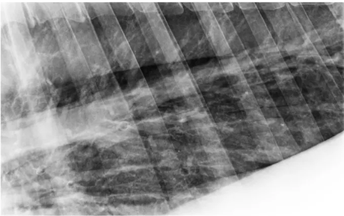
Fig. 2 – Lung x-ray imaging of an asthmatic horse during disease exacerbation. Note the bronchial interstitial pattern accompanied by thickening of bronchial walls.
6.4. Respiratory endoscopy
Respiratory endoscopy allows the assessment of mucosal oedema and/or hyperaemia, mucopurulent secretions, bronchospasm and foreign bodies, making its use commonplace in equine practice in cases presenting with respiratory disease (Hare and Viel, 1998; Kutasi et al., 2011).
The use of endoscopy, by itself or in association with other techniques (bronchoalveolar lavage and tracheal lavage), in severe EAS allows the evaluation of airway inflammation, and also contributes to the exclusion of other differentials (Couëtil et al., 2016).
An association between upper and lower airway inflammation has been extensively documented in human medicine, giving rise to the expression “one airway, one disease” and asthmatic humans will often present with rhinitis as well as lower airway inflammation (Jeffery & Haahtela, 2006). However, no association was found in the horse and the results from upper and lower airway examination should be interpreted independently (Koblinger et al., 2011).
18
Endoscopic evaluation of severe EAS horses is characterised by the presence of mucus or mucopurulent secretions in the tracheal and bronchial lumen, due to hypersecretion, decreased clearance and changes in mucus rheological properties (Gerber et al., 2000; Gerber et al., 2003; Robinson et al., 2003). Depending on its quantity, the mucus exudate can form a small pool at the lowest point of the trachea, or if it becomes more profuse form rafts or streams along the tracheal wall (Gerber et al., 2004 a, b).
As a result, several endoscopic scoring systems have been reported in the literature for assessing mucus quantity and quality and airway oedema/thickness, having been used in the evaluation of the asthmatic horse (Dixon et al., 1995; Hare and Viel, 1998; Gerber et al., 2004b; Koch et al., 2007; Tilley et al., 2012c).
Mucus accumulation was found to correlate well with clinical scores for severe EAS (Costa, Seahorn & Moore, 2000), as well as with cough frequency (Robinson et al., 2003). A correlation between tracheal mucus and lower airway inflammation has also been reported (Koblinger et al., 2011) and airway obstruction due to mucus presence can persist in severe asthmatic horses in remission (Gerber et al., 2004a).
In 2004, Gerber and co-workers developed a mucus scoring system for quantitative and qualitative classification of secretions observed in the tracheal lumen which has been extensively used in clinical practice and which presented a correlation with cytological indicators of airway inflammation (Gerber et al., 2004b).
An increase in the thickness of the tracheal septum (carina), a consequence of airway wall inflammation, is commonly observed in severe EAS (Feutz, Riley, Adamec & Couëtil, 2011). However, Koch et al (2007) reported no correlation between septum thickness and severe EAS clinical, endoscopic or cytological features.
In 2012, building on the works by Gerber et al (2004b) and Koch et al (2007), the team of investigators used an endoscopy scoring system, which evaluated airway mucosal hyperaemia, septum thickness and tracheal mucus quantity and its characteristics. Only the mucus scores had a high correlation with clinical and inflammatory features of severe EAS and were included in a proposed model of severe EAS staging (Tilley et al., 2012c).
However, mucus accumulation cannot be considered a sensitive indicator of severe EAS, since it is not an exclusive feature of this disease and has been observed in cases of mild and moderate EAS (formerly IAD) (Couëtil et al., 2016). Also, in severe asthmatic horses in remission little or no differences were found in endoscopic mucus scores compared to healthy horses (Gerber et al., 1998; Rettmer et al., 2015).Therefore, mucus grading systems should not be used as a sole indicator of severe EAS (Pirie, 2014; Couëtil et al., 2016).
19
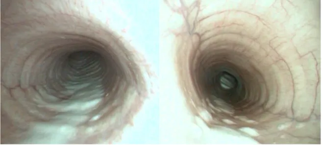
Fig. 3 – Respiratory endoscopic examination of an asthmatic horse.
Fig. 4 – Tracheal endoscopy of asthmatic horses. Note the increased accumulation of mucus on the ventral aspect of the trachea.
20
6.5. Tracheal wash (TW) cytology
The tracheal wash (TW) allows sampling of secretions found in the asthmatic horses’ trachea, thus providing information on airway inflammation.
The technique may be performed either percutaneously or transendoscopicaly, the latter being a less invasive option (Allen & Franklin, 2007; Couëtil & Hawkins, 2013). Saline is infused into the tracheal lumen and the loosen secretions are aspirated for cytological analysis (Robinson et al., 2001).
Despite being relatively easy to perform, TW has not been included in the guidelines for severe EAS diagnosis (Couëtil et al., 2016). This is mainly due to reports of poor correlation between neutrophil counts in the TW and in the BALF, which may indicate that the cellular population found in the tracheal lumen is not representative of the lower airways (Derksen, Brown, Sonea, Darien & Robinson, 1989; Malikides, Hughes, Hodgson & Hodgson, 2003; Fraipont et al 2011). Also, a large variability of inflammatory cells can be observed in the TW cytology of healthy horses, making results’ interpretation challenging (Robinson, 2001; Allen & Franklin, 2007). However, Rossi and co-workers reported a good correlation between neutrophil percentage in TW and BALF cytology in healthy horses and in horses with respiratory disease (Rossi et al., 2018).
Nonetheless, TW neutrophilia has been associated with cough and mucus (Derksen et al., 1989; Christley et al., 2001; Robinson et al., 2006), but not with poor performance (Holcombe et al., 2006).
6.6. Bronchoalveolar lavage (BAL)
Bronchoalveolar lavage fluid (BALF) cytology is considered the reference technique for the diagnosis and monitoring of generalised lung disease, enabling the assessment of lower airway inflammation characteristic of severe EAS (Hewson & Viel, 2002; Jean, Vrins, Beauchamp & Lavoie, 2011; Pirie, 2014).
Unlike TW, BAL provides direct insight of the small airways and alveoli, being a better representative of potential gas exchange impairment and thus allowing the diagnostic of severe EAS (Hoffman, 1999; Hoffman & Mazan, 1999; Couëtil et al., 2016).
BAL can be easily performed in the standing lightly sedated horse and in field conditions (Hoffman, 1999; Mazan & Hoffman, 2003). The technique can be executed using a flexible endoscope or ‘blindly’ with an equine BAL catheter equipped with an inflatable cuff (Couëtil & Hopkins, 2013). The catheter or endoscope is advanced through the nasopharynx and into the trachea, until it is firmly wedged in a small calibre bronchus, which depends on the diameter of the catheter/endoscope (Hoffman, 2008; Tilley et al., 2012c).