A LINK BETWEEN TGF-β SIGNALLING PATHWAYS AND
NUCLEOLUS FUNCTION IN DROSOPHILA
MELANOGASTER
ANDREIA CLÁUDIA PEREIRA CORREIA
Dissertação de Mestrado em Bioquímica
Universidade do Porto
Faculdade de Ciências
Instituto de Ciências Biomédicas Abel Salazar
ANDREIA CLÁUDIA PEREIRA CORREIA
A LINK BETWEEN TGF-β SIGNALLING PATHWAYS AND
NUCLEOLUS FUNCTION IN DROSOPHILA MELANOGASTER
Dissertação de Candidatura ao grau de
Mestre em Bioquímica da Universidade do
Porto
Orientador – Torcato Martins
Categoria – Investigador Doutorado
Integrado
Afiliação – Instituto Biologia Molecular e
Celular, Universidade do Porto
A viagem não acaba nunca. Só os viajantes acabam. E mesmo estes podem prolongar-se em memória, em lembrança, em narrativa. Quando o visitante sentou na areia da praia e disse:
“Não há mais o que ver”, saiba que não era assim. O fim de uma viagem é apenas o começo de outra. É preciso ver o que não foi visto, ver outra vez o que se viu já, ver na primavera o que se vira no verão, ver de dia o que se viu de noite, com o sol onde primeiramente a chuva caía, ver a seara verde, o fruto maduro, a pedra que mudou de lugar, a sombra que aqui não estava. É preciso voltar aos passos que foram dados, para repetir e para traçar caminhos novos ao lado deles. É preciso recomeçar a viagem. Sempre.
Agradecimentos
Enfim, mais uma viagem quase terminada. Chegou o momento em que olhamos para trás e começamos a ser invadidos por aquela nostalgia típica de quando sentimos que o tempo passou demasiado depressa, sem nos ter deixado saborear cada momento como gostaríamos…mas as memórias perduram, e a recordação do que passou e do que aprendi nestes dois anos de Mestrado é um tesouro que ficará sempre guardado.
O Mestrado de Bioquímica trouxe-me para esta cidade maravilhosa que é o Porto há precisamente dois anos. Devo bastante ao Ricardo que, mesmo na minha ausência, fez sempre força para integrarmos o Mestrado, apesar de todas as barreiras que nos foram colocadas. Assim, agradeço à Universidade do Porto e à comissão do Mestrado por nos ter acolhido, e espero sinceramente o nosso esforço e dedicação tenham de alguma forma contribuído para que seja dado maior valor aos alunos de Genética e Biotecnologia vindos da UTAD. Queria também agradecer em especial à Professora Maria João Saraiva pela sua posição como minha tutora e por toda a orientação e disponibilidade, na concretização deste projeto.
Durante o ano curricular foram muitos os momentos que nunca vou esquecer, passados com pessoas também elas inesquecíveis. Aulas, trabalhos, jantares, festas, e principalmente muitas horas de estudo no velho ICBAS. Quero agradecer muitos desses momentos a várias pessoas que de desconhecidos se tornaram amigos, como o Jorge, a Juliana, a Rita, o Bruno, o Filipe, a Diana, a Leninha e o Finger. E claro, um obrigado muito especial a vocês. À Cátia, por toda a ajuda e orientação ao início, pelas conversas e desabafos, por aquele abraço. À Je porque sendo vitoriana é uma verdadeira guerreira. Obrigado por me ouvires, obrigado por tudo. Ao Elísio, pelas palavras e pelos conselhos, pelo teu ombro. E à Andreia, pelo momento mais awkward da minha vida no 200, pelos risos, pelos desabafos, pelo atum, por todas as conversas. Adoro-vos.
Chegado o segundo ano de Mestrado voltei para a minha paixão: a Genética. E não podia ter corrido melhor. O “Developmental Biology Lab”, na altura constituído apenas pelo Torcato e pelo Paulo, recebeu-me de braços abertos. Foi neste laboratório que eu dei os primeiros passos na verdadeira Ciência. E é do fundo do coração que agradeço a todos. À Lígia pela simpatia, pelas conversas, pelos conselhos. Ao safadão do Traquete pelos desabafos e pelas conversas que tanto me ajudaram, desejo-te toda a sorte do mundo no teu futuro. À Sarinha porque dá mais luz ao laboratório. Obrigado por todo o apoio, pelas palavras no momento certo, por todos os risos. Ao Paulo, por toda a disponibilidade e orientação. Pelo incomparável
raciocínio, que nos deixa sempre a pensar. Um sincero obrigado. E finalmente ao Torcato. Nunca vou esquecer tudo o que fizeste por mim. Toda a paciência, toda a dedicação, todos os conselhos. Mais do que um professor foste um amigo. Transmitiste-me essa enorme paixão que tens pela Ciência e pela Genética, ajudaste-me sempre com um sorriso, com uma palavra amiga. Obrigado por tudo, obrigado por nunca desistires de mim, obrigado por este maravilhoso projeto, obrigado por me teres contagiado com este bichinho da ciência, do querer saber, de descobrir, de questionar…obrigado por tudo o que me ensinaste, não só como cientista mas também como pessoa.
Deixo também um obrigado a todas as pessoas que de alguma forma contribuíram para a construção deste trabalho. O Rui e a Elsa que nos aturaram tanto tempo na microscopia, a Filipa, Francisco, Paula Sampaio. Também um grande obrigado à companhia de todos os dias na sala das moscas. Sofia obrigada por todo o apoio e simpatia, Joni obrigado pelas dicas nas francesinhas, Eurico e Sara, pelas histórias e pelos sorrisos.
Também à Ana e à Telma um Obrigado. À Ana viveu comigo durante estes dois anos. És uma pessoa muito especial. Simples, carinhosa, boa ouvinte. Obrigado por todas a conversas, pelos jantares à luz da vela, pelos risos, pelo companheirismo. A Telma, porque esteve sempre lá, porque continua a estar. Obrigado. E, como prometido, ao Café Vera Cruz, porque foi onde escrevi as primeiras e as últimas palavras da minha tese. Obrigado por toda a simpatia e pelas discussões futebolísticas.
Além da Ciência foi também no Porto que descobri o Pedro, tornaste-te tão importante na minha vida. Obrigado por tudo o que passámos, por cada palavra, por cada sorriso, por cada gesto. És o meu porto de refúgio. Contigo é tudo mais fácil, é tudo melhor. Obrigado por seres quem és. Obrigado por existires na minha vida e a tornares mais colorida.
Um grande obrigado a todos os meus amigos que estando longe continuam sempre por perto. Em especial à Mary pela amiga incansável e pela pessoa maravilhosa que é.
Por fim, e mais importante, à minha família. Aos meus pais. Porque sem vocês não seria o que sou. Obrigado por terem acreditado em mim, e por lutarem todos os dias pelo meu futuro. Obrigado pelo apoio incondicional, pela preocupação, pela vossa constante presença mesmo sem estarem perto. Ao Alexandre e à Inês, porque são o meu sorriso de todos os dias. Amo-vos.
Table of Contents
AGRADECIMENTOS ... vii LIST OF FIGURES ... xi ABSTRACT ... xiii RESUMO ... xiv LIST OF ABBREVIATIONS ... xv INTRODUCTION ... 1How genes drive development? ... 3
Growth control: a complex crosstalk between different pathways ... 4
1. TGF-β signalling pathway ... 4
1.1 BMP (Dpp) signalling ... 6
1.2 Activin signalling ... 7
2. Fat-Hippo pathway ... 8
3. Myc induces growth through Viriato ... 9
Nucleolus: a sensorial centre in the cell ... 11
1. Ribosome biogenesis ... 12
Drosophila melanogaster: a small fly that solves big questions ... 15
1. Life cycle ... 15
2. Drosophila imaginal discs: models to study growth ... 16
2.1 Eye-antennal imaginal disc development ... 17
3. Salivary glands ... 20
4. Drosophila genetic tools ... 21
4.1 GAL4-UAS system ... 21
4.2 RNA interference ... 21
Aims of the thesis ... 23
MATERIAL AND METHODS ... 25
Fly strains and Genotypes ... 27
Immunostaining ... 27
Antibodies ... 28
Transmission Electron Microscopy (TEM) ... 28
Size measurements and statistics ... 29
RESULTS ... 31
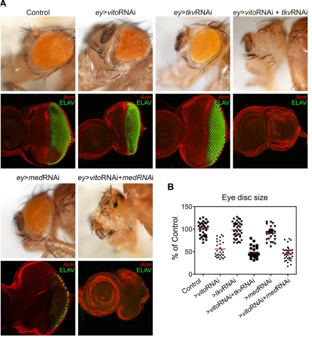
Vito genetically interact with TGF-β signalling pathway during eye growth and differentiation ... 33
The progression of the Morphogenetic Furrow in the eye requires the correct nucleolar function ... 36
Disruption of TGF-β signalling has a direct effect on nucleolar integrity and salivary glands growth ... 38
TGF-β signalling controls the recruitment of specific nucleolar components ... 42
Disruption of TGF-β signalling results in mis-localization of ribosomal proteins ... 44
Disruption of TGF-β signalling results in defective ribosome biogenesis ... 48
putRNAi2 phenotype is mainly due to downregulation of Put levels ... 52
TGF-β signalling disruption impairs regulation of the miRNA Bantam ... 56
DISCUSSION AND FINAL REMARKS ... 61
REFERENCES ... 71
List of Figures
I
NTRODUCTIONFigure 1- TGF-β signalling pathways. ... 5
Figure 2- Fat-Hippo pathway interacts with Dpp signalling to stimulate growth. ... 9
Figure 3- Myc signalling in vertebrates. ... 11
Figure 4- Ribosome biogenesis.. ... 13
Figure 5- Life cycle of Drosophila melanogaster.. ... 16
Figure 6- The eye-antennal imaginal discs develop in the fly head organs.. ... 17
Figure 7- Development of the eye imaginal disc. ... 19
Figure 8- RNA interference approach in Drosophila melanogaster. ... 22
R
ESULTS Figure 9- A eye-targeted double RNAi genetic screen show a strong interaction between Vito and members of TGF-β signalling pathway. ... 34Figure 10- Vito and TGF-β signalling interaction is required for retinal differentiation. ... 35
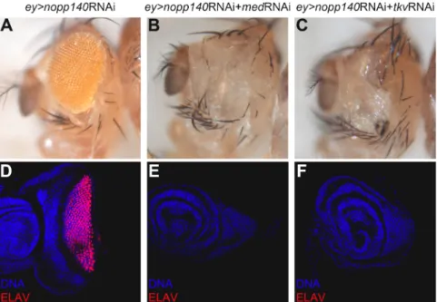
Figure 11- Expression of an RNAi for Nopp140 alters the differentiation process. ... 36
Figure 12- Severity of different RNAis targeting Put. ... 37
Figure 13- The nucleolar morphology is highly dynamic in response to TGF-β signalling. ... 38
Figure 14- RpL41 expression pattern in the eye disc for the indicated genotypes. ... 39
Figure 15- Absence of Put results in nucleolar accumulation of RpL41-YFP.. ... 40
Figure 16- TGF-β signalling directly regulates the ratio between nucleolar and nuclear areas. ... 40
Figure 17- Put levels directly affect salivary glands size. ... 41
Figure 18- Nucleolar levels of Nopp140 decrease in absence of TGF-β signalling.. ... 42
Figure 19- TGF-β signalling regulates the recruitment of Fibrillarin to nucleolus. ... 43
Figure 20- Absence of TGF-β signalling results in ectopic accumulation of RpS6 at nucleolus.. ... 45
Figure 21- RpS9 levels in salivary gland cells decrease when TGF-β signalling is disrupted.. ... 45
Figure 23- RpL11 levels in salivary gland cells decrease in absence of TGF-β signalling. 47 Figure 24- RpL26 intensity in salivary gland cells is not altered in the absence of TGF-β
signalling. ... 47
Figure 25- The fluorescence intensity of UAS-GFP6xmyc also decreases in salivary glands expressing Put RNAi. ... 48
Figure 26- Disruption of TGF-β signalling results in nucleolar aggregates of rRNA, which co-localize with Vito.. ... 49
Figure 27- Absence of TGF-β signalling results in a decrease of mature rRNA.. ... 50
Figure 28- TGF-β signalling regulates nucleolar structure. ... 51
Figure 29- Put gene, transcripts and RNAis targeting sequence. ... 53
Figure 30- Expression of putRNAi3 in a mutant background increase the Put silencing efficacy. ... 54
Figure 31- putRNAi3-expressing cells have an increased ratio between nucleolar and nuclear areas ... 55
Figure 32- Put depletion results in expanded nucleolus, in relation to nuclear and cellular areas. ... 56
Figure 33- Bantam activity depends on TGF-β signalling.. ... 58
Figure 34- The expression patterns of ban are modified in the absence of TGF-β signalling. ... 59
Figure 35- Overexpression of Bantam rescues the retina formation in putRNAi2-expressing eye discs.. ... 60
D
ISCUSSION Figure 36- Absence of Put results in nucleolar stress. ... 69Abstract
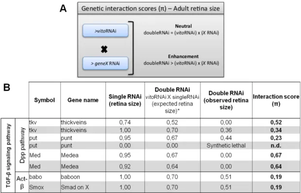
Animal organogenesis requires the establishment of a highly regulated interplay between cell growth, proliferation and differentiation. Cell growth and proliferation are critically dependent on an efficient ribosome production, to sustain high protein-synthesis levels. Ribosome biogenesis and maturation takes place in the nucleolus, a dynamic subnuclear organelle that has also been characterized as a regulatory compartment involved in important cellular processes as cell-cycle control, apoptosis and cellular stress response.Previous studies in our laboratory have shown that the fruit fly Nol12 homologue Viriato (Vito) is a key determinant of nucleolar structure that is required for tissue growth and cell survival during Drosophila development. Moreover, we have identified a strong genetic interaction between Vito and TGF-β signalling pathway members, and demonstrated that Vito is required for TGFβ-dependent tissue growth and photoreceptor neuronal differentiation. These results strongly support a novel signalling branch where nucleolar events contribute positively in the transmission of TGF-β signalling.
The main aim of this work is to understand if the described function of TGF-β in tissue growth is based on a possible role of this pathway in nucleolar function. We observed that expression of a strong RNAi targeting Put, the shared type II receptor for both branches of TGF-β signalling, affects nucleolar retention of structural proteins, Fibrillarin and Nopp140, which are also crucial in rRNA processing. In addition, ribosomal proteins involved in ribosome biogenesis were found to change its nucleolar localization and levels. We also observed a strong increase in nucleolar rRNA levels, suggesting that pre-rRNA processing and thus ribosome biogenesis may be compromised. In fact, putRNAi-expressing salivary glands have decreased amounts of mature rRNA subunits, and a more detailed analysis by transmission electron microscopy also showed that those nucleoli display nuclear accumulation of groups of small circular structures that might represent retained pre-ribosomes. Importantly, alteration of Put levels directly affects the nucleolar architecture and salivary glans growth, therefore supporting a regulation of nucleolar function by TGF-β signalling.
Putting all together, our results unveil a new growth mechanism, in which TGF-β signalling regulates nucleolar structure and function, opening new perspectives in the understanding of the developmental networks that govern animal organogenesis.
Resumo
A organogénese animal é um processo complexo, que requer o estabelecimento de uma interação altamente regulada entre crescimento, proliferação e diferenciação celulares. O crescimento e proliferação celulares dependem fortemente de uma produção eficiente de ribossomas, de forma a suster elevados níveis de síntese proteica. A biogénese e maturação ribossomal ocorrem no nucléolo, um organelo subnuclear que também tem sido considerado como um compartimento regulador. Estudos anteriores no nosso laboratório demonstraram que o homólogo de Nol12 na mosca-da-fruta, Viriato (Vito), é um regulador chave da estrutura nucleolar, necessário para crescimento e sobrevivência celular durante o desenvolvimento. Identificámos também uma forte interação genética entre Vito e membros da via de sinalização TGF-β, que mostrou ser essencial não só para o crescimento do tecido do olho mas também para a diferenciação neuronal.Assim, o principal objetivo deste trabalho consiste em perceber se a função da via TGF-β no crescimento de tecidos é baseada numa possível interação com a função nucleolar. Expressão do RNAi mais forte para Put, o receptor tipo II comum para os dois ramos da via TGF-β, afeta a retenção de proteínas nucleolares estruturais, também cruciais no processamento do RNA ribossomal (rRNA), Fibrilarina e Nopp140. Os níveis e localização de várias proteínas ribossomais também sofrem alterações ao nível do nucléolo. Por outro lado, expressão do RNAi resulta em acumulação nucleolar ectópica de rRNA, sugerindo que o processamento do rRNA, e consequentemente a biogénese ribossomal, poderá estar comprometido. De facto, estas glândulas salivares possuem subunidades maduras de rRNA em quantidades reduzidas, e uma análise mais detalhada por Microscopia Eletrónica de Transmissão revelou que estes nucléolos apresentam acumulação de partículas de rRNA imaturas, retidas no nucleoplasma. Alteração dos níveis de Put mostrou afetar diretamente a morfologia do nucléolo, assim como o crescimento das glândulas.
Em conclusão, os resultados obtidos com este trabalho revelam uma nova interação na qual a função nucleolar contribui positivamente para a sinalização TGF-β, abrindo novas perspetivas na compreensão da rede complexa de interações que governam a organogénese animal.
List of abbreviations
Actβ - Activin β Ago - Argonaute Ato - Atonal Babo - Baboon Ban - BantamBMP - Bone Morphogenetic Proteins Brk - Brinker
Dac - Dachshund
Dad - Doughters against dpp DAPI - 4’,6’ – diamidino-2-phenylindole
Daw - Dawdle
DFC - Dense Fibrillar Component Diap-1 - Drosophila inhibitor of apoptosis 1
dm- diminutive
dMyc - Drosophila Myc DP - Disc proper
Dpp – Decapentaplegic
dsRNA- Double-stranded RNA dTOR- Drosophila target-of-rapamycin
ETS - External Transcribed Spacer Ey - Eyeless
Eya - Eyes absent Eyg - Eyegone
FC - Fibrillar component FMW - First mitotic wave Gbb - Glass Bottom Boat GC - Granular component
GFP - Green fluorescent protein Hh – Hedgehog
Hid- Head involution defective hpRNA - Hairpin RNA
Hth – Homothorax i.e.- Id Est
IGS - Intergenic spacer
ITS - Internal Transcribed Spacer JAK/STAT - Activated Kinase/Signal Transducer and Activator of
Transcription
Mad - Mother against dpp Mav - Maverick
Med - Medea
MF - Morphogenetic Furrow Myo - Myoglanin
NoD - Nucleolar detention sequence NOR - Nucleolar organizer region NPC- Nuclear pore complex Omb - Optomotor-blind PE - Peripodial epithelium pMad- phosphorylated Mad Pol I- RNA polymerase I Pol II- RNA polymerase II Pol III- RNA polymerase III
Pre-rRNA- precursor ribosomal RNA Put – Punt
RD - Retinal determination RFP - Red fluorescent protein
RISC - RNA-induced silencing complex
RNAi - RNA interference Rp - Ribosomal protein RpL - Large subunit Rp RpS - Small subunit Rp rRNA - Ribosomal RNA Sax - Saxophone Scw - Screw Sd - Scalloped Shn - Shnurri
siRNA - Small interference RNA SMW - Seconde mitotic wave snoRNA - Small nucleolar RNA So - Sine oculis
TEM- Transmission Electron Microscopy
TGF-β - Transforming Growth Factor Beta
Tkv - Thickveins
TOP - Terminal oligopyrimidine Toy - Twin of eyeless
UAS- Upstream activating sequence Upd - Unpaired
UTR - Untranslated region Vito – Viriato
Wg - Wingless
Wit - Wishful Thinking
YFP - Yellow fluorescent protein Yki - Yorkie
How genes drive development?
How a single cell gives rise to a collection of organs with the correct architecture to build a highly complex multicellular organism is one of the critical questions of developmental biology. Animal organogenesis demands for a tightly regulated interplay between cell growth, proliferation, differentiation and apoptosis. Extremely complex signalling networks regulate cellular genetic profiles, modifying cell behaviours to precisely arrange cells in time and space, and thus ensure a correct organ patterning and growth (Lecuit and Le Goff, 2007). Besides external signals, such as diet or temperature, also physiological (e.g., hormones) and organ-intrinsic signals are combined to manage these signalling networks (Edgar, 2006b; Neto-Silva et al., 2009).The first observations of tissue-intrinsic regulation of growth arose from polyploid salamanders. Although tetraploid salamanders have the same organ size than diploid salamanders, their cell number is reduced by half (Fankhauser, 1945). Therefore, cell size is proportional to cell ploidy but the balance between cell growth and division is finely controlled in order to reach the correct final organ size. In other experiments in which salamander’s limbs were transplanted between different sized species of salamanders, they grew until reach the donor organ adult size, suggesting the existence of an organ-intrinsic growth programme (Twitty and Schwind, 1931). Likewise, after grafting many thymus pieces on the same developing mice each graft grew independently and reached the typical adult size, suggesting that thymus growth is mainly regulated by organ-intrinsic signals (Metcalf, 1963). Conversely, spleen grafts transplanted to a splenectomised mouse grew until the total mass of transplanted tissue attained the mass of a normal adult spleen, implying that spleen growth is mainly regulated by organ-external signals (Metcalf, 1964).
Therefore, tissue growth and patterning are dependent on the proliferation rate, mass accumulation and cell survival of the composing cells. These parameters are finely coordinated by tissue intrinsic and extrinsic signals that restrict cells correctly arranged within the limits of the tissue target size. Growth and patterning are key developmental processes intimately related. Although little is known about the mechanisms that control organ growth, some conserved signalling pathways involved in patterning are also linked to growth regulation, such as TGF-β signalling.
Growth control: a complex crosstalk between different
pathways
Regulation of tissue growth requires a highly complex interplay between several signalling pathways to keep the correct balance between growth positive and negative signals, thereby ensuring that the correct tissue size is achieved. An imbalance of this homeostatic process could result in impaired cell growth and tumorigenesis.1. TGF-β signalling pathway
The transforming growth factor β (TGF-β) superfamily is highly conserved along metazoans (Padgett et al., 1987; Huminiecki et al., 2009) and comprises a group of secreted proteins that control key cellular functions during development. Members of this family are expressed in precise chronological and spacial patterns to ensure a proper organogenesis and adult tissue homeostasis, by directing cell growth, differentiation, and apoptosis (Massagué, 1998; Moustakas and Heldin, 2009).
Signal transduction is initiated by the binding of the dimeric ligand to a heteromeric receptor complex that consists in two units of each two distinct type I and type II serine/threonine protein kinases. Upon receptor complex formation, the type II receptor, which is constitutively active, phosphorylates the dormant type I receptor, activating its kinase domain. The signal is internalized with the recognition and activation by Type I receptor of a group of transcription factors known as receptor-activated SMADs (R-SMADs), which in turn assembles in a trimeric complex with a common SMAD (Co-SMAD) (Massagué, 1998; Shi and Massagué, 2003; Moustakas and Heldin, 2009). This complex, composed by two activated R-SMADs and one Co-SMAD, is translocated to the nucleus, where it associates with transcriptional co-factors to regulate gene expression (Massagué et al., 2000; Feng and Derynck, 2005).
Phosphorylation and subsequent activation of particular R-SMADs by type I receptor induces distinct transcriptional cell responses, and thus type I receptors coordinate specificity of TGF-β signal transduction (Feng and Derynck, 2005). In this way, TGF-β family is generally divided into two functional signalling
subfamilies, the TGF-β/Activin branch and the bone morphogenetic proteins (BMP) branch, depending on which type I receptor is activated.
Since the discovery of the first ligand of TGF-β signalling pathway in
Drosophila melanogaster, Decapentaplegic (Dpp) (Padgett et al., 1987), a total of
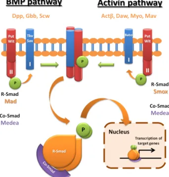
seven ligand family members were identified in this organism: Dpp, Screw (Scw) and Glass bottom boat (Gbb) belonging to BMP family, and Activin-β (Actβ), Dawdle (Daw), Maverick (Mav) and Myoglianin (Myo) belonging to TGF-β/Activin family (Parker et al., 2004). In BMP branch, Thickveins (Tkv) and Saxophone (Sax) are the specific type I receptors, which transduce the signal through phosphorylation of R-SMAD Mother against dpp (Mad). The single type I receptor in TGF-β/activin branch, Baboon (Babo) signals through Smad2 (Smox). Whereas type I receptors confers branch specificity, the type II receptors, Punt (Put) and Wishful thinking (Wit), are shared between the two branches of TGF-β signalling. R-SMADs of both branches also associate with the common Co-SMAD, Medea (Figure 1) (Moustakas and Heldin, 2009).
Figure 1- TGF-β signalling pathways. In Drosophila TGB-β signalling comprises two branches, BMP and Activin. Decapentaplegic (Dpp), Glass Bottom Boat (Gbb) and Screw (Scw) are the BMP pathway ligands, whereas Activin β (Actβ), Dawdle (Daw), Myoglanin (Myo) and Maverick (Mav) are the Activin branch ligands. After binding of the dimeric ligands to the heteromeric receptor complex, two activated R-SMADs (Mad in BMP branch or Smox in Activin branch), compose a trimeric complex with the common Co-SMAD Medea that translocates to the nucleus, and activates the transcription of target genes.
1.1 BMP (Dpp) signalling
The BMP branch of TGF-β signalling has been extensively studied, particularly the Dpp ligand. Besides its role in the establishment of the dorsal fate during embryogenesis (Ferguson and Anderson, 1992), it is well described the importance of Dpp to the Anterior/Posterior compartments organization in the
Drosophila’s wing imaginal disc (Affolter and Basler, 2007). In this epithelial
bilayer that gives rise to the adult wing, dpp is expressed in a restricted range of cells nearby the A/P axis, from which it diffuses throughout the wing disc in a graded concentration to perform a long-range activity (Basler and Struhl, 1994; Nellen et al., 1996; Entchev et al., 2000). This gradient is essential for the proper patterning of the wing disc, as ectopic expression of dpp results in adult flies exhibit abnormal wings with altered patterns, such as symmetric pattern duplications or presence of extra-tissue, the “winglets” (Capdevila and Guerrero, 1994; Zecca et al., 1995). Furthermore, ectopic clones expressing dpp induce the expression of Dpp target genes, such as spalt and optomotor-blind (omb) in the surrounding cells, opposing to the cell-autonomous behaviour observed in clones expressing the constitutively active form of the Dpp receptor Tkv (TKVQD) (Burke and Basler, 1996; Nellen et al., 1996). Therefore, Dpp exerts a morphogen action in the surrounding tissue by modelling cellular behaviour in a positional-dependent fashion. In response, Dpp-receiving cells assume particular genetic profiles and acquire different cell fates depending on its distance to the Dpp diffusion point (Affolter and Basler, 2007; Dekanty and Milán, 2011).
Besides its role on patterning it was also observed that flies with a reduction in dpp activity in the wing display “no wing” phenotype (Zecca et al., 1995), whereas ubiquitous expression of dpp results in overgrowth of the wing disc (Nellen et al., 1996), supporting a strong connection between Dpp and growth. Although several models have been emerged in an attempt to explain how graded morphogenes control the uniform tissue growth, this question remains extremely controversial (Schwank and Basler, 2010). Some evidence suggests that such Dpp-induced tissue growth is positional-independent. In fact, it was demonstrated that the slope of Dpp expands together with the tissue and thus is preserved during wing development (Wartlick et al., 2011). In addition, uniform activation of Dpp pathway by expressing TKVQD in the medial area of the wing disc does not prevent proliferation in the growing tissue, further indicating that the Dpp slope is not a requisite to induce proliferation in this region
(Schwank et al., 2008). Therefore, Brinker (Brk) seems to be a key mediator between Dpp signalling and growth control, by creating an inverse gradient within the wing disc, repressing transcription of Dpp target genes (Müller et al., 2003). Their complementary activity proved to be crucial to define cell fates and to ensure that the growing tissue does not exceed the expected size (Affolter and Basler, 2007; Schwank et al., 2008).
1.2 Activin signalling
In Drosophila the Activin signalling branch has been less characterized than the BMP branch. Nevertheless, besides the described role of this pathway in neuronal plasticity and axon guidance (Zheng et al., 2003, 2006), it was shown that babo mutant larvae display smaller imaginal discs, whereas the ubiquitous expression of a constituvely active form of Babo results in tissue overgrowth (Brummel et al., 1999). Importantly, loss of babo and smox increases cyclin A levels, with subsequent delay in M phase exiting during cell cycle. Thereby,
babo/smox mutant larvae are not capable to undergo pupariation and die in late
larval or early pupal stages, displaying small brain lobes and deficient targeting of photoreceptor’s axons. These defects involve a decrease in the number of brain precursor cells, due to its impaired proliferation (Brummel et al., 1999; Zhu et al., 2008). Therefore, these experiments strongly suggest a role of Activin pathway in tissue growth, although it seems not affect tissue patterning.
Depending on the developmental context, the two branches could perform agonistic or antagonistic functions and therefore their balance would affect the organogenesis. In Drosophila melanogaster both branches are required during development and several reports have emerged describing cross-pathway activity. In the context of the Drosophila’s wing it was observed that depletion of smox results in the ectopic growth of vein tissue around the L5 vein, the same phenotype displayed by gain-of-function of Mad. In addition, smox depleted clones exhibit elevated levels of phospho-Mad (pMad), indicating an increased Dpp signalling pathway activation. Hence these experiments demonstrated a developmental context in which Mad and Smox have a epistatic activity (Sander et al., 2010). Recently, it was observed that both the Drosophila and the mammalian Activin type I receptors can phosphorylate Mad and Smox in vitro, demonstrating
a highly conserved cross-pathway activity. Moreover, studies in the Drosophila wing showed that ubiquitous expression of babo in the expression domain of a Dpp target gene, vestigial (vg), results in blistered and crumpled adult wings. This phenotype is suppressed with depletion of mad expression or with overexpression of smox, suggesting that the R-SMADs compete for Baboon phosphorylation in vivo. In fact, when smox expression is depleted, pMad levels are increased in the babo-expressing domains, whereas simultaneous depletion of babo and smox does not alter the normal pMad expression pattern. Therefore, Mad is phosphorylated by Babo in a Smox level-dependent fashion in vivo, suggesting a potential signal switching system mediated by Smox levels that mediates cross-pathway activity during developmental processes (Peterson et al., 2012).
2. Fat-Hippo pathway
In the last few years another conserved signalling network strongly related with growth control has been unveiled. Such network named Fat-Hippo comprises a set of tumour suppressor proteins that limit the activity of a growth promoter, Yorkie (Yki), retaining it at the cytoplasm by phosphorylation (Edgar, 2006a; Reddy and Irvine, 2008; Oh and Irvine, 2010). Loss-of-function mutations in these proteins results in hyperactivation of Yki and consequent tissue overgrowth. Surprisingly, it was shown that this overgrowth phenotype results not only from an increase in cell proliferation but also from a failure of cells to undergo apoptosis. Accordingly, yki mutant clones do not proliferate and go through apoptosis, failing to survive within the tissue (Harvey et al., 2003; Udan et al., 2003; Huang et al., 2005; Thompson and Cohen, 2006).
Many Yki targets have been identified, including cell-cycle regulators, such as Cyclin E, apoptotic inhibitors, such as the Drosophila inhibitor of apoptosis 1 (Diap-1) and the microRNA Bantam (Ban) (Huang et al., 2005; Thompson and Cohen, 2006). It is well described that bantam expression induces cell proliferation and inhibits the pro-apoptotic gene head involution defective (hid) (Brennecke et al., 2003). Nevertheless, Yki does not bind directly to the DNA, so its activity requires other DNA-binding proteins to induce target genes transcription, such as Scalloped (Sd) and Homothorax (Hth) (Halder and Johnson, 2011).
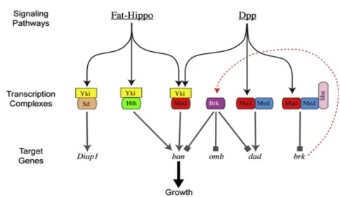
Recently, it was shown that in clones expressing Brk ban expression is repressed, even in the presence of Yki, and this effect is only reversed with simultaneous activation of Yki with Mad and TkvQD. Therefore, Mad can form a transcriptional complex with Yki to induce ban transcription, independently of Medea, whereas Brk represses ban expression by directly repressing its enhancer (Martín et al., 2004; Oh and Irvine, 2011). In addition, in other study has proposed the “opposing growth pathways model”, by showing that uniform proliferation rates along the A/P boundary during wing development are achieved by complementary growth regulation of Dpp/Brk signalling in the wing lateral areas and Fat signal in the medial region (Schwank et al., 2011). These results unveil a new interplay between different signalling pathways, in which Dpp and Fat-Hippo modulate growth possibly through ban (Figure 2).
Figure 2- Fat-Hippo pathway interacts with Dpp signalling to stimulate growth. Yorkie (Yki) is the downstream target of the Fat-Hippo pathway and cooperates with different partners to induce growth by transcription of many target genes. One of those partners is Scalloped (Sd), that together with Yki induces the expression of apoptosis inhibitor Diap-1, and other partner is Homothorax (Hth). Dpp, ligand of the TGF-β pathway, activates the transcriptional factor Mad that can form a complex with Medea to directly activate the transcription of target genes, such as daughters
against Dpp (dad). In addition, Mad forms a transcriptional complex with Medea and Schnurri (Shn) to repress brk,
thereby de-repressing Dpp target genes such as omb. Besides their independent role in growth control, both pathways can cooperate by the formation of a transcriptional complex that comprises Yki and Mad, and subsequent activation of the microRNA bantam (ban). Adapted from (Schwank et al., 2011).
3. Myc induces growth through Viriato
An important player in growth regulation is the Drosophila Myc (dMyc), a transcription factor ortholog of c-Myc oncogene in mammals (de la Cova and Johnston, 2006). It was shown that hypomorphic dMyc mutant flies are smaller
than wild-type flies, whereas flies with dMyc overexpression are bigger than controls (Johnston et al., 1999). Additionally, null mutants for dMyc codifying gene, diminutive (dm), display a strong developmental delay in larval phases, exhibiting small nucleus that fail to reach normal DNA content in endoreplicating tissues, such as fat cells and salivary glands (Pierce et al., 2004). On the other hand, cells overexpressing dMyc reveal a large nucleus and nucleolus, with increased rRNA content and higher ploidy (Pierce et al., 2004; Grewal et al., 2005).
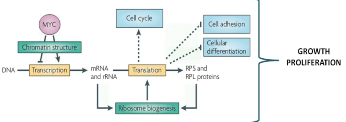
In vertebrates it is well known that Myc regulates several processes by controlling the expression of several genes related with growth and proliferation, including the pre-rRNAs and ribosomal proteins (Figure 3), and its action as transcription factor is mediated by chromatin remodelling. In Drosophila, besides its role in controlling cell cycle regulators, it was shown that dMyc stimulates rRNA synthesis, having an important advantage in the homeostatic process of cell competition (Grewal et al., 2005; Pierce et al., 2008; van Riggelen et al., 2010). Cell competition takes place when cells within the same growing tissue have different metabolic rates, originating an environmental stress in which cells interact with each other. Winner cells have a growth advantage, which is conferred by the capacity of protein synthesis. Thus, these cells continue to proliferate and survive, whereas the loser ones undergo apoptosis and are eliminated (Johnston, 2009). When clones of cells overexpressing dMyc are present in a wild-type tissue they act as super-competitors, thereby eliminating the wild-type loser cells by inducing Hid-mediated apoptosis, without affecting the target tissue size (Moreno et al., 2004; de la Cova et al., 2004; Johnston, 2009).
Other experiments have shown that in a tissue without dMyc expression Yki is not capable to induce growth, whereas clones overexpressing Yki display some increase in dMyc levels. Such clones induce apoptosis in neighbouring cells, exhibiting competitor behaviour, suggesting that Yki induces dMyc expression to promote growth. In fact, a model was proposed in which Yki and dMyc coordinate the action of each other. Thus, Yki forms a transcriptional complex with Sd to induce dMyc expression, which in turn repress yki, in an equilibrated network that ensure the correct tissue size (Neto-Silva et al., 2010; Ziosi et al., 2010).
Figure 3- Myc signalling in vertebrates. Myc stimulates cell growth and proliferation by transcriptional activation of several genes, including rRNA and ribosomal proteins (RPS and RPL) (adapted from van Riggelen et al., 2010).
Recently, a study performed at the host laboratory has characterized the functional viriato (vito), a novel gene identified in a screen for genes required for tissue growth in Drosophila. This gene is mainly expressed in the anterior proliferating cells of the eye imaginal disc and codifies for a nucleolar protein, the Drosophila homologue of the conserved Nol12 protein family. Flies mutants for vito display a developmental delay, whereas its depletion in the eye-antennal imaginal disc results in reduction of the eye size. Moreover, vito was shown to regulate the recruitment of Fibrillarin to the nucleolus and to maintain proper nucleolar structure. In addition, null dm mutants display decreased levels of vito, and in salivary glands Vito was shown to be necessary for the dMyc-induced growth, although it is not needed for the transcription of other dMyc target genes. These results strongly suggest that Vito is required to mediate nucleolar response of dMyc-induced growth (Marinho et al., 2011).
Nucleolus: a sensorial centre in the cell
The nucleolus is an extremely dynamic nuclear body that surrounds the tandemly repeated ribosomal RNA (rRNA) genes assembled in nucleolar organizer regions (NORs), in eukaryotic acrocentric chromosomes. The most important nucleolar function consists in creating the appropriate environment for an efficient ribosome biogenesis, essential to maintain cell metabolism. Nevertheless, the nucleolus has been described as multifunctional, as it is involved in several cellular functions, such as cell cycle and apoptosis control, DNA repair or stress response (Raska et al. 2004; Boisvert et al. 2007).
Since the nucleolus is a non-membrane organelle, its structure is maintained by the interaction between resident nucleolar proteins and several other proteins that are in continuous exchange between the nucleolus and the nucleoplasm. This nucleolar trafficking, in which several proteins are retained and released, occurs in response to cellular demands, thereby reflecting the physiological state of the cell (Raska et al., 2004; Andersen et al., 2005). Recently, it was found that the nucleolar targeting of proteins can be mediated by intergenic spacer (IGS) regions, present between the rDNA clusters (described in detail in the next chapter). Therefore, in response to cell stress stimuli these highly repeated DNA sequences are transcribed in long non-coding RNAs that are able to capture and retain proteins containing a specific peptidic motif, NoDS (Nucleolar Detention Sequence) (Audas et al., 2012; Prasanth, 2012).
The transient nature of the nucleolar proteome ensure the sensitivity of nucleolus to external changes, making possible the interaction between different proteins with related functions in a cellular context-dependent fashion (Boisvert et al., 2007). Importantly, the highly dynamic assemble of nucleolar components provides the necessary pool of proteins in the correct stoichiometry to support ribosome biogenesis, the main function of nucleolus (Emmott and Hiscox, 2009).
1. Ribosome biogenesis
Cell growth and proliferation are critically dependent on an efficient ribosome production, to sustain high protein synthesis levels. As ribosome production is a highly energy-consuming process, it needs to be rigorously controlled.
Each rDNA cluster repeat presented in the nucleolus is composed by an array of genes codifying for rRNAs, two external transcribed spacers (ETS) and two internal transcribed spacers (ITS) (Figure 4). Therefore, ribosome biogenesis is initiated with transcription of rRNA genes by RNA polymerase I (pol I), which originates a long rRNA precursor 47S. This polycistronic rRNA goes through a stepwise processing in which is successively cleaved by endo- and exonucleases to remove the ITS and ETS sequences, and to give rise to the 18S, 5.8S and 28S pre-rRNAs. A fourth pre-rRNA 5S is transcribed at the nucleus, by the RNA polymerase III (pol III), and imported to the nucleolus to undergo maturation (Lafontaine and Tollervey, 2001; Raska et al., 2004; Xue and Barna, 2012b).
During the ribosomal maturation, small nucleolar RNAs (snoRNAs) associate with nucleolar proteins, such as Gar-1 and Fibrillarin, to form small nucleolar ribonucleoproteins (snoRNPs), which drive site-specific rRNA modifications (Wang et al., 2000; Reichow et al., 2007). Simultaneously, several ribosomal proteins (Rps) are transcribed at the nucleus by RNA polymerase II (pol II), translated at the cytoplasm and quickly imported to the nucleolus, where they assemble with the pre-rRNAs to compose the mature ribosome subunits. The large 60S subunit comprises the 5.8S, 28S and 5S pre-rRNAs and nearly 47 ribosomal proteins named RPLs, whereas the small 40S subunit is composed by the 18S pre-rRNA and about 32 RPS ribosomal proteins (Raska et al., 2004; Lam et al., 2007; Xue and Barna, 2012b) (Figure 4).
Figure 4- Ribosome biogenesis. The nucleolus comprises an array of rRNA gene clusters, which are transcribed by RNA polymerase I in the 5.8S, 28S and 18S pre-rRNAs. The fourth 5S pre-rRNA is transcribed by RNA polymerase III at the nucleoplasm and imported to the nucleolus. Processing, maturation and assembly of pre-rRNAs to form the ribosome subunits requires ribonucleases (endo- and exonucleases), ribosomal proteins, small nucleolar ribonucleoproteins (snoRNPs) and other accessory factors. Ribosomal proteins are transcribed at the nucleus by RNA polymerase II, translated at the cytoplasm and imported to the nucleolus. The mature small 40S and large 60S subunits are translocated to the cytoplasm in a late maturation process, and assemble with mRNA to form functional 80S ribosomes.
Both subunits are subsequently translocated from the nucleolus to the nucleoplasm and then to the cytoplasm, in a late maturation process involving
several ribosomal and non-ribosomal proteins that assist the nuclear export. One of these proteins is RpL10, which interacts with the non-ribosomal 60S-associated protein NMD3, to export 60S ribosomal subunit through the nuclear pore complex (NPC). At the cytoplasm the mature subunits assemble with mRNA to form 80S functional ribosomes (Gadal et al., 2001; Lafontaine and Tollervey, 2001; Tschochner and Hurt, 2003; van Riggelen et al., 2010).
In mammalian cells the distinct processes that comprise ribosomal biogenesis take place in different nucleolar regions characterized by the presence of different protein combinations. Thus, rRNA transcription occurs in the border between the fibrillar centre (FC) and the dense fibrillar component (DFC), processing mostly occurs at DFC and assembly at the granular component (GC) (Olson et al. 2000; Raska et al. 2004; Boisvert et al. 2007). In Drosophila this tripartite organization was not observed, being the nucleolus a homogeneous structure (Orihara-Ono et al., 2005).
Ribosomal proteins are crucial for all steps of ribosome biogenesis, and the constant flux of these proteins between the nucleolus and cytoplasm along with the constant formation of new proteins and degradation of those unassembled, provide equilibrated amounts of RPs and pre-rRNAs, required for the efficient ribosome biogenesis process (Andersen et al., 2005; Lam et al., 2007). Therefore, defects in the normal balance between RPs and rRNA amounts results in a dramatic change in the normal ribosome activity and subsequent alteration of protein synthesis. Besides their fundamental role in ensuring cellular ribosome availability, RPs have many extra-ribosomal functions, thereby enhancing their requirement for the correct cellular function (Lindström, 2009; Warner and McIntosh, 2009). One good example is the mammalian RpL11, which in normal conditions is maintained at the nucleolus, but during nucleolar stress it accumulates at the nucleoplasm and mediates the stabilization of the tumour-suppressor protein p53 (Zhang and Lu, 2009; Boulon et al., 2010). Accumulation of RpL11 at the cytoplasm is also important to prevent Myc-mediated transcription of target genes as it inhibits the recruitment of its Myc co-activator, thereby limiting Myc-induced proliferation during cell response to oncogenic signals (Dai et al., 2007; Lindström, 2009; van Riggelen et al., 2010). Accordingly, several human disorders have been linked to alterations of many RPs, including cancer. Studies point to a neoplasic transformation model in which
alterations in the normal ribosome activity results in qualitative and quantitative changes in the normal protein translation (Montanaro et al., 2008).
In Drosophila, loss of one copy of several RPs is sufficient to result in a phenotypic class of dominant mutations named Minute. These mutants have a developmental delay and in the adult stage are characterized for possessing poor viability and fertility as well as for displaying short and thin bristles. These phenotypic effects are a consequence of suboptimal protein synthesis, which arises from an impaired ribosome function (Marygold et al., 2007).
Drosophila melanogaster: a small fly that solves big questions
Drosophila melanogaster represents an ideal system to study the
mechanisms that underlie growth. Thomas Morgan was the first biologist studying Drosophila early in the 20th century to explore the basis of heredity. He placed the small fruit fly in the vanguard of genetic research and since then, along with the emerging of a large range of genetic and molecular tools,
Drosophila has been considered a powerful model organism for many reasons
(Roberts, 2006; Arias, 2008a). Its small genome size, with around 14000 genes, short generation time and ease of culture and manipulation are some of the characteristics that make the fruit fly so attractive (Arias, 2008a). In addition, its genome was completely sequenced (Adams et al., 2000) and a large range of genetic tools that enables its manipulation are available. More importantly, several key developmental mechanisms and signalling pathways were highly conserved between Drosophila and mammals during evolution. The surprising finding that more than 70 per cent of the human disease-related genes have homologues in Drosophila genome (Reiter et al., 2001) revealed new perspectives for using this organism as model to study human diseases.
1. Life cycle
The duration of Drosophila’s life cycle critically depends on the temperature. Therefore, whereas at 18ºC the cycle takes approximately twenty days, at 25ºC an egg turns into an adult fly in no more than ten days (Figure 5). After mating, the cycle initiates with deposition of the eggs in the food. An egg develops to an embryo during the process of embryogenesis, and after 24 hours first larvae
hatch. The larval phase is divided into 3 instars separated by molts, L1 (24 hours), L2 (24 hours) and L3 (48 to 72 hours), and is characterized by a drastic accumulation of mass, by which larvae accumulate nearly 200-fold in weight. During this period larvae are feeding, and shortly before the end of L3 they leave the food source and search for a dry place appropriate to enter in pupariation (wondering phase). The final phase of metamorphosis occurs within the pupal case and takes approximately 5 days. Upon eclosion adults become receptive for mating after a few hours (Arias, 2008b).
Figure 5- Life cycle of Drosophila melanogaster. Life cycle takes approximately 10 days at 25ºC. After mating, eggs deposited in the food undergo embryogenesis for 24 hours. The larval phase initiates after larvae hatch, and takes approximately 5 days. In the final phase larvae move to a dry place (wondering phase) and form the pupa, where metamorphosis takes place. Adapted from (Arias AM, 2008).
2. Drosophila imaginal discs: models to study growth
Drosophila imaginal discs comprise the epithelial sac-like tissues present
in the larvae that give rise to the body appendages in the adult fly, such as eye, antenna, or wing. These structures emerge from collections of cells that acquire identity by the input of positional-dependent signals during embryogenesis, and are composed by two epithelial layers with different properties. Indeed, the main epithelium or disc proper (DP) is formed by columnar cells, and is surrounded by the squamous cells of the peripodial epithelium (PE). Such layers are separated by
a lumen but remain in close communication during development (Atkins and Mardon, 2009; Neto-Silva et al., 2009).
During larval phases these small clusters of cells undergo faster proliferation rates, and the imaginal discs suffer a dramatic increase in size. During this process, tissue growth is coordinated with tissue patterning, and differentiation occurs before the beginning of metamorphosis. Within each imaginal disc, different cell lineages acquire divergent fates, becoming progressively determined to assemble in organ-specific regions, known as compartments. These developmental fields act as independent units of growth and are composed by cells expressing the same combination of the regulatory selector genes (Domínguez and Casares, 2005; Neto-Silva et al., 2009).
Therefore, the structural simplicity of imaginal discs is transcended by the complex regulatory networks that orchestrate tissue development, which ensures the achievement of the exact organ size and shape. As a result, Drosophila imaginal discs provide perfect developmental models to study growth and pattern control during organogenesis.
2.1 Eye-antennal imaginal disc development
The eye-antennal imaginal disc develops into the adult eye, antenna, head cuticle and head structures, such as the ocelli and maxillary palps (Figure 6).
Figure 6- The eye-antennal imaginal discs develop in the fly head organs. The organs in the adult exhibit the same colours of the correspondent organ-primordia, in the eye-antennal imaginal disc. Adapted from (Domínguez and Casares, 2005).
The fly retina is composed approximately by 800 eye units, called ommatidia. Each ommatidium is a cell cluster comprising 8 photoreceptors (R1-8), and non-neuronal cells, such as clone and pigment cells.
Specification of the eye territory happens early in the first larval instar, with uniform expression of the mammal PAX6 Drosophila homologues, eyeless (ey) and its paralogue twin of eyeless (toy). Ectopic expression of these selector genes induces the formation of ectopic eyes, and thus ey and toy have a key role in conferring the eye identity (Halder et al., 1995; Czerny et al., 1999).
During the second larval stage, cells are proliferating asynchronously, and a range of different developmental signals emerges. At this stage, the disc has grown enough to unlock the mutual repressive action between Wingless (Wg) and Dpp, and thus, posterior cells exposed only to Dpp signal started to express retinal determination (RD) genes, eyes absent (eya), sine oculis (so) and
dachshund (dac) (Kenyon et al., 2003; Domínguez and Casares, 2005; Amore and
Casares, 2010). Wg, the Wnt-1 Drosophila homologue, is expressed in the ventral and dorsal margins of the disc and inhibits retinal specification by a repressive effect on Dpp signalling. Thus, Wg signalling restricts the retina formation to the posterior region, and maintains the cells in a proliferative state at the anterior region of the disc, by inducing the TALE-type homeodomain protein Homothorax (Hth) (Treisman and Rubin, 1995; Pichaud and Casares, 2000; Kenyon et al., 2003). Evidences suggest that Hth, in turn, sustain the anterior proliferative state by interacting with Yki and the zinc finger transcription factor Teashirt (Tsh) to form a transcriptional complex that induces the expression of bantam (Bessa et al., 2002; Peng et al., 2009).
The differentiation begins at the posterior margin of the disc, induced by the Dpp and the Hedgehog (Hh) signals, and cross the disc in a wave-like mode. The front of the differentiation wave is marked by the morphogenetic furrow (MF), a constriction of the epithelium in which cells arrest in G1 phase of the cell cycle, to posterior differentiation in photoreceptors (Domínguez and Casares, 2005) (Figure 7). Hh is a small-range signal secreted by the differentiated photoreceptors and directs the progression of the MF by inducing the expression of Dpp in the furrow (Greenwood and Struhl, 1999). As MF progresses in the disc, Dpp signal accompanies its movement. As Hh is also implicated in the MF progression at medial region, Dpp signal becomes essential for the progression of the MF at the margins of the disc, where it sustains its own expression.
Depletion of dpp in the third instar discs results in smaller discs exhibiting a delay mainly in the marginal progression of the MF. In these discs the retinal differentiation is restricted to the central area, where the Hh signal drive the furrow progression (Treisman and Rubin, 1995; Chanut and Heberlein, 1997).
At the anterior region, the progenitor cells that receive the long-range signal of Dpp from the MF enter a pre-proneural state, in which hth expression is lost and RD genes are up-regulated. In addition, these cells synchronize their cell cycles by going through a few rounds of cell cycle divisions, called the first mitotic wave (FMW). Following synchronization, cells arrest in G1 phase at the MF and acquire neuronal competence by expression of atonal (ato), induced by Hh. Cells expressing ato are selected to be the founder cells (R8), thereby successively recruiting the other neuronal and non-neuronal cells required to form the ommatidial clusters (Tomlinson, 1985; Domínguez and Casares, 2005). Cells that do not differentiate after the FMW undergo in a second mitotic wave (SMW), behind the MF. It was shown that the inhibition of hth expression by the Dpp long-range activity is crucial to get cells ready to cell cycle arrest and for the retinal fate acquisition (Bessa et al., 2002; Domínguez and Casares, 2005; Lopes and Casares, 2010).
Figure 7- Development of the eye imaginal disc. During the third instar an apical constriction, the morphogenetic furrow (MF), drive the differentiation across the eye disc, from the posterior to the anterior region. With the approximation of the MF, anterior proliferating cells synchronize their cycles and arrest in G1, to differentiate in photoreceptors. Hedgehog (Hh) is a small range signal expressed by differentiated cells, inducing the expression of Decapentaplegic (Dpp) in the MF. Dpp is a long-range signal, inducing the expression of retinal determination genes in the anterior cells, thereby stimulating cells to arrest their cycles. Wingless (Wg) is secreted by the ventral and dorsal edges of the disc, in the region that develops in the head cuticle, limiting the retina formation to the posterior region. Adapted from (Silver and Rebay, 2005).
Therefore, at L3 phase the eye imaginal disc represents a highly ordered system, comprising layers of cells with different levels of commitment, which respond to distinct combinations of signals. The MF represents a changing point, dividing the anterior cells in asynchronous proliferation, from the differentiated photoreceptors at the posterior region.
3. Salivary glands
Drosophila salivary glands are polarized epithelia comprising two extended
secretory tubes, composed by an epithelial layer of cells surrounding an inner lumen. Salivary glands produce and secrete several proteins, such as the salivary gland glue proteins that allow the adherence to solid substrates, essential to larvae undergo pupariation (Myat, 2005).
During embryogenesis, salivary cells invaginate from the ventral ectoderm of the embryo, by apical surface constriction. This process results in the formation of the tubular structure and is finely regulated to control the final size and shape of the salivary gland. After internalization, distal cells in contact with the visceral mesoderm migrate to the posterior region, elongating their apical membrane in the direction of the migration, along the Proximal/Distal axis (Hogan and Kolodziej, 2002; Myat, 2005).
Several genes are involved in the salivary gland development. The transcription factor Forkhead has a central role in the invagination process, whereas Hairy, Huckebein, Crumbs and Ribbon are important to the apical growth and migration. Recently it was found that the GTPase Rho1 is necessary to the apical and cell rearrangement, controlling the lumen size by regulation of the actin cytoskeleton and Moesin (Hogan and Kolodziej, 2002; Myat, 2005; Xu et al., 2011).
Terminally differentiated cells of Dosophila salivary glands lack the ability to divide and undergo successive cycles of endoreplication, characterized by consecutive rounds of S phases of DNA synthesis and gap phases, without occurring cell division. The resultant polyploid cells acquire a high metabolic output, which allow them to support the larval growth, thereby suffering a dramatic increase in cellular size (Smith and Weaver, 1991; Edgar and Orr-weaver, 2001). The final ploidy of these cells is regulated by external factors,
pathways. In fact, loss of the Drosophila serine/threonine kinase, target-of-rapamycin (dTOR), a growth regulator that links the nutritional signalling with protein synthesis, decrease the ability of cells to endoreplicate (Zhang et al., 2000). On the other hand, overexpression of dMyc in salivary glands results in remarkable increase of DNA content (Pierce et al., 2004).
As tubular organs, salivary glands of Drosophila have been used as model to study the organogenesis of important tubular systems, such as the mammalian kidneys or lungs (Hogan and Kolodziej, 2002; Myat, 2005).
4. Drosophila genetic tools
4.1 GAL4-UAS system
The GAL4-UAS binary system was introduced in Drosophila by Brand and Perrimon (Brand and Perrimon, 1993). GAL4 is a potent transcriptional activator of Saccharomyces cerevisiae, being expressed in transgenic lines under the control of endogenous regulatory sequences. The upstream activation sequence (UAS) is a GAL4-responsive enhancer, and is positioned upstream of a gene of interest, regulating its expression (Arias, 2008c). The two regulators of the system, GAL4 and UAS, are maintained at separate parental lines. Therefore, when the driver line expressing GAL4 is crossed with the responsive line containing the UAS-dependent transgene, induces the interaction between the GAL4 with the UAS sequence, which activates the expression of the gene of interest in the progeny (Brand and Perrimon, 1993; Duffy, 2002).
Several GAL4 enhancer trap lines exist, to express the GAL4 transcription factor in different tissues or patterns of interest. The main advantages of this technique are the simplicity and ease of manipulate the expression of target genes, at a defined time and space (Brand and Perrimon, 1993; Duffy, 2002; Arias, 2008c).
4.2 RNA interference
The first experiments with double stranded RNA (dsRNA) and the subsequent discovery of the RNA interference (RNAi) in Caenorhabditis elegans
resulted in a Nobel Prize in Medicine to Craig Mello and Andrew Fire, in 2006 (Fire et al., 1998).
RNA interference consists in the mechanism of silencing specific target mRNAs as a response to its hybridization with its complementary sequence, and occurs naturally in eukaryotes (Zamore and Haley, 2005). In Drosophila, the RNAi can be regulated by the GAL4-UAS system, thereby making possible to down-regulate endogenous target genes in a restricted time and space. This control is achieved by expression of the UAS-dsRNA targeting the gene of interest and a specific GAL4 driver that lead to the degradation of the endogenous target gene mRNA in the target tissue (Duffy, 2002; Dietzl et al., 2007) (Figure 8).
Figure 8- RNA interference approach in Drosophila melanogaster. A hairpin RNA (hpRNA) is expressed when GAL4 expressing lines are crossed with a UAS line. The hpRNA is recognized and cleaved by Dicer, in small interference RNAs (siRNAs). The siRNA guide strand is incorporated in the complex RISC driving it to degrade the complementary endogenous target mRNA.
In this approach, the GAL4-UAS system drives the expression of a sequence containing inverted repeats connected by a linker sequence. When transcribed, this sequence gives rise to a double stranded RNA in a hairpin structure perfectly base-paired (hpRNA), recognized by the RNase III Dicer, which cleaves it in double-stranded small interference RNAs (siRNA). One of the siRNA strand is
complementary to the target mRNA, known as the guide strand, whereas the other, named passenger strand, is subsequently degraded. The guide strand is incorporated into the RNA-induced silencing complex (RISC), driving it to the target mRNA. After base-pairing, the RISC protein Argonaute (Ago) mediates the mRNA degradation (Winter et al., 2009).
The creation of the first Drosophila genome-wide RNAi transgenic library has allowed the construction of several RNAi screens to study gene function (Dietzl et al., 2007). Coupling of GAL4-UAS system with the RNAi mechanism provides a simple and robust approach in which it is possible to target a specific gene in a temporal- and tissue-specific manner. Moreover, RNAi does not eliminate in absolute the target mRNA levels, thereby creating a hypomorphic condition. All these characteristics allow us to overcome the lethality resultant from mutations in the most essential genes.
The main disadvantage of this approach is the potential off-target effect, resultant from non-specific interactions of the RNAi mechanism with non-desired mRNAs. In fact, it is established that 19 nucleotides of perfect match between the dsRNA and non intended mRNAs is the critical threshold to origin pleotropic phenotypic effects and thus false-positive results (Kulkarni et al., 2006).
Aims of the thesis
Although it is well described the crucial role of TGF-β signalling in patterning, it remains very controversial how this signalling pathway regulates tissue growth.
Based on recent studies in which the Drosophila NOL12 homologue Viriato was shown to genetically interact with TGF-β signalling members, the main aim of this thesis is to understand whether the described function of TGF-β in tissue growth is mediated by regulation of nucleolar function.
As Put is the shared type I receptor for both TGF-β pathways, I will use a strong RNAi targeting Put to further investigate the role of TGF-β signalling in nucleolar morphology, integrity and function. Finally I will try to prove if the phenotypic effects caused by the RNAi targeting Put are, in fact, target gene-specific.
Fly strains and Genotypes
The following stocks (described in FlyBase, unless stated otherwise) were
used: w1118, ey-Gal4, UAS-lacZ, UAS-putRNAi1 (Vienna Drosophila RNAi
Center,VDRC, #849) UAS-putRNAi2 (VDRC #37279), UAS-putRNAi3 (Transgenic RNAi Project, TRiP, #27514), UAS-putRNAi4 (TRiP, #35195), UAS-putRNAi5 (TRiP, #35701), putRNAi6KK (VDRC, #107071), medRNAi (VDRC, #19688),
UAS-tkvRNAi (VDRC, #3059), UASnopp140RNAi (VDRC, #45583), RpS9-YFP/TM6c
(CPTI-000493, Flannotator), RpL10Ab-YFP/TM6c (Cambridge Protein Trap YFP insertions, #115-462), RpL41-YFP/SM6c (Cambridge Protein Trap insertions, #115-344), Bantam sensor JB20/TM6B (gift from Marco Millan), w;;ban 5’-lacZ
(gift from Wei Du), UASBantam (gift from Stephen Cohen), put88ry/MKRS,Ser1,
St1put135e1/TM3,Ser1 (#3100), w1118; Df(3R)BSC841/mwh1kni 1i-1
snk4red1e1TL3ca1/TM6c,Sb1cu1. Hsp70GFPNopp140-True, UAS-RFPRpL26, and
UASGFPRpL11 were gifts from Patrick Di Mario. All crosses were raised at 25ºC under standard conditions, unless stated otherwise.
To observe the adult eye phenotype resultant from the expression of different RNAis targeting Put, the flies were examined under a stereomicroscope
(Stemi 2000, Zeiss) equipped with a digital camera (Nikon Digital Sight DS-2Mv).
Representative pictures for each RNAi targeting Put were taken.
Immunostaining
Eye-antennal imaginal discs and salivary glands were dissected in cold Phosphate Buffer Saline (PBS) for 20 minutes. After that, imaginal discs and salivary glands were washed three times with PBT (PBS with 0.1% Triton X100) during 10 minutes, and immunostained during 3 hours with the primary antibodies in PBT, at room temperature. Subsequently imaginal discs and salivary glands were washed three times during 10 minutes with PBT, at room temperature, and immunostained with the secondary antibodies in PBT during 2 hours. After incubation, imaginal discs and salivary glands were washed three times during 10 minutes at room temperature, and stored in 50% Glycerol/PBS, at 4ºC.