Identification of lamb flocks susceptible and resistant
against Brachiaria poisoning
1Rayane C. Pupin2, Gleice K.A. Melo2, Rubiane F. Heckler2, Tatiane C. Faccin3, Camila C.B.F. Ítavo4, Carlos E. Fernandes5, Danilo C. Gomes4 and Ricardo A.A. Lemos4*
ABSTRACT.- PupinR.C.,Melo G.K.A., Heckler R.F., Faccin T.C., Ítavo C.C.B.F., Fernandes C.E, Gomes D.C. & Lemos R.A.A. 2016. Identification of lamb flocks susceptible and resistant against Brachiaria poisoning.Pesquisa Veterinária Brasileira36(5):383-388. Laboratório de Anatomia Patológica, Faculdade de Medicina Veterinária e Zootecnia, Universidade Fe-deral de Mato Grosso do Sul, Av. Senador Filinto Müller 2443, Campo Grande, MS 79074-460, Brazil.E-mail: ricardo.lemos@ufms.br
This study was designed to assess the influence of genetic resistance against brachiaria poisoning in sheep. Two groups of sheep, one identified as susceptible (formed by two ewes and one ram) and the other as resistant against brachiaria poisoning (formed by
three ewes and one ram) were selected. Sheep considered susceptible were those that presented clinical signs of brachiaria poisoning at any time of their life; resistant sheep were those that even raised on Brachiaria spp. pastures, did not developed any sign of the poisoning during their life. The offspring of the two flocks (15 lambs from the sensi
-tive flock and 9 lambs from the resistant flock) were placed into brachiaria pasture (ini -tially Brachiaria decumbens and B. brizantha,and only B. decumbens after weaning) and followed up during two years (2013-2014). The determination of protodioscin levels in B. decumbens pasture was performed only in 2014 and revealed significant amounts of the toxic principle. Eleven lambs of the susceptible group were affected to some degree of brachiaria poisoning and six died; no lamb of the resistant group was affected. Clinical signs consisted of varying degrees of subcutaneous edema of the face and, erythema and loss of hair of the ears, crusts on the skin of ears, around the eyes and on planum nasale, scar deformation of the ears, and bilateral ocular discharge; affected lambs also sought for shadowy shelters and they were poor doers. Several sheep recovered from the
condi-tion and then relapsed. Necropsy findings in six lambs included pale mucous membranes,
emaciation, dermatitis, scar deformation of the ears, large yellow livers with marked lo-bular pattern, and moderate infestation by Haemonchus contortus. Histologically the liver lesions were similar in all necropsied lambs but with varying degrees of severity; they were consistent with brachiaria poisoning and included architectural disruption of hepa-tocellular trabecula, clusters of foamy macrophages occasionally forming multinucleated giant cells, swollen and vacuolated hepatocytes, crystals or negative images of crystals
in the biliary system, bilestasis, bile duct proliferation and lymphoplasmacytic infiltrate
in portal triads. The skin lesions were those of photodermatitis and included epidermal
necrosis, hyperkeratosis and dermal neutrophilic infiltrate. The results of this study allow
to conclude that there is a genetic related resistance to brachiaria poisoning in sheep since
the progeny of resistant sheep did not manifest the poisoning. The use of resistant flocks
1 Received on September 9, 2015.
Accepted for publication on March 7, 2016.
2 Programa de Pós-Graduação em Ciência Animal, Universidade Federal de Mato Grosso do Sul (UFMS), Av. Senador Filinto Müller 2443, Campo Grande, MS 79070-900, Brazil.
3 Programa de Pós-Graduação em Medicina Veterinária, Universidade Federal de Santa Maria (UFSM), Av. Roraima 1000, Santa Maria, RS 97105-000, Brazil.
4 Faculdade de Medicina Veterinária e Zootecnia (FAMEZ), Universidade Federal de Mato Grosso do Sul (UFMS), Av. Senador Filinto Müller 2443, Campo Grande, MS 79070-900, Brazil. *Corresponding author: ricardo. lemos@ufms.br
RESUMO.- [Identificação de rebanhos de cordeiros suscetíveis e resistentes a intoxicação por braquiá-ria.] Este estudo avaliou a resistência genética na ocor-rência de intoxicação por braquiária em ovinos. Foram
se-lecionados dois grupos de ovinos, um identificado como suscetível (formado por duas ovelhas e um carneiro) e o outro como resistente (formado por três ovelhas e um
carneiro). Foram considerados suscetíveis ovinos que apresentaram sinais de intoxicação por Brachiaria spp. em algum ponto de suas vidas e resistentes aqueles ovi-nos que, mesmo criados em pastagem de braquiária, nun-ca desenvolveram qualquer sinal da intoxinun-cação. A
progê-nie desses dois grupos (15 cordeiros do grupo suscetível
e 9 no grupo resistente) foi colocada numa pastagem de
braquiária (inicialmente Brachiaria decumbens e B. bri-zantha e, após o desmame, apenas B. decumbens) e
acom-panhada durante dois anos (2013-2014). A determinação
dos níveis de protodioscina em B. decumbens foi realizada apenas em 2014 e foram encontradas quantidades
sig-nificativas do princípio tóxico. Onze cordeiros do grupo
suscetível foram afetados por algum grau de intoxicação por braquiária; nenhum cordeiro do grupo resistente foi
afetado. Os sinais clínicos consistiam de graus variáveis
de edema subcutâneo da face e eritema e alopecia da pele das orelhas, crostas na pele das orelhas e ao redor dos olhos e no plano nasal, retração cicatricial das orelhas, fo-tofobia e corrimento ocular bilateral. Três cordeiros apre-sentaram desenvolvimento retardado. Vários cordeiros se recuperaram da condição, mas posteriormente quando foram colocados na pastagem apresentaram recidivas. Achados de necropsia em seis cordeiros incluíam muco-sas pálidas, pobre condição corporal, dermatite,
deforma-ção cicatricial das orelhas, fígado aumentado de volume,
amarelo e com padrão lobular evidenciado e graus mode-rados de infestação por Haemonchus contortus. Histolo-gicamente, as lesões hepáticas eram semelhantes em to-dos os cordeiros necropsiato-dos, mas apresentavam vários graus de intensidade; eram consistentes com as lesões de intoxicação por braquiária e consistiam de desorganiza-ção da arquitetura trabecular dos hepatócitos, agregados de macrófagos espumosos ocasionalmente formando cé-lulas gigantes multinucleadas, hepatócitos tumefeitos e vacuolizados, cristais ou imagem negativa de cristais no sistema biliar, bilestase, proliferação de ductos biliares
e infiltrado linfoplasmocitário nas tríades portais. As le -sões da pele eram de fotodermatite e incluíam necrose da
epiderme, hiperqueratose e infiltrado neutrofílico na der
-me. Os resultados desse estudo permitem concluir que há
uma resistência genética à intoxicação por braquiária em ovinos, uma vez que a progênie dos ovinos resistentes não
manifestou a intoxicação. O uso de rebanhos resistentes
em pastagens e braquiária é sugerido como uma valiosa opção para prevenir a intoxicação por braquiária em ovi-nos.
TERMOS DE INDEXAÇÃO: Plantas tóxicas, Brachiaria spp.,
sapo-ninas, doenças de ovinos, resistência genética, intoxicação por plantas.
INTRODUCTION
Large areas of cultivated pastures in Brazil consist of Bra-chiaria spp. (Tokarnia et al. 2012). The consumption of this forage by different farm animal species is associated with
toxicosis being sheep and cattle the most susceptible (Le -mos et al. 2011) and younger stock are more susceptible
than mature animals (Albernaz et al. 2010, Castro et al.
2011, Mustafa et al. 2012). Furthermore, sheep from her-ds that never had contact with brachiaria species are more susceptible than sheep raised with free access to the plant
(Lemos et al. 1996, Castro et al. 2007, 2011, Riet-Correa et al. 2011, Oliveira et al. 2012, Faccin et al. 2014). It is pos -sible that animals raised in brachiaria pastures develop greater capacity in metabolize and degrade the steroidal
saponin, the toxic principle of this type of grass (Castro et al. 2007) and/or development of a special ruminal flora (Albernaz et al. 2010) that make them able to reduce the
effects of the toxin.
The typical clinical manifestation of Brachiaria spp. poi-soning is photodermatitis. Affected animals show apathy, anorexia, icterus, skin pruritus, photophobia, serous ocular discharge, subcutaneous edema in the face and ears; in the skin of poorly pigmented areas there are lesions characte-rized by erythema, hair loss, and crusts. These areas evolve to necrosis and sloughing of patchy areas of affected skin, associated in many cases to deformity of the ears due to
scarring of these lesions (Brum et al. 2007, Saturnino et al.
2010, Albernaz et al. 2010, Castro et al. 2011, Lemos et al.
2011, Mustafa et al. 2012, Oliveira et al. 2012, Faccin et al.
2014).
Main necropsy findings include icterus, enlarged yellow
to orange discolored liver with marked lobular pattern, and
distended gall bladder (Brum et al. 2007, Castro et al. 2007,
2011, Albernaz et al. 2010, Lemos et al. 2011, Mustafa et al.
2012, Oliveira et al. 2012, Faccin et al. 2014).
Histopathologically there is disruption of the
hepa-tocyte trabecular arrangement with infiltration of foamy
macrophages and, that occasionally coalesce to form mul-tinucleated giant cells, hepatocytic swelling, individual
hepatocellular necrosis, and bilestasis. In the portal triads there is periportal fibrosis, acicular birefringent crystals
within bile ducts, bile duct proliferation and
lymphoplas-macytic inflammatory infiltrate (Brum et al. 2007, Castro
et al. 2007, 2011, Albernaz et al. 2010, Lemos et al. 2011,
Riet-Correa et al. 2011, Mustafa et al. 2012, Oliveira et al.
2012, Faccin et al. 2014). Skin lesions are characterized by hyperkeratosis and parakeratosis, epidermal coagulative necrosis, necrotic debris, bacterial clumps, neutrophilic
inflammatory infiltrate and necrosis of the dermal vessels (Lemos et al. 1996, Faccin et al. 2014).
in brachiaria pastures is suggested as a valuable option for the prevention of brachiaria poisoning in sheep.
Brachiaria decumbens is the most toxic species of this genus; it may contain a concentration of more than 2% of
the steroidal saponin, protodioscin (Riet-Correa et al. 2011,
Barbosa-Ferreira et al. 2011).
Several measures to prevent or reduce the severity of the poisoning by brachiaria species in farm animals have been proposed. Among these are use as forage of less
to-xic (lower content of saponins) species o Brachiaria, like B. brizantha and B. humidicola (Riet-Correa & Medeiros 2001, Riet-Correa et al. 2011) in which the concentrations of pro-todioscin are respectively 0,84% ±0,28 and 0,11% ±0,02
(Barbosa-Ferreira et al. 2011). However this is not comple -tely safe since B. brizantha may, at times, accumulate con-centrations of saponins high enough to cause intoxication
in sheep (Albernaz et al. 2010, Faccin et al. 2014). An alter
-native measure of control is the identification of animals
resistant to the intoxication by Brachiaria spp. (Riet-Correa & Medeiros 2001, Riet-Correa et al. 2011) since it has alre-ady been reported that within one given herd there may be
animals with different degrees of susceptibility (Castro et
al. 2007, 2011, Saturnino et al. 2010).
Although conclusive data on the existence of genetic ba-sed resistance or susceptibility to Brachiaria spp. are still lacking, epidemiological evidences and experimental re-sults indicated that the genetic make up could play a
funda-mental role in the resistance to this toxicosis (Melo 2014).
The importance of a genetic associated resistance to a toxi-cosis was reported in association with the so called facial eczema, the mycotoxicosis caused by sporidesmin produ-ced by Pithomyces chartarum (Morris et al. 1991)
The current study was design to assess the influence of
genetic resistance on the occurrence of Brachiaria sp. poiso-ning in two flocks of sheep, one identified as susceptible and
the other as resistant to the poisoning by Brachiaria spp.
MATERIALS AND METHODS
During the years of 2013 and 2014 two groups of lambs were ob-served. These two groups were the offering of a sheep herd con-sisting of susceptible and resistant animals and both were raised in pastures of Brachiaria brizantha and B. decumbens; there were histories of occurrence of outbreaks of photosensitization in this pasture. Sheep considered susceptible were those that presented clinical signs of brachiaria poisoning at any point of their lives; re-sistant sheep were those that even raised in Brachiaria spp. pas-tures, did not developed any sign of the intoxication at any time of their lives.
During the breeding season the herd was divided in two groups: one susceptible and one resistant. The original suscep-tible herd was formed by two suscepsuscep-tible ewes and a suscepsuscep-tible ram, and the resistant original herd by three resistant ewes and one resistant ram. Reproduction was by natural breeding keeping the rams with the ewes the whole time the breeding season. In the two years of the study the lamb produced by these breeding were kept in pastures of Brachiaria spp. with history of intoxica-tion. Until weaning the lambs were kept in pastures consisting of
B. decumbens and B. brizantha and after weaning (at 50 days of
age) exclusively in B. decumbens.
During the experimental period, every 30 days, the number of eggs per gram of feces (EPG) were determined according to the te -chnique Gordon and Whitlock (1939). The sheep whose EPG was above 500 were dewormed with levamisole (Ripercol®).
Those lambs which presented any manifestation of clinically appreciable photosensitization were withdraw from the brachia-ria pasture and held until clinical recover in stalls protected from the sun; during this time they were fed commercial ration for sheep, chopped Tifton-85 (Cynodon dactylon) hay and water ad libitum.
The lambs that spontaneously died or were euthanatized in the terminal phases or due to marked incapacity to reach an adequate corporal development were necropsied. Euthanasia was perfor-med by intravenous injection of 4 mg/kg of body weight (bw) of ketamine associated with 0.2mg/kg/bw of xylazine hydrochlori-de followed intravenous potassium chlorihydrochlori-de. Fragments of several organs were sampled, fixed in 10% formalin, processed routinely for histopathology and stained with hematoxilin and eosin (HE).
The determination of protodioscin in the pastures were made only in 2014. Samples from the grass were harvested at 28 days intervals from May to November, following the pattern of simu-lated animal grazing. To accomplish this pattern of consumption by the sheep was followed observing the heights and parts of the plant consumed, aiming to obtain a sample more closely to what the animal would spontaneously consume. To this end, the grass was grasped manually, in order to somewhat simulate the move-ment of the lips of lambs during grazing and then, a minimum of 200g of grass were cut with a jack knife in a height similar to na-turally cut by a grazing lamb (adapted from Cook 1964). The total sample obtained was placed in a forced-air ventilation stove at 55oC, for 96 hours. The dried leaves harvest were passed through
a Lab Willey grinder, with a sieve cloth mesh of 1mm and send to the Chemistry laboratory of the Universidade Federal de Mato Grosso do Sul (UFMS) for protodioscin extraction and quantifica -tion by high-performance liquid chromatography (HPLC) using a evaporative light scattering detector (ELSD). Data obtained were evaluate statistically by the G Test with a confidence margin of P<0.05.
The experiment was approved by the UFMS Ethics Committee on the Experiments with animals and register under the number 400/2012.
RESULTS
During 2013, six lambs were born in the susceptible group – two of which developed clinical signs of brachiaria
poiso-ning – and five in the resistant group, in which no cases of
the toxicosis developed. During 2014, nine and four lam-bs were born respectively in the susceptible and resistant groups and there were eight cases of the toxicosis in former and no cases in the latter. All cases of brachiaria toxicosis occurred after weaning.
Overall, there were 15 lambs born in the susceptible
group, of which ten developed clinical signs of photosen-sitization, eight of which presented relapses, two of which
died. One of the lambs died without presenting photoder -matitis and was heavily parasitized by Haemonchus contor-tus; however, this lamb had severe hepatic lesions of bra-chiaria poisoning. The morbidity rate was 73.33% and the mortality rate 20%. Another three lambs on the susceptible group that had clinical signs and survived, were poor doers and were euthanatized at the end of the experiment.
No case of brachiaria poisoning was detect in any of the nine lambs from the resistant group. This is translated in a
statistically significant difference (p=0,006) in the inciden -ce of the toxicosis between the two groups.
and 150 days, that is, 7 to 90 days after weaning. These in-cluded varying degrees of subcutaneous edema of the face
and ears (8 out of 10 lambs), reddening and loss of hair of the ears (6/10), crusts in the ears and planum nasale (5/10), scar deformation of the ears (3/10), bilateral ocu
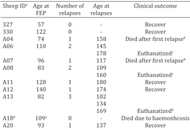
-Table 1. Poisoning by Brachiaria decumbens in lambs from
the susceptible group that developed the toxicosis. Age (in days) of the lamb at first episode of photodermatitis (FEP),
at the occurrence of relapses and clinical outcome Sheep IDa Age at Number of Age at Clinical outcome FEP relapses relapses
327 57 0 - Recover
330 122 0 - Recover
A04 74 1 158 Died after first relapseb
A06 110 2 145
178 Euthanatizedc
A07 96 1 117 Died after first relapseb
A08 83 2 109
160 Euthanatizedc
A11 128 1 180 Recover
A12 140 1 174 Recover
A13 82 3 102
134
169 Euthanatizedb
A18d 109e 0 - Died due to haemonhcosis
A20 93 1 137 Recover
aIdentification; b submitted to liver biopsy; ceuthanasia at the end of the experiment; din spite of lack of clinical signs of the intoxication, hepatic lesions of similar type and intensity as those of lambs(AO4, AO7) with clinical signs of photodermatitis were found in this lamb; eage of death.
6 Centro de Monitoramento do Tempo, do Clima e dos Recursos Hídricos de Mato Grosso do Sul, Agência de Desenvolvimento Agrário e Extensão Rural.
Table 2. Brachiaria decumbens poisoning in lambs from susceptible flocks. Necropsy findings in six lambs that died or were
euthanatized due to effects of the toxicosis
Lamb ID Nutricional Pale mucous Crust in the Scar Liver: yellow, enlarged Icterus Haemonchus contortus score membranes skin (ears and deformation and with marked specimens in abomasum
around the eyes) of the ears lobular pattern
A04a P + ++ - + + ++
A06b I +++ - - - - ++
A07c P ++ +++ +++ + ++ ++
A08b I ++ ++ +++ - - ++
A13b I ++ +++ - + -
A18d P +++ - - + ++ -e
aSpontaeous death; beuthanatized as poor doers; ceuthanatized in extremis due to advanced manifestation of the toxicosis; ddeath from haemonchosis associated with severe hepatic lesions attributed to brachiaria toxicosis; ethe absence of Haemonchus contortus specimens in abomasum of this lamb is explained be the deworming carried out just prior to death; P = proper body development for the age; I = inappropriate body development for the age; - = absence of lesion, += mild lesion; ++ = moderate lesion; +++= marked lesion.
Table 3. Brachiaria decumbens poisoning in lambs from susceptible flocks. Histopathological findings in six lambs that died
or were euthanatized due to effects of the toxicosis
Liver Skin
Lamb ID Architectural Foamy Swollen Crystals or Mutinucleated Bilestasis Bile Lymphoplas- Epidermal Hyperke- Neutrophilic disruption of macro- and negative image foamy duct mocytic infil- necrosis ratosis infiltrate hepatocellular phages vacuolated of crystals giant prolife- trate in portal
tabeculae hepatocytes in the biliary cells ration triads
system
A04a ++ +++ - + + - ++ - ++ + ++
A06b - +++ ++ - +++ - - - - -
A07c ++ +++ +++ ++ - + - - - +++ +
A08b - - ++ - + - +
A13b - + ++ - - - - + +++ ++ ++
A18d +++ ++ + ++ - + - - - -
-aSpontaeous death; beuthanatized as poor doers; ceuthanatized in extremis due to advanced manifestation of the toxicosis; ddeath from haemonchosis associated with severe hepatic lesions attributed to brachiaria toxicosis. - = absence of lesion, += mild lesion; ++ = moderate lesion; +++= marked lesion.
lar secretion (2/10); affected lambs seek for shadowy shel
-ters (2/10).
There were no clinical relapses detected in 2013. Ho-wever, during 2014 all the lambs that got sick relapsed at some point. Three lambs had only one episode of relapse in
34-84 days after the first outburst of clinical disease. The
three euthanatized lambs had clinical relapses, two of the-se lambs prethe-sented two relapthe-ses each within intervals of
26 to 52 days after the first outburst of clinical signs; the
other lamb had three relapses with intervals of 20 to 35
days after the first clinical outburst. Another two lambs died after one relapse (Table 1). One lamb died from hae
-monchosis (A18 in Table 1); in spite of lack of clinical signs
of the intoxication in this lamb, similar lesions in type and
severity as those described above for the two lambs (AO4, AO7) with photodermatitis were observed.
Altogether six lambs died during the experiment.
Ne-cropsy findings in these six lambs are summarized in Table 2.
Histologically the lesions observed in the livers from lambs that died were similar but with varying degrees of
severity (Table 3).
No determination of protodioscin levels was carried out in the pasture during 2013. Levels of protodioscin found in the pasture during 2014 are expressed in Figure 1.
precipita-tion during May-November varied from 17.20mm and 225.60mm. Minimum and maximum values of rain preci-pitation were observed respectively on August and Novem-ber. There was no correlation observed between the pro-todioscin levels in the pastures and the total monthly rain precipitation.
DISCUSSION
Eleven out of the 15 lambs selected as part of the suscepti-ble group developed some degree of Brachiaria decumbens poisoning while none of the nine lambs of the resistant
group was affected, which had a statistically significance
between the two groups suggesting a genetic induced re-sistance to brachiaria poisoning in sheep. Although there are circumstantial evidences that genetics play an
impor-tant role in the resistance to this toxicosis (Riet-Correa et
al. 2011) and different degrees of individual susceptibility
have been demonstrated (Castro et al. 2007, 2011, Saturni -no et al. 2010, Faccin et al. 2014), up to date there were -no reported studies comparing descendants lambs from sus-ceptible and resistant progenitors in relation to Brachiaria spp. poisoning. Studies evaluating the importance of gene-tic on the resistance to other weed induced toxicosis were done in relation to facial eczema. The inheritance of
resis-tance to facial eczema (mycotoxicosis induced by sporides -min from Pithomyces chartarum) in cattle was already
de-monstrated (Morris et al. 1991). The greater vulnerability
in the susceptible group in the current study was stressed by the occurrence of relapses in eight out of 10 lambs that showed clinical signs of photodermatitis.
Our findings support the hypothesis of inherited resis -tance to Brachiaria decumbens poisoning, since the time elapsed between relapses is more likely enough for the cle-arance of the toxic principle and complete recover of the hepatic tissue. Although there are no reports about period necessary to eliminate protodioscin avoiding a possible ac-cumulative effected, in none of the lambs of this study that relapsed were there chronic hepatic lesions in the
histopa-thological exam. Out of 11 lambs that got sick in the suscep -tible group, six died naturally or were euthanatized as poor
doers, and five recovered. This set of results suggests that
adaptation of susceptible lambs may occur as the
consump-tion of the plant goes on. It is interesting to point out that
the lambs of the current study were observed continuously and were withdrawn from the brachiaria pasture
immedia-tely after the onset of clinical signs, which may influence
in decreasing the lethality rate, inversely to which occur in
flocks raised extensively in which the identification of the onset of clinical signs is difficult, decreasing de likelihood
that poisoned animals survive.
Death of susceptible animals lead to a natural selection
process allowing for the emergence of resistant flocks as
the duration of their permanence in the infested Brachiaria spp. pasture increases, as has been the case in sheep and
cattle herds of Midwestern Brazil (Riet-Correa et al. 2011,
Faccin et al. 2014). However it should be pointed out that high mortality rates may occur in lambs after the
introduc-tion of susceptible ram in a resistant flock (Melo 2014).
The clinic-pathological presentation of the affected lam-bs of this study lamlam-bs were close similar to what has been reported for Brachiara spp. poisoning (Brum et al. 2007, Castro et al. 2007, 2011, Saturnino et al. 2010, Albernaz et
al. 2010, Lemos et al. 2011, Mustafa et al. 2012, Oliveira et
al. 2012, Faccin et al. 2014).
The lack of correlation between the protodioscin levels in the pasture and the rain precipitation rates differs to what
was found by others (Faccin et al. 2014). Concentrations of
protodioscin in May-July, when higher rates of rain preci-pitation occurred were marked lower than those observed
from August to October, when rain precipitation rates were
lower and was when the clinical cases occurred. Besides the higher levels of protodioscin in the pastures, the weaning may have contributed to the occurrence of the toxicosis as after weaning B. decumbens - which is considered the most
toxic of the genus (Riet-Correa et al. 2011, Tokarnia et al.
2012) - was the only choice for the lambs to graze. However, it must also be considered that poisoning in the lambs of the current study occurred only after the separation of the lambs from the ewes which induced intake greater amounts of B. decumbens by the lambs. The onset of clinical signs oc-curred respectively seven and 24 days after weaning, while
during 2013 and 2014. We were unable to find any specific
information in the literature on the relation of photosensi-tization and weaning in sheep; however, studies carried out in cattle mention a greater incidence of cases of brachiaria poisoning after weaning and introduction of the weaned calves to pastures that were not grazed for certain amount
of time before the introduction of the calves (Tokarnia et al.
2012), due to greater ingestion of brachiaria after the
dis-continuation of suckling (Fagliari et al. 1994).
One of the lambs of the susceptible group died with the
diagnosis of haemonchosis, but had severe histopathologi-cal hepatic changes, compatible with brachiaria poisoning.
It was considered that the hepatic lesions have contributed
to this lamb dismay. Another three were euthanatized due to poor thriving. Thus, in addition to the loss by death of animals, brachiaria poisoning may be the and underneath factor related to increased susceptibility to another disea-ses and loss of productivity, since some lambs, able to sur-vive the toxicosis, become poor doers that are not economi-cal feasible to raise. Although there are reports that animals susceptible to the poisoning by certain plants demonstrate Fig.1. Determination of the amounts of protodioscin in Brachiaria
decumbens samples during 2014. The samples were obtained
lesser development degrees and productivity and
repro-duction, and decreased resistance to verminosis (Oliveira
Júnior et al. 2011), there are no reports of this latter aspect and brachiaria poisoning. Cases of wasting associated with
brachiaria poisoning are reported in cattle (Riet-Correa et
al. 2002, Souza et al. 2010). The loss of weight in these ca-ses were attributed to a malabsorption syndrome induced
by the infiltration of foamy macrophages in the intestinal mucosa (Riet-Correa et al. 2002) which was not corrobora
-ted by other authors (Souza et al. 2010). In the current stu -dy the cause of poor development in three lambs remains undetermined since these lambs did not have neither gross nor histopathological lesions that could explain this clinical
presentation, such as diffuse hepatic fibrosis or foamy ma
-crophages infiltrating in the intestine.
Although there were no determination of protodioscin levels during 2013, the occurrence of two cases - one in June and another in September of this year - in the same pasture where the cases of the next year occurred, allow the inference that there was toxic concentrations of proto-dioscin in the pasture. The methodology for the evaluation of concentrations of protodioscin in the pasture in 2014 proved to be adequate, however it is no possible to
com-pare the efficiency with other studies (Brum et al. 2007,
Castro et al. 2007, 2011, Faccin et al. 2014) since they used different methods for the evaluation.
In conclusion, the lambs of this study had a genetic re -sistance to brachiaria poisoning since the progeny of re-sistant sheep did not manifest the toxicosis, indicating the
investment in genetically resistant flocks may be a valuable
option for prevent poisoning by Brachiaria spp. in sheep.
Acknowledgements.- This study was funded by grants of the Conselho Nacional de Desenvolvimento Científico e Tecnológico (CNPQ, grant num -ber 14/2011, 483211), Instituto Nacional de Ciência e Tecnologia para o Controle das Intoxicações por Plantas (INCT/CNPq, 573534/2008-0), by the Fundação de Apoio ao Desenvolvimento do Ensino, Ciência e Tecno-logia do Estado de Mato Grosso do Sul (FUNDECT/CNPq, grant number 15/2014 – PRONEM – MS).
REFERENCES
Albernaz T.T., Silveira J.A.S., Silva N.S., Oliveira C.H.S., Reis A.S.B., Oliveira C.M.C., Duarte M.D. & Barbosa J.D. 2010. Fotossensibilização em ovinos associada à ingestão de Brachiaria brizantha no estado do Pará. Pesq. Vet. Bras. 30:741-748.
Barbosa-Ferreira M.B., Brum K.B., Oliveira N.M.R., Valle C.B., Ferreira V.B.N., Garcez V., Riet-Correa F. & Lemos R.A.A. 2011. Concentração da saponina esteroidal protodioscina em diferentes espécies e cultivares de Brachiaria spp. Vet. Zootec., 18 (suppl. 3):98-113.
Brum K.B., Haraguchi M., Lemos R.A.A., Riet-Correa F. & Fioravante M.C.S. 2007. Crystal-associated cholangiopathy in sheep grazing Brachiaria decumbens containing the saponin protodioscin. Pesq. Vet. Bras. 27:39-42. Castro M.B., Moscardini A.R.C., Reckviegel G.C., Novaes E.P.F., Mustafa V.S.,
Guedes K.M.R., Paludo G.R., Borges J.R. & Riet-Correa F. 2007. Suscetibili-dade de ovinos a intoxicação por Brachiaria decumbens. Proceedings 5o Congresso Latino Americano de Especialistas en Pequeños Rumiantes y Camélideos Sudamericanos, Mendoza, Argentina, p.57-59.
Castro M.B., Santos Jr. H.L., Mustafa V.S., Gracindo C.V., Moscardini A.C.R., Louvandini H., Paludo G.R., Borges J.R.J., Haraguchi M., Ferreira M.B. & Riet-Correa F. 2011. Brachiaria spp. poisoning in sheep in Brazil: ex-perimental and epidemiological findings, p.110-117. In: Riet-Correa F., Pfister J., Schild A.L. & Wierenga T. (Eds), Poisoning by Plants, Mycotox -ins and Related Tox-ins. CAB International, Wallingford, UK.
Cook C.W. 1964. Symposium on nutrition of forages and pastures: collect-ing forage samples representative of collect-ingested material of grazcollect-ing ani-mals for nutritional studies. J. Anim. Sci. 23:265-270.
Faccin T.C., Riet-Correa F., Rodrigues F.S., Santos A.C., Melo G.K.A., Silva J.A., Ferreira R., Ítavo C.C.B. & Lemos R.A.A. 2014. Poisoning by Brachiaria brizantha in flocks of naïve and experienced sheep. Toxicon 82:1-8. Fagliari J.J., Okuda H.T., Kuchembuck M.R.G. & Curi P.R. 1994. Intoxicação
natural de bovinos pela micotoxina esporidesmina. I. Aspectos epide -miológicos. Arq. Bras. Med. Vet. Zootec. 45:263-274.
Gordon H.M.C.L. & Whitlock H.V. 1939. A new technique for counting ne-matode eggs in sheep faeces. JSIR 12:50-52.
Lemos R.A.A., Ferreira L.C.L., Silva S.M., Nakazato L. & Salvador S.C. 1996. Fotossensibilização e colangiopatia associada a cristais em ovinos em pastagem com Brachiaria decumbens. Ciência Rural 26:109-113. Lemos R.A.A., Nogueira A.P.A., Souza R.I.C., Santos B.S., Carvalho N.M., Aniz
A.C.M. & Freitas P.C. 2011. Brachiaria spp. poisoning in ruminants in Mato Grosso do Sul, Brazil, p.129-132. In: Riet-Correa F., Pfister J., Schild A.L. & Wierenga T. (Eds), Poisoning by Plants, Mycotoxins and Related Toxins. CAB International, Wallingford, UK.
Melo G.K.A. 2014. Desempenho de cordeiros lactentes suplementados em cocho privativo em pastagem de Brachiaria spp. Dissertação de Mestra-do, Programa Ciência Animal, Universidade Federal de Mato Grosso do Sul, Campo Grande, MS. 62p.
Morris C.A., Towers N.R., Smith B.L. & Southey B.R. 1991. Progeny testing bulls for susceptibility to facial eczema. N.Z. J. Agric. Res. 34:413-417. Mustafa V.S., Moscardini A.R.C., Borges J.R.J., Reckziegel G.C., Riet-Correa F.
& Castro M.B. 2012. Intoxicação natural por Brachiaria spp. em ovinos no Brasil Central. Pesq. Vet. Bras. 32:1272-1280.
Oliveira R.S., Silva R.M.M., Dutra P.A., Ferreira E.A., Pinheiro E.E.G., Macêdo J.T. & Pedroso P.M.O. 2012. Intoxicação espontânea por Brachiaria de-cumbens em ovinos no estado da Bahia. Arq. Pesq. Anim. 1:58-63. Oliveira Júnior C.A., Riet-Correa F., Duarte M.D., Cerqueira V.D., Araújo C.V.
& Riet-Correa G. 2011. Sinais clínicos, lesões e alterações produtivas e reprodutivas em caprinos intoxicados por Ipomea carnea subsp. fistu -losa (Convolvulaceae) que deixaram de ingerir a planta. Pesq. Vet. Bras.
31:953-960,
Riet-Correa F. & Medeiros R.M.T. 2001. Intoxicações por plantas em rumi -nantes no Brasil e no Uruguai: importância econômica, controle e riscos para a saúde publica. Pesq. Vet. Bras. 21:38-42.
Riet-Correa G., Riet-Correa F., Schild A.L. & Driemeier D. 2002. Wasting and death in cattle associated with chronic grazing of Brachiaria decum-bens. Vet. Human Toxicol. 44:179-180.
Riet-Correa B., Castro M.B., Lemos R.A.A., Riet-Correa G., Mustafa V. & Riet-Correa F. 2011. Brachiaria spp. Poisoning of ruminants in Brazil. Pesq. Vet. Bras. 31:183-192.
Saturnino K.C., Mariani T.M., Barbosa-Ferreira M., Brum K.B., Fernandes C.E.S. & Lemos R.A.A. 2010. Intoxicação experimental por Brachiaria de-cumbens em ovinos confinados. Pesq. Vet. Bras. 30:195-202.
Souza R.I.C., Riet-Correa F., Brum K.B., Fernandes C.E., Barbosa-Ferreira M. & Lemos R.A.A. 2010. Intoxicação por Brachiaria spp. em bovinos no Mato Grosso do Sul. Pesq. Vet. Bras. 30:1036-1042.