Disciplina de Neurologia Infantil - Departamento de Neurologia - Faculdade de Ciências Médicas da Universidade Estadual de Campinas (FCM/UNICAMP) Campinas SP, Brasil: 1Mestre - Ciências Biomédicas; 2Professora Assistente, 3Professora Titular.
Received 13 October 2003, received in final form 5 April 2004. Accepted 27 May 2004.
Dra. Maria Valeriana L. Moura Ribeiro - Departamento de Neurologia - FCM-UNICAMP - 13081-970 Campinas SP - Brasil. E-mail: [email protected]
ISCHEMIC CEREBROVASCULAR
DISEASE IN CHILDHOOD
Cognitive assessment of 15 patients
Sônia D. Rodrigues
1, Sylvia M. Cíasca
2, M. Valeriana L. Moura-Ribeiro
3ABSTRACT - The aim of this study was to evaluate and to compare the cognitive function of children with ischemic cerebrovascular disease (ICVD). Fifteen children, 7 girls and 8 boys, aged 7.9 to 16.1 years, were evaluated by Piaget’s clinical method. The control group was composed by fifteen children whose ages, sex and socioeconomic conditions were similar to those of the ICVD group. The cognitive function evalu-ation of the ICVD group showed that most of the children (10/15) were under their age group. The SPECT was performed on 14 children with ICVD and the results showed that 8/9 children with hypoperfusion pre-sented a poor cognitive estimation and 3/5 children with normal perfusion demonstrated an adequate per-formance. We conclude that ICVD in childhood may compromise cognition; therefore, it needs a follow up of acquisitions during alI the stages of development.
KEY WORDS: cerebrovascular disease, stroke, childhood, cognitive function, SPECT .
Doença cerebrovascular isquêmica na infância: avaliação cognitiva de 15 pacientes
RESUMO - O objetivo deste estudo foi avaliar e comparar as funções cognitivas de crianças acometidas por doença cerebrovascular isquêmica (DCV-I). Quinze crianças com idade entre 7,9 e 16, 1 anos foram ava-liadas pelo método clínico de Piaget, sendo 8 do sexo masculino. Outras 15 crianças, com idade, sexo e ní-vel sócio-econômico similares aos do grupo propósito compuseram o grupo controle. A avaliação das funções cognitivas mostrou que a maioria das crianças (10/15) do grupo DCV-I apresentou defasagem para a faixa etária. Catorze crianças do Grupo DCV-I realizaram o SPECT. Comparando-se o resultado deste exame com a avaliação cognitiva, verificou-se que a maioria das crianças com hipoperfusão (8/9) apresentou déficit na avaliação cognitiva e 3/5 com perfusão normal tiveram desempenho adequado. Concluímos que a DCV-I na infância pode comprometer a cognição da criança, indicando a necessidade do acompanhamento evo-lutivo das aquisições em todas as etapas do desenvolvimento.
PALAVRAS-CHAVE: doença cerebrovascular, ictus, infância, funções cognitivas, SPECT.
Cerebrovascular disease (CVD) in childhood is considered rare. Its incidence was reported to be 1.2 to 2.5 per 100000/year in children under 15 years of age1-3. The technological, laboratory and
imag-ing advances have been allowimag-ing increase precise diagnoses and it has been motivating a growing interest in researchers to increase their knowledge on CVD. If this is true for clinical and neurological aspects, it does not happen regarding to further investigation of cognitive function, with rare publi-cations about this aspect3-8.
The majority of the studies that had investigat-ed the cognitive aspect of children with CVD had
used isolated psychologic tests and none of them had added the evaluation of cognitive functional proposed by Piaget, which is considered an impor-tant diagnoses in clinical psychology. One of the first works, in this line of research was developed by Inhelder in 19639, when she investigated the
log-ical reasoning in mental impairment patients. Aju-riaguerra and Tissot10reported the vality of the
me-thod to evaluate the cognitive functions. In the same way, Lefèvre11, in 1992, suggested that this
clinical method, it is possible to identify the child’s logical reasoning and, consequently, the stage of development are he/she situated12, sensori-motor
stage (from birth up to 2 years of age); pre-oper-ational stage (from 2 up to 6-7 years of age); con-crete operational stage (from 6-7 up to 11-12 years of age) and formal operational stage (from 11-12 up to 15-16 years of age).
The purpose of this study is to evaluate and to compare the performance of children with CVD of ischemic type (ICVD) in six operative tests.
METHOD
The ICVD group consisted of 15 patients (7 females and 8 males, aged from 7.9 to 16.1 years), treated at Faculdade de Ciências Médicas (FCM) of the Universidade Estadual de Campinas (UNICAMP), during the period from June 2000 to May 2002. Children evaluation was performed through: a) neurological examination; b) electroencephalogram (EEG); c) computadorized tomography of the skull (CT); d) single photon emission computed tomography (SPECT). The first evaluation was performed at the acute phase of the disease and afterwards during the evolution in the
Table 1. Ischemic cerebrovascular disease: clinical characteristics in 15 children.
Subject Gender Onset age Age at Damaged CT - changes in acute stage Vascular Neurological
installation hemisphere territory evolution
1 F 9 y 3m 7 y 8m B L: Insula Capsule (L > R) PB Hp R
8 y 5m R: Capsule Insula Frontal (R < L) PB / MCA (brachial)*
2 M 10 y 9m 4 y 11m B R: Superior parietal MCA Hp L
5 y L: Brain stem PCA (crural)*
3 M 9 y 6m 6 y 11m R Brain stem PCA Hp L
4 M 11 y 6m 3 y 1m R Capsule PB Hp L
5 M 14 y 6m 5 y 4m L Temporal Parietal Frontal Capsule MCA / PB Hp L, Lgg dis
6 M 8 y 1m 4 y 10m L Frontal Parietal Temporal Insula MCA / PB Hp R Basal nucleus
7 F 12 y 8m 10 y L Basal nucleus (corona radiata) PB Hp R
8 M 8 y 3m 4 y 7m-I L Brain stem BA Hp R,
4 y 8m-I Cerebellum Lgg dis
4 y 8m-H
9 M 13 y 3 y 11m B Occipital Parietal Frontal R ** MCA / PB Hp L,
Occipital L visual inv
10 F 13 y 1m 10 y 7m R Basal nucleus PB Normal
11 F 8 y 5 y 2m B B: Parietal R Occipital Parietal L** MCA / PB Hp L, visual,
5 y 4m PCA renal cardiac
inv
12 M 7 y 9m 7 y R Frontal Temporal** MCA / PB Hp L, visual
inv
13 F 12 y 4m 7 y B Temporal Parietal L** MCA / PB Hp R
7 y 6m Occipital Parietal R** 8 y
14 F 9 y 9m 1 y 1m L Basal nucleus PB Normal
15 F 16 y 1m 6 y 6m R Temporal Parietal** MCA/PB Hp L
RESULTS
Fifteen patients were evaluated. Their school grades were from first to 8thgrade (primary school),
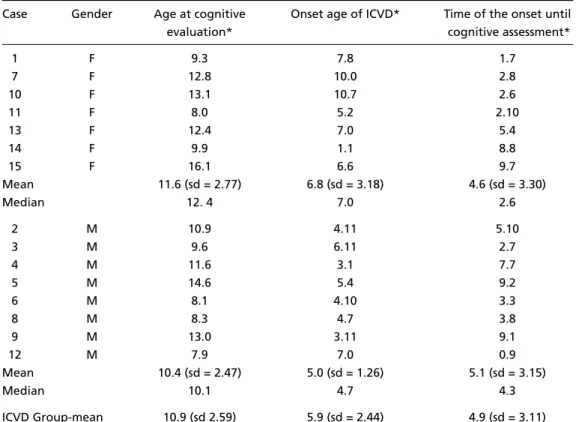
most of them were public school (11/14). The mean age at the time of the ICVD was 5.9 years (range 1.1 to 10.7 years). ln the boys the mean age of the ICVD was 5.0 (range 3.1 to 7.0 years), while the girl’s mean age was 6.8 (range 1.1 to 10.7 years).
The mean age of cognitive function assessment of the ICVD group was 10.9 years (range to 7.9 to 16.1 years). ln the boys the mean age was 10.4 (ran-ged 7.9 to 14.6 years), while in the girls the mean age was 11.6 (range 8.0 to 16.1) The mean age bet-ween acute vascular lesion onset and cognitive function evaluation was 4.9 years (range 9 months to 9.7 years), summarized in Tables 1 and 2.
The period between the onset until cognitive assessment was similar for both gender, with the mean of 5.1 for boys (range 9 months to 9.2 years) and 4.6 for girls (range 1.7 years to 9.7) (p=0.6943). The left hemisphere was affected in five pa-tients, while the right hemisphere was affected in five patients and bilateral lesions were found in five other patients. In 9/15 patients, the lesion simulta-neously affected the cortical and subcortical areas and in the remaining patients it only had affected
Children and Adolescent CVD Ambulatory Research Unit. The researcher design was approved by Ethical Committee on research of FCM/UNICAMP.
The control group consisted of 15 children with sim-ilar characteristics of the ICVD group (age, sex and so-cioeconomic conditions). They were alI enrolled in pub-lic schools and according to their teachers they had no learning difficulties.
The cognitive function of the ICVD group and con-trol group were evaluated by Piaget’s clinical method, which consisted of six operative tests conservation of number; conservation of the quantity of water, conser-vation of the quantity of plasticine, two tasks involving arrangement of objects in class; seriation of objects). The criteria used for scoring the tests were: correct response - one point; responses indicating typical transition - 0.5; incorrect response - none. Hence, the maximum score that could be achieved was 6 points (the PROEPRE Pro-tocol - Laboratório de Psicologia Genética - Faculdade de Educação/UNICAMP).
The SAS System for Windows (version 8.02)and the
SPSS for Windows (version 10.0.5)were used to analyze the statistics and to chose the test (Fisher, Mann-Whitney, Kruskal Wallis or Wilcoxon) and in order to evaluate the results it was performed an analysis according to the type of variable that was being analyzed (categorical or continuous).The significance leveI adopted was p<0.05.
Table 2. Characteristics of the ICVD Group.
Case Gender Age at cognitive Onset age of ICVD* Time of the onset until
evaluation* cognitive assessment*
1 F 9.3 7.8 1.7
7 F 12.8 10.0 2.8
10 F 13.1 10.7 2.6
11 F 8.0 5.2 2.10
13 F 12.4 7.0 5.4
14 F 9.9 1.1 8.8
15 F 16.1 6.6 9.7
Mean 11.6 (sd = 2.77) 6.8 (sd = 3.18) 4.6 (sd = 3.30)
Median 12. 4 7.0 2.6
2 M 10.9 4.11 5.10
3 M 9.6 6.11 2.7
4 M 11.6 3.1 7.7
5 M 14.6 5.4 9.2
6 M 8.1 4.10 3.3
8 M 8.3 4.7 3.8
9 M 13.0 3.11 9.1
12 M 7.9 7.0 0.9
Mean 10.4 (sd = 2.47) 5.0 (sd = 1.26) 5.1 (sd = 3.15)
Median 10.1 4.7 4.3
ICVD Group-mean 10.9 (sd 2.59) 5.9 (sd = 2.44) 4.9 (sd = 3.11)
the subcortical areas. The middle cerebral artery (MCA) and its perforating branches (PB) were the most affected. Five of 15 patients had recurrence ICVD and three of them were females (Table 1).
Thirteen children presented hemiparesis, and fi-ve of them had also presented disturbance of mo-tor speech (subjects 5 and 8), visual involvement (subjects 9 and 12) and visual, renal and cardiac involvement was seen in one patient (subject 11).
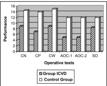
Information about the cognitive functions of both ICVD group and control group is summarized in Table 3. The ICVD group showed a perform-ance significantly below (p=0.002) compared to the control group (Fig 1). No significant difference was observed in patients who had recurrence and those who had been affected only once.
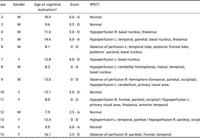
Finally, the cognitive results were compared with the SPECT, obtained in 14/15 children of the ICVD group: 8/9 patients with hypoperfusion showed a performance below normal for age group, while 3/5 with normal perfusion, present-ed an adequate performance (Table 4).
DISCUSSION
There are no published studies at the literature which had assessed the cognitive evolution of
chil-dren with CVD by Piaget’s clinical method. In this way, are results are important because they can add data about the evolutive cognitive aspect of chil-dren with ICVD.
Nicolaides & Appleton13mention that at CVD
there is no preference in terms of which gender is affected, a fact that is also verified in our study. These same authors and also Moura-Ribeiro et al.14had conclude that children with inferior age
of 2 years old were more often affected by CVD. In our research, the mean age was 5.9 years old. Therefore, it should be considered that in this search the casuistic is small and it does not include the patients from CVD Research Out-Patient. The majority of the patients (9/15) had simultaneous attacks of subcortical and cortical areas involving MCA and PB, a fact that was also mentioned by the literature. Schryner et al.3 described two
recur-rences in 35 patients and at this present study it was seen 5 recurrences in 15 patients. Perhaps this fact should be related to the features of the out-patients, which is referred unit in this area of the country.
Regarding to the cognitive assessment, the expec-tiveness is that all the children between 7-8 years of age (maximum 9 of age) had reached success at
Table 3. Performance of ICVD and Control Group in operative tests.
ICVD Gender Age* Score Control Sex Age* Score
1 F 9.3 4 1-c F 9.3 4.0
2 M 10.9 6 2-c M 10.7 6
3 M 9.6 0.5 3-c M 9.3 3.5
4 M 11.6 5 4-c M 11.6 6
5 M 14.6 6 5-c M 14.10 6
6 M 8.1 0 6-c M 8.1 5
7 F 12.8 4 7-c F 12.4 6
8 M 8.3 0 8-c M 8.3 5
9 M 13.0 0 9-c M 12.7 6
10 F 13.1 5 10-c F 12.9 6
11 F 8.0 0 11-c F 7.8 6
12 M 7.9 3.5 12-c M 7.4 3.5
13 F 12.4 0 13-c F 11.7 6
14 F 9.9 6 14-c F 9.9 6
15 F 16.1 3.5 15-c F 15.3 6
Score 43.5 Score 81
ICVD Group (mean) 2.9 Control Group (mean) 5.4
the six operative tasks12. Under this perspective, 11
children from ICVD group should have received maximum score (6 points). Although, only three children had achieved this score (Table 3). It is impor-tant to point out that 6/12 children (subjects 4,7,9,10,13 and 15) that not achieved the expected 6 points at the score, were the ones with ages above 9 years old (from 11.6 to 16.1 years of age), a fact that indicates a clear discrepancy at the cognitive development. The remaining children with a score below 6 (subjects 1, 3, 6, 8, 11 and 12) were between 7.9 and 9.6 years of age. The cultural and environ-mental factors can contribute to the delay on acqui-sition of the operative thought12. So, the children
from this age period could be in a transitory phase and could only have success in some of the tests. At this present study only subjects 1 and 12 were in this phase and also 5/10 children (subjects 1, 2, 5, 12 and 14) from ICVD group had satisfactory performance. Accounting for the discrepancy hypothesis at the per-formance of ICVD it was considered the onset age, the site and lesion length.
Regarding to the onset age, ICVD had affected the children in different stages of their development
(from 1.1 to 10.7 years of age), difficulting the per-formance analysis of the subjects at operative tests. The children with satisfactory performance (subjects 1, 2, 5, 12 and 14) had cerebrovascular lesion between 1.1 to 7.8 years of age and the others who had dissatisfactory performance (subjects 3, 4, 6, 7, 8, 9, 10, 11, 13 and 15) were between 3.1 and 10.7 years of age. So, the onset age of ICVD did not influ-ence the children’s performance.
Regarding to hemispheric localization of ICVD, it was verified that five subjects had bilateral lesion, while five had lesion on the right hemisphere and the others, on the left hemisphere. Knowing befo-rehand that the left hemisphere is related to the speach arrangement and with superior mental ways linked to it, it was hypothezed that the children with lesion on the left hemisphere should have a less performance. Therefore, considering the chil-dren who had dissatisfactory performance (subjects 3, 4, 6, 7, 8, 9, 10, 11, 13 and 15) it was seen that four of these subjects had the right hemisphere affected (subjects 3, 4, 10 and 15), three of them had the left hemisphere affected (subjects 6, 7, and 8) and the other three children had bilateral lesions.
Table 4. Performance of ICVD and results from the SPECT.
Case Gender Age at cognitive Score SPECT evaluation*
2 M 10.9 6.0 - A Normal
3 M 9.6 0.5 - D Normal
4 M 11.6 5.0 - D Hypoperfusion R: basal nucleus, thalamus
5 M 14.6 6.0 - A Hypoperfusion L: temporal, parietal, basal nucleus, thalamus 6 M 8.1 0 - D Absence of perfusion L: temporal lobe, posterior frontal lobe,
posterior parietal, basal nucleus 7 F 12.8 4.0 - D Hypoperfusion L: basal nucleus
8 M 8.3 0 - D Hypoperfusion L: cerebellar hemispheres, mesial, temporal, basal nucleus
9 M 13.0 0 - D Absence of perfusion R: hemisphere (temporal, parietal, occipital); Hypoperfusion L: cerebellum, primary visual area.
10 F 13.1 5.0 - D Normal
11 F 8.0 0 - D Hypoperfusion B: frontal, parietal, occipital / Hypoperfusion L: primary visual area, thalamus, anterior temporal
12 M 7.9 3.5 - A Normal
13 F 12.4 0 - D Hypoperfusion L: temporal, parietal / Hypoperfusion R: parietal, occipital
14 F 9.9 6.0 - A Normal
15 F 16.1 3.5 - D Absence of perfusion R: parietal, frontal, temporal
So, the hemisphere localization of ICVD had not influenced the performance of the patients.
In the same way as age and the hemisphere lo-calization, the lesions on subcortical areas or cor-tical and subcorcor-tical areas together were not deter-minant on the performance of the evaluated sub-jects, because among the 10 subjects who had pre-sented dissatisfactory performance, 5 had subcor-tical lesion (subjects 3, 4, 7, 8 and 10) and in 5 pa-tients the lesion was cortical and subcortical (sub-jects 6, 9, 11, 13 and 15). The cognitive discrepan-cy of the studied group is significantly inferior (p= 0.002) to the control group, confirming the hypo-thesis previously seen15,16. At the control group 10/15
subjects had reached the maximum score and the remaining children (5/15) that did not reach suc-cess in all the 6 tests (subjects 1c, 3c, 6c, 8c and 12c) were between 7.4 and 9.3 years of age and they also were in transitory phase, showing an ade-quated performance for the age period. So, all the 15 children from this group had presented a satisfactory performance (Fig 1).
Regarding to the findings on the SPECT, it was verified a high correlation between the perfusion and the result of the cognitive evaluation: 8 chil-dren with hypoperfusion had a performance
infe-rior to the expected one while 3 with normal per-fusion had an adequated performance. These re-sults suggest that new studies should be done and also they restrengthen the importance of neuroi-mage resources at the clinical and functional investi-gation for the understanding of the cerebral patho-physiology.
It was concluded that children with ICVD asses-sed evolutionally by Piaget’s clinical method had presented a performance significantly inferior when compared to the control group, a rare verifi-cation in children with ICVD.
REFERENCES
1. Powell FC, Hanigan WC, McCluney KW. Subcortical infarction in children. Stroke 1994;25:117-121.
2. Giroud M, Lemesle M, Gouyon JB, Nivelon JL, Milan C, Dumas R. Cerebrovascular disease in children under 16 years of age in the city of Dijon, France: a study of incidence and clinical features from 1985 to 1993. J Clin Epidemiol 1995;48:1343-1348.
3. Schryner LLM, Kappelle LJ, Jennekens-Schinkel A, Peters ACB. Prognosis of ischemic stroke in childhood: a long-term follow-up study. Develop Med Child Neurol 2000;42:313-318.
4. Ciasca SM, Alves HL, Guimarães IE. Avaliação neuropsicológica em menina com doença cerebrovascular bilateral (moyamoya) antes e após a intervenção cirúrgica. Arq Neurpsiquiatr 1999;57:1036-1040. 5. Ganesan V, Isaac E, Kirkham FJ. Variable presentation of
cerebrovas-cular disease in monovular twins. Develop Med Child Neurol 1997; 39:628-631.
6. Ganesan V, Hogan A, Shack N, Gordon A, Isaacs E, Kirkham FJ. Outcome after ischaemic stroke in childhood. Develop Med Child Neurol 2000; 42:455-461.
7. Guimarães IE, Ciasca SM, Moura-Ribeiro MVL. Neuropsychological eva-luation of children after ischemic cerebrovascular disease. Arq Neuro-psiquiatr 2002;60:386-389.
8. Koelfen W, Freund M, Konig S, Varnholt V, Rohr H, Schultze C. Results of parenchymal and angiographic magnetic resonance imaging and neu-ropsychological testing of children after stroke in neonates. Eur J Pediatr 1993;152:1030-1035.
9. Inhelder, B. Le diagnostic du raisonnement chez les débiles mentaux. 3.Ed, Suisse: Delachaux et Niestlé, 1963:249-286.
10. Ajuriaguerra J, Tissot R. Aplicación clínica de la psicologia genética. In Inhelder B, et al (eds). Psicologia y epistemologia geneticas. Tradução Hugo Acevedo. Buenos Aires: Editorial Proteo, 1970:273-277. 11. Lefèvre B. Avaliação neuropsicológica. Rev de Psicologia Hospitalar
1992;1:60-63.
12. Piaget J. Seis estudos de Psicologia. 23.Ed. Tradução Maria Alice Magalhães D’Amorim e Paulo Sérgio Lima Silva. Rio de Janeiro: Forense Universitária, 1998:13-136.
13. Nicolaides P, Appleton RE. Stroke in children. Develop Med Child Neurol 1996;38:172-180.
14. Moura-Ribeiro MVL, Ferreira LS, Montenegro MA, et al. Doença cére-bro vascular na infância: aspectos clínicos em 42 crianças. Arq Neuropsiquiatr 1999;57:594-598.
15. Rodrigues SD, Guimarães IE, Oliveira LS, Ciasca SM, Moura-Ribeiro MVL. Doença cerebrovascular na infância: avaliação das estruturas cognitivas em 12 pacientes (Abstr) Arq Neuropsiquiatr 2002;60:93. 16. Rossini SDR, Guimarães IE, Oliveira LS, Ciasca SM, Moura-Ribeiro MVL.
Children’s cognitive structures after isquemic cerebrovascular acci-dent (Abstr). J Int Neuropsych Soc 2001;7:1431.