• Nenhum resultado encontrado

Study of in vivo interactions between penicillin-binding proteins of Staphylococcus aureus

N/A
N/A
Protected

Academic year: 2019

Share "Study of in vivo interactions between penicillin-binding proteins of Staphylococcus aureus"

Copied!
77
0
0

Texto

(1)

Rita de Sá Martins Pinto Gonçalves

Licenciatura em Biologia pela Faculdade de Ciências da Universidade de Lisboa

Study of

in vivo

interactions between

penicillin-binding proteins of

Staphylococcus

aureus

Dissertação para obtenção do Grau de Mestre em Genética Molecular e Biomedicina da FCT/UNL

Orientador: Mariana Gomes de Pinho, Professora Associada

Instituto de Tecnologia Química e Biológica/ UNL

Júri:

Presidente: Prof.a Dr.a Ilda Santos Sanches

Arguente: Prof. Dr. Luís Jaime Mota Vogal: Prof.a Dr.a Mariana Gomes de Pinho

(2)

Study of in vivo interactions between penicillin-binding proteins of Staphylococcus aureus Rita Gonçalves

(3)

Rita de Sá Martins Pinto Gonçalves

Licenciatura em Biologia pela Faculdade de Ciências da Universidade de Lisboa

Study of

in vivo

interactions between

penicillin-binding proteins of

Staphylococcus

aureus

Dissertação para obtenção do Grau de Mestre em Genética Molecular e Biomedicina da FCT/UNL

Orientador: Mariana Gomes de Pinho, Professora Associada

Instituto de Tecnologia Química e Biológica/ UNL

Júri:

Presidente: Prof.a Dr.a Ilda Santos Sanches

Arguente: Prof. Dr. Luís Jaime Mota Vogal: Prof.a Dr.a Mariana Gomes de Pinho

(4)

iv

Study of in vivo interactions between penicillin-binding proteins of Staphylococcus aureus

Copyright em nome de Rita de Sá Martins Pinto Gonçalves, da FCT, UNL e da UNL.

(5)

v

ACKNOWLEDGMENTS

(6)
(7)

vii

RESUMO

Staphylococcus aureus (S. aureus) é um organismo patogénico humano de grande relevância que adquiriu resistência a praticamente todas as classes de antibióticos, sendo responsável por um elevado número de infeções resistentes principalmente a antibióticos β-lactâmicos a nível hospitalar e comunitário. Os β -lactâmicos têm como alvo as proteínas de ligação à penicilina (PBPs) e estas são responsáveis por catalisar os últimos estágios de síntese do principal componente da parede celular, o peptidoglicano. Tal como em

Escherichia coli, foi sugerido que S. aureus sintetiza a parede celular através de um complexo multi-proteico. Na presença de β-lactâmicos, as proteínas PBP2A e PBP2 sintetizam a parede celular, garantindo a sobrevivência celular. A PBP2 coopera com a PBP4 nas para estabelecer as ligações peptidicas entre os polímeros da parede celular. Como tal, a investigação da interação entre estas proteínas e a sua localização

in vivo é de grande interesse, pois fornece novas pistas acerca da maquinaria de síntese da parede celular em S. aureus. O objetivo deste trabalho foi desenvolver um sistema de Split-GFPP7 para determinar as

interações entre a PBP2 e a PBP4. Dividiu-se o GFPP7 num local estratégico e fizeram-se fusões de proteínas

de interesse a ambas as porções resultantes. Quando se expressou uma fusão contendo o fragmento de GFPP7

ligado a determinada proteína, não foi detetada fluorescência na célula. Pelo contrário, quando ambos os fragmentos de GFPP7 em fusão com proteínas de síntese do peptidoglicano (PBP2 e PBP4) ou de divisão

celular (FtsZ e EzrA) foram expressos na mesma célula, detetou-se fluorescência celular a nível do septo. Contudo, análises posteriores revelaram que este resultado se deve à auto-associação do GFPP7. Deste modo,

interpretamos os resultados com base neste acontecimento e fornecemos novas pistas que possam ser úteis para o melhoramento deste sistema.

(8)
(9)

ix

ABSTRACT

Staphylococcus aureus (S. aureus) is a major human pathogen that has acquired resistance to practically all classes of β-lactam antibiotics, being responsible of Multidrug resistant S. aureus (MRSA) associated infections both in healthcare (HA-MRSA) and community settings (CA-MRSA). The emergence of laboratory strains with high-resistance (VRSA) to the last resort antibiotic, vancomycin, is a warning of what is to come in clinical strains. Penicillin binding proteins (PBPs) target β-lactams and are responsible for catalyzing the last steps of synthesis of the main component of cell wall, peptidoglycan. As in

Escherichia coli, it is suggested that S. aureus uses a multi-protein complex that carries out cell wall synthesis. In the presence of β-lactams, PBP2A and PBP2 perform a joint action to build the cell wall and allow cell survival. Likewise, PBP2 cooperates with PBP4 in cell wall cross-linking. However, an actual interaction between PBP2 and PBP4 and the location of such interaction has not yet been determined. Therefore, investigation of the existence of a PBP2-PBP4 interaction and its location(s) in vivo is of great interest, as it should provide new insights into the function of the cell wall synthesis machinery in S. aureus. The aim of this work was to develop Split-GFPP7 system to determine interactions between PBP2 and PBP4.

GFPP7 was split in a strategic site and fused to proteins of interest. When each GFPP7 fragment, fused to

proteins, was expressed alone in staphylococcal cells, no fluorescence was detectable. When GFPP7

fragments fused to different peptidoglycan synthesis (PBP2 and PBP4) or cell division (FtsZ and EzrA) proteins were co-expressed together, fluorescent fusions were localized to the septum. However, further analysis revealed that this positive result is mediated by GFPP7 self-association. We then interpret the results

in light of such event and provide insights into ways of improving this system.

(10)
(11)

xi

TABLE OF CONTENTS

ACKNOWLEDGEMENTS --v

RESUMO vii

ABSTRACT ix

TABLE OF CONTENTS xi

FIGURES AND TABLES INDEX xiii

SYMBOLS AND ABBREVIATIONS xv

1. INTRODUCTION 1

1.1 Staphylococcus aureus: Importance as a pathogen 1

1.2 Mechanisms of Cell Growth in bacteria 2

1.2.1 Cell Division 2

1.2.2 Cell Wall Synthesis 4

1.3 Penicillin-binding proteins (PBPs) 8

1.3.1 S. aureus PBPs 9

1.4 Evidence for multienzyme complexes formed by PBPs 10

1.5 Divisome and cell wall synthesis machinery of S. aureus 11

1.6 Evidence for cooperative interaction between FtsZ and EzrA 13

1.7 Evidence for cooperative functioning of PBP2 and PBP4 in S. aureus 14

1.8 Methods for detection of protein-protein interactions 15

1.8.1 BiFC: A fluorescence protein complementation assay 15

1.8.2 Applications of BiFC 16

1.8.3 GFP scaffold and possible split-sites 17

1.9 Aim of the thesis 17

2. MATERIAL AND METHODS 18

2.1 Bacterial strains, plasmids and growth conditions 18

2.2 Molecular biology 19

2.2.1 Purification of plasmid DNA from E. coli 19

2.2.2 Isolation of genomic DNA from S. aureus 19

2.2.3 Polymerase Chain reaction (PCR) 19

2.2.4 Sequencing 20

2.2.5 Restriction digestion and ligation of DNA 20

2.2.6 Transformation of chemically competent E. coli cells 21

(12)

xii

2.2.8 Transduction of S.aureus cells 22

2.3 Construction of plasmids and strains 23

2.3.1 Construction of split-gfpp7 fusions to gene of interest 23

2.3.1.1 Construction of ftsZ-c-gfpp7 and ftsZ-n-gfpp7 fusions 27

2.3.1.2 Construction of ezrA-c-gfpp7and ezrA-n-gfpp7fusions 28

2.3.1.3 Construction of pbp4–c-gfpp7 fusion 29

2.3.1.4 Construction of n-gfp-pbp2p7 fusion 29

2.3.1.5 Construction of n-gfpp7 fusion 30

2.3.1.6 Construction of c-gfpp7fusion 30

2.3.2 Construction of S. aureus strains expressing two fusions 30

2.3.2.1 Strain expressing FtsZ and EzrA fusions 30

2.3.2.2 Strain expressing PBP2 and PBP4 fusions 31

2.3.2.3 Strains expressing PBP2 and EzrA or PBP2 and FtsZ fusions 31

2.3.2.4 Strain expressing N-GFPP7 and C-GFPP7 fusions 31

2.3.2.5 Strains expressing C-GFPP7 and N-GFPP7-PBP2 fusions or N-GFPP7 and PBP4-C- GFPP7 fusions 32

2.4 Fluorescence microscopy 32

2.5 Fluorescence quantification and statistical analysis 32

3. RESULTS 34

3.1 GFPP7 split-site selection 34

3.2 Choice of type of fusions based on protein topology 35

3.3 Protein interaction studies of single split-GFPP7 protein fusions 36

3.4 Protein interaction studies of combined split-GFPP7 protein fusions 39

3.4.1 FtsZ-EzrA interaction as a positive control 40

3.4.2 Analysis of FtsZ-EzrA split-GFPP7 fusions 40

3.4.3 Analysis of PBP2-PBP4 split-GFPP7 fusions 42

3.4.4 Analysis of PBP2-EzrA and PBP2-FtsZ split-GFPP7 fusions 42

3.5 Analysis of additional split-GFPP7fusions 45

3.5.1 Analysis of C-GFP, N-GFPand C-GFP + N-GFPfusions 46

3.5.2 Analysis of C-GFPP7 + N-GFPP7-PBP2 and N-GFPP7 + PBP4-C-GFPP7 fusions 48

3.5.3 Analysis of C-GFPmut1 + N-GFPmut1 fusions 49

4. DISCUSSION 51

REFERENCES 54

(13)

xiii

FIGURES AND TABLES INDEX

Figure 1.1 Divisome assembly in B. subtilis 4

Figure 1.1 Primary structure of S. aureus peptidoglycan 5

Figure 1.3 Peptidoglycan biosynthesis 7

Figure 1.4 Hypothetical murein-synthetizing machinery formed by gram-negative bacteria 10

Figure 1.5 First model of cell wall synthesis in S. aureus 12

Figure 1.6 Two models of cell wall synthesis in S. aureus 13

Figure 1.7 Model for cooperative functioning of PBP2 and PBP4 14

Figure 1.8 Principle of Bimolecular fluorescence complementation assay (BiFC) 16

Table 2.1 Primers used in this study 23

Table 2.2 Plasmids used in this study 24

Table 2.3 Bacterial strains used in this study 25

Figure 3.1 GFPP7 split-site selection based on GFPP7 topology and sequence comparison with YFP 34

Figure 3.2 Schematic representation of the constructed fusions and respective sizes 35

Figure 3.3 Microscopy analysis of cells expressing single split-GFPP7 protein fusions 38

Figure 3.4 Fluorescence quantification of single split-GFPP7 fusions 39

Figure 3.5 Microscopy analysis of cells expressing FtsZ-EzrA split-GFPP7 fusions 40

Figure 3.6 Microscopy analysis of cells expressing PBP2-PBP4 split-GFPP7 fusions 42

Figure 3.7 Microscopy analysis of cells expressing PBP2-EzrA and PBP2-FtsZ split-GFPP7 fusions 43

Figure 3.8 Fluorescence quantification of combined split-GFPP7 fusions 45

Figure 3.9 Microscopy analysis of cells expressing C-GFP P7, N-GFPP7and C-GFPP7 + N-GFPP7 fusions 47

Figure 3.10 Fluorescence quantification of C-GFP, N-GFP and C-GFPP7 + N-GFPP7fusions 47

Figure 3.11 Microscopy analysis of cells expressing N-GFPP7 + PBP4-C-GFPP7 and C-GFPP7 + N-GFPP7-PBP2 fusions 49

Figure 3.12 Microscopy analysis of cells expressing C-GFPP7 + N-GFPP7fusions and C-GFPmut1 + N-GFPmu1fusions 50

Figure 3.13 Fluorescence quantification of C-GFPmut1 + N-GFPmut1 fusions 50

(14)
(15)

xv

SYMBOLS AND ABBREVIATIONS

% Percentage

°C Degrees Celsius

~ Approximately Amp Ampicillin

B. subtilis Bacillus subtilis

BTH Bacterial two-hybrid bp Base pair

BiFC Bimolecular fluorescence complementation assay CdCl2 Cadmium Chloride

Cm Chloramphenicol DNA Deoxyribonucleic acid DNAse Deoxyribonuclease

dNTP Deoxyribonuclotide triphosphate

E. coli Escherichia coli

EDTA Ethylenediaminetetraacetic acid Erm Erythromycin

eYFP Enhanced yellow fluorescent protein FastAP Thermosensitive Alkaline Phosphatase FP(s) Fluorescent protein(s)

GFP Green fluorescent protein

GFPP7 Superfast green fluorescent protein

h Hour(s)

IPTG Isopropyl β-D-thiogalactosidase KAc Potassium acetate

Kan Kanamycin kb Kilo base pair LA Luria-Bertani agar LB Luria-Bertani broth

M Molar

min Minute(s)

MnCl2·4H2O Manganese(II) chloride tetrahydrate

MOPS 3-(N-morpholino)propanesulfonic acid MRSA Methicilin-resistant Staphylococcus aureus MSSA Methicilin-sensitive Staphylococcus aureus

NaCl Sodium chloride Neo Neomycin

OD600 Optical density at 600 nm

PBP(s) Penicillin-binding protein(s) PCR Polymerase chain reaction PG Peptidoglycan

RbCl2 Rubidium Chloride

Rpm Revolutions per minute

(16)

xvi VISA Vancomycin-intermediate Staphylococcus aureus

VRSA Vancomycin-resistant Staphylococcus aureus

WT WTA

Wild-type

Wall teichoic acid(s)

(17)

1

INTRODUCTION

1.1 Staphylococcus aureus: Importance as a pathogen

Staphylococcus aureus is a prominent pathogen well known for its virulence and antibiotic resistance in both community and nosocomial settings. Virulence is mediated by innumerous factors, such as secreted or cell surface-associated proteins that compromise the host immune system to help colonization (Foster, 2005). The major importance of this pathogen increased with the development of resistance to most classes of antibiotics.

Penicillin was an effective antimicrobial agent that protected against S. aureus infections. Soon after the first use of penicillin in 1942 in the U.S, penicillinase-producing S. aureus emerged in the clinical setting and its prevalence increased dramatically within a few years, mostly due to wide use of penicillin. Since then, Penicillin-resistant S. aureus (PRSA), phage-type 80/81 clone, become pandemic and in the late 1950s, a new antibiotic, methicillin, was used to select against PRSA strains. Shortly after the introduction of methicillin, in 1961, the first cases of methicillin- resistant S. aureus (MRSA) strains were reported. MRSA emerged in the U.S in the early 1960s and have since spread among hospital and health care settings worldwide (Deleo and Chambers, 2009). More recently, in the 1990s, MRSA strains have spread into the community, among healthy individuals, giving rise to new epidemic waves of community acquired S. aureus

(CA-MRSA) (Udo et al., 1993; Deleo and Chambers, 2009). MRSA is resistant to practically all available β-lactams, which include penicillin, amoxicillin, oxacillin, methicillin, and others (Deleo and Chambers, 2009).

(18)

2 (Voyich et al., 2005). These strains express Panton-Valentine leukocidin (PVL) toxin, which is often used as a marker for CA-MRSA. (Wardenburg et al., 2009).

Vancomycin is a glycopeptide antibiotic used as an alternative to β-lactams for treatment of MRSA infections as it inhibits synthesis of S. aureus cell wall by a different mechanism from these antibiotics (Walsh and Howe 2002). However, according to some authors, it showed reduced efficacy due to weak antimicrobial activity and poor tissue penetration comparing to other β-lactams. The poor therapeutic effect along with the rapid reduction of S. aureus susceptibility to vancomycin has led to questioning its effectiveness in the long run (Deresinski, 2007). After the first clinical isolate of MRSA S. aureus with decreased susceptibility to vancomycin, vancomycin-intermediate S. aureus (VISA) strains spread across several countries (Howe et al., 2004). A thickened cell wall contributes to the vancomycin resistance and is a common phenotype in these isolates (Cui et al., 2000). This altered composition is related to a decrease in highly cross-linked muropeptides that compose the PG, along with an increase in muropeptides monomers and dimers (Sieradzki and Tomasz, 1997; Sieradzki et al., 1999), which is in turn associated to defects in penicillin-binding protein 4 (PBP4) activity (Sieradzki et al., 1999). A few years later, strains highly resistant to vancomycin, designated vancomycin-resistant S. aureus (VRSA) have emerged and vancomycin efficacy as an antistaphylococcal antibiotic is threatened (Bartley, 2002; Deresinski, 2007).

1.2 Mechanisms of Cell Growth in bacteria

Cell division and cell wall synthesis are two essential processes for bacterial growth that are carried out by multiprotein complexes. Escherichia coli (E. coli) and Bacillus subtilis (B. subtilis) are rod-shape bacteria which have been used as model organisms to understand the mechanisms of cell division, namely the localization and order by which proteins are recruited to the division site (reviewed in Errington et al., 2003)

1.2.1 Cell Division

(19)

3 and Addinall, 1997). The Z-ring serves as scaffold for the recruitment of later divisome proteins and directs synthesis, location and shape of the division septum. B. subtilis and E. coli are rod-shaped bacteria frequently used as model organisms to study the mechanisms of cell division. In these bacteria, the site of division is the mid-cell. B. subtilis and E. coli divide through a medial plane. Unlike these bacteria, coccoid shaped bacteria S. aureus divides by three perpendicular planes in 3 phases of the cell cycle (Pinho et al. 2013).

In E. coli several proteins are involved in assembly of Z-ring. The divisome components target the division site in a co-ordinate manner to form the septal Z-ring (Errington et al., 2003; Weiss, 2004). FtsZ filaments tethering to the membrane involves two proteins, FtsA and ZipA, which bind to its short highly conserved C-tail. These proteins are composed by a transmembrane domain and a cytoplasmic domain that are connected by a flexible linker (Pichoff et al, 2012). It has been shown that either FtsA or ZipA is required to form and stabilize the preformed Z-rings (Pichoff and Lutkenhaus 2002). Importantly, FtsA location to the division site is needed for recruitment of the later-assembling membrane-bound division proteins. Therefore, recruitment of nine essential proteins, designated Fts proteins (FtsE/X, FtsK, FtsQ/L/B, FtsW/I) and PBP1b to the divisome is dependent on the amount of monomeric FtsA at the Z ring. Fts proteins assemble to the Z-ring in a defined order, which is still not well known. Non-essential proteins ZapA-D are then recruited to the Z ring to promote its integrity (Lutkenhaus et al., 2012). It is suggested that septal synthesis is activated when PBP3 is recruited to the septum by the lipid II flippase FtsW to interact with the ternary FtsQLB complex, PBP1B and FtsN. FtsN is an essential protein that triggers septation. AmiB and AmiC hydrolases are recruited by EnvC and NlpD respectively, participate in splitting of the septum to separate daughter cells. The Tol-Pal complex enables invagination of the outer membrane, to finally drive cell separation (Buddelmeijer and Beckwith, 2004; Typas et al., 2012; Lutkenhaus et al., 2012).

(20)

4 2012a). Unlike E. coli, later divisome proteins are targeted to the division site, in an interdependent manner (Errington et al., 2003). DivIB, DivIC and FtsL have similar structures and are likely to interact to form a ternary complex (DivIB-DivIC-FtsL), which is equivalent to the FtsQLB complex in E. coli. It has been proposed that within this subcomplex, DivIC interacts with FtsL and DivIB binds to the DivIC-FtsL heterodimer. PBP2B is then recruited, binds to DivIB and directs the PG synthesis machinery to the division site (Graumman, 2012a). It is suggested that the DivIB-DivIC-FtsL trimeric complex may have a role in PG metabolism at the septum through interactions with PBP2B (Rowland et al., 2010). Although it is not well known which proteins are involved in PBP1 late recruitment to the divisome, some authors suggest dependence on DivIB, DivIC, and PBP2B (Scheffers and Errington, 2004), while others defend that GspB/YspB complex acts together with EzrA to shuttle PBP1 to the divisome (Claessen et al., 2008; Tavares

et al., 2008).

Figure 1.1 Divisome assembly in B. subtilis. The formation of the divisome starts with the recruitment of the early divisome proteins (FtsA, EzrA, ZapA and SepF) to the Z-ring by FtsZ. Later, the later divisome proteins (DivIB,

DivIC, FtsW, FtsL, PBP2B, GspB, PBP1) are assembled onto the Z-ring in a concerted and interdependent manner, as

opposed to the linear mode of recruitment of these proteins in E. coli (Adams and Errington, 2009).

1.2.2 Cell Wall Synthesis

(21)

N-5 acetyl-glucosamine (GlcNAc) and N-acetyl-muramic acid (MurNAc) subunits (Fig.1.2). The glycan chains have few variations between different bacterial species, but there is considerable variation in the composition of stem peptides that are linked to the carboxyl group of MurNac (Schleifer and Kandler, 1972; Scheffers and Pinho, 2005). The length of glycan chains is also different between bacterial species, since in

S. aureus PG strands are shorter than in E. coli and B. subtilis. The length of the peptide chains can also differ (tri-, tetra-, pentapeptide) (Vollmer and Bertsche, 2008). Despite of its stiffness, PG is an elastic structure, which can reversible expand and shrink without breaking (Koch and Woeste, 1992). The cell wall stability implies a fine control of the insertion and removal of wall material by enzymes capable of synthesizing or hydrolyzing the PG layers.

In all of these bacteria stem peptides are pentapepide chains, which only differ in the dibasic amino acid composition. Whereas mesodiamoinopimelic acid (m-A2pm) is present in E. coli and B. subtilis, L

-Lysine (L-Lys) is the dibasic amino acid in S.aureus. Therefore, the composition of a newly synthetized

stem peptide is L-Ala–D-Glu–m-A2pm/L-Lys–D-Ala–D-Ala, containing L-Ala bounded to the MurNac.

Additionally in S.aureus, L-lys is attached to a pentapeptide of Glycines (Gly), forming a flexible

pentaglycine cross bridge, which is responsible for the high degree of cross-linking observed in these bacteria. PG is formed via two basic processes: assembly of glycan chains (transglycosylation) and peptide cross bridge formation (transpeptidation) (Scheffers and Pinho, 2005). Unlike gram-positives B. subtilis and

(22)

6 Figure 1.2 Primary structure of S. aureus peptidoglycan. The overall bacterial peptidoglycan is composed of linear glycan chains linked through pentapeptide cross-bridges. N-acetyl-glucosamine (GlcNAc) and N-acetyl-muramic acid (MurNAc) disaccharide subunits are bound together by a β-1,4 linkage. Each pentapeptide, known as stem peptide, is composed of L-Ala–D-Glu–DA–D-Ala–D-Ala. The carboxyl group of MurNAc is linked to the L-Ala of a stem peptide. Thedibasic amino acid (DA) present in S. aureus is L-Lys, which is attached to a pentaglycine bridge and confers a high level of PG cross-linkage. Adapted from Scheffers and Pinho, 2005.

PG biosynthesis occurs in three sequential stages: i) Synthesis of the PG precursor by specific enzymes and linkage to the carrier lipid (undecaprenyl-phosphate or bactoprenol), ii) Flipping across the membrane, iii) Incorporation of the precursor into the cell wall (Vollmer & Bertsche, 2008).

In the first stage, the intermediates of PG precursor synthesis Uridine diphosphate-N-acteyl-glucosamine (UDP-GlcNAc) and uridine diphosphate-N-acteyl-muramic acid (UDP-MurNAc), are formed in the cytoplasm (Fig.1.3). UDP-MurNac is originated from UDP-GlcNac. L-Ala, D-Glu, m-A2pm and a D

-Ala–D-Ala dipeptide are successively added to the peptide side chain (linked to UDP-MurNAc) by

ATP-dependent ligases MurC, MurD, MurE and MurF, respectively. The D-amino acids are generated from L-amino acids by racemases. Before transport of hydrophilic precursors across the cytoplasmic membrane, a lipid carrier (bactoprenol) is attached by MraY to form lipid I (undecaprenyl pyrophosphoryl-MurNAc-pentapeptide). Then, GlcNAc is added to lipid I by MurG, yielding lipid II (undecaprenyl pyrophosphoryl- GlcNAc-β-(1, 4)-MurNAc-pentapeptide). The coupling of a disaccharide precursor (hydrophilic substrate) to a lipid molecule facilitates its translocation through the hydrophobic membrane (van Heijenoort, 2001; Vollmer and Bertsche, 2008).

During the second stage, Lipid II is transported across the cytoplasmic membrane to the periplasmic site at the outer membrane by a flippase or specific translocase (Vollmer and Bertsche, 2008). It is though that the integral membrane homologues, FtsW and RodA (or mrdB) function as translocases of lipid precursors across the cytoplasmic membrane, which may then be directed to their cognate PBPs (Scheffers and Pinho, 2005). In E. coli lipid II is translocated to the periplasmic site by FtsW, localized at the septum. It is likely that S. aureus lipid II is translocated by an homologue of FtsW (Pinho et al., 2013).

(23)

7 the next stem peptide (van Heijenoort, 2001; Scheffers and Pinho, 2005). PG is concomitantly formed and degraded by specific enzymes, named PG synthases and murein hydrolases. PG synthases with a TPase activity are termed Penicillin-binding proteins (PBPs), due to their capacity of binding Penicillin and other β-lactams, which are structurally similar to the D-Ala-D-Ala termini of the stem peptides (Vollmer and Bertsche, 2008).

Figure 1.3 Peptidoglycan biosynthesis. In the first stage, the intermediates of PG precursor synthesis UDP-GlcNac and UDP-MurNac are assembled in the cytoplasm via addition of UDP precursors and lipid intermediates. The

cytoplasmic enzymes MurA to MurF catalyze the formation of the MurNAc-pentapeptide precursor from

UDP-GlcNAc. MraY catalyzes the transfer of the phospho-MurNAc- pentapeptide moiety of UDP-MurNAc-pentapeptide

(24)

8 second stage, lipid II precursor is transported through the hydrophobic membrane to the periplasmic site by a flippase

or specific translocase, where it is incorporated into nascent peptidoglycan. In the third stage, lipid II precursors are

incorporated into the pre-existing cell wall, by murein synthases or PG synthases, known as PBPs, which have

transpeptidation and transglycosylation activities. Adapted from van Heijenoort, 2001.

B. subtilis and E. coli are proposed to have two modes of cell wall synthesis, i) through elongation of the lateral wall and ii) through formation of a division septum (Scheffers and Pinho, 2005). More recently, another model for cell wall synthesis, which divides the first mode (elongation) into two, dispersed elongation and pre-septal elongation, was proposed. Dispersed elongation occurs firstly with the insertion of PG in several sites of the lateral wall; the tubulin homologue FtsZ locates then to the mid-cell to drive “preseptal” elongation (Typas et al., 2012). S. aureus cell wall synthesis is discussed below.

1.3 Penicillin-binding proteins (PBPs)

PBPs are classified as acyl serine transferases and are divided into high-molecular-weight (HMW) PBPs, low-molecular-weight (LMW) PBPs, and β-lactamases. HMW PBPs contain two domains, a C-terminal domain, located in the periplasm and a short N-C-terminal transmembrane domain that anchors the protein to the membrane. They are grouped into Class A and Class B PBPs, bifunctional or monofunctional enzymes, depending on the composition of the N-terminal domain. In Class A PBPs, the C-terminal domain is a penicillin-binding domain (PB module) with TP activity (responsible for cross-linking between peptides) linked to an N-terminal domain that contains TG activity (participates in glycan strands polymerization). Class B PBPs contain a C-terminal PB module with TP activity and an N-terminal non-penicillin binding (n-PB) module with presumable functions as a chaperone, in cell morphogenesis, or in control of cell division (Ghuysen, 1991; Scheffers and Pinho, 2005).LMW PBPs are monofunctional DD-peptidases, mostly DD-carboxypeptidases that catalyze the hydrolysis of the carboxyl terminal D-ala–D-ala of a stem peptide. Although, some, have TPase (such as S. aureus PBP4) or endopeptidase activities (Scheffers & Pinho, 2005). Besides PBPs, another class of proteins, called monofunctional transglycosylases (MGTs), contains TG activity.

The analysis of mutants containing mutations in several PBPs and the localization of PBPs by fluorescence microscopy has provided new information about the function of these enzymes and the identification of the place where PG synthesis occurs.

(25)

9 fact that most of these PG synthesizing enzymes have redundant functions makes the study of their functions a difficult task (Scheffers and Pinho, 2005; Pinho and Errington, 2005).

1.3.1 S. aureus PBPs

S. aureus seems to be a better candidate to study the role and possible interactions between PBPs, as it has only 4 to 5 PBPs. Four native PBPs, PBP1, PBP2, PBP3 and PBP4 are present in MSSA strains and an extra PBP2A is only present in MRSA strains and is responsible for β-lactam resistance, due to its low affinity to these antibiotics (Scheffers and Pinho 2005; Hartman and Tomasz, 1984). PBP2 is a class A PBP, containing both TPase and TGase activities (Goffin and Ghuysen, 1998; Murakami et al., 1994).

In the presence of oxacillin, PBP2 TPase domain becomes acylated and thus is unable to bind to its pentapeptide substrate. Moreover, PBP2 loses its ability to localize to the division septum and perform its role in cell wall synthesis. Under these conditions, cells exhibit a dispersed mode of cell wall synthesis in confined spots at the periphery of the cell (Pinho and Errington, 2005). Unlike methicillin sensitive S.aureus

strains, MRSA strains contain an extra PBP, PBP2A that is able to replace PBP2 function as a TPase in presence of β-lactams (Pinho et al., 2001b). However, PBP2 is still required for cell survival as its TGase domain seems to cooperate with the TPase domain of PBP2A (Pinho et al., 2001). Moreover, in these strains PBP2 is not delocalized from the septum suggesting that PBP2A maintains acylated PBP2 at that place (Pinho and Errington, 2005).

PBP1 is a class B HMW monofunctional TPase essential for cell growth and survival in MRSA and MSSA strains. In the absence of PBP1, cells are unable to divide and lose their viability (Okonog et al., 1995; Wada and Watanabe, 1998; Pereira et al., 2007). PBP1 localizes mainly to the division septum (Pereira et al., 2007). Loss of the functional TPase domain of PBP1 resulted in minor alterations in PG composition, and in a process that may repress the expression of autolytic genes and thus, prevent cell separation (Pereira et al., 2009). Therefore, it is suggested that the essential activity of PBP1 as a TPase is related to the control of cell division instead of having a direct role in PG cross-linking (Pereira et al., 2007; Pereira et al., 2009).

PBP4 is a LMW non-essential TPase involved in secondary cross-linking of PG (Wyke et al., 1981). As it is required for synthesis of highly branched peptidoglycan, depletion of PBP4 leads to a decrease in PG cross-linking (Wyke et al., 1981; Sieradzki et al., 1999; Memmi et al., 2008). Moreover, together with

PBP2A, PBP4 contributes to β-lactam resistance in CA-MRSA (but not HA-MRSA strains), since loss of

(26)

10 PBP4 is delocalized (becomes dispersed over the cell membrane) and is not functional; ii) Synthesis of WTA was shown to be required for PBP4 recruitment to the division septum (Atilano et al., 2010).

PBP3 is a non-essential monofunctional TPase which localization remains unknown (Pinho et al., 2000; Scheffers and Pinho, 2005).

1.4 Evidence for multienzyme complexes formed by PBPs

Many studies have provided support for the existence of multienzyme complexes formed by PBPs in several bacteria (Bhardwaj and Day,1997; Alaedini and Day, 1999; Figge et al., 2004; Simon and Day, 2000). Based on previous reported interactions between murein synthases and hydrolases in E.coli, it has been proposed that this organism forms two multienzyme complexes, which play a role in cell wall synthesis either during cell elongation or cell constriction, called murein-synthetizing machinery. They are similar but have different specificities based on the presence of PBP2 (elongation complex) or PBP3 TPases (constriction complex). This model of PG assembly has been identified in other gram-negative bacteria, being in agreement with the “three-for-one” model that states that insertion of three new glycan strands in the cell wall is concomitant with removal of one strand (Fig.1.4) (Höltje, 1996; Höltje, 1998; Scheffers and Pinho, 2005).

Figure 1.4 Hypothetical murein-synthetizing machinery formed by gram-negative bacteria. It is proposed that a multiprotein complex is responsible for i) synthesizing three new cross-linked glycan strands (shown in gray). A

bifunctional PBP, PBP1A or PBP1B, with TPase and TGase (TP/TG) activities dimerizes and joins a pre-existing PG

strand to form a murein triplet, which is connected to the strand by transpeptidation, catalyzed by a dimer of either

(27)

11 sides of the peptide bridges (middle gray strand) and degrading the docking strand by action of hydrolases. A dimeric

endopeptidase (EP; PBP4 or PBP7) splits the peptide cross-bridges of the old strand while a lytic TGase (LT; Slt70,

MltA, or MltB) depolymerizes the glycan strand. Strands shown in white represent preexisting strands (Höltje, 1998).

In B. subtilis, isolation of PBPs by chromatography has revealed that these enzymes form two multi-protein complexes, composed of either three or seven PBPs (Simon and Day, 2000).

Importantly, in S. aureus, EzrA was found to interact with several divisome proteins, including PBP1, PBP2 and PBP3, suggesting that these proteins form a complex that plays a role in cell division (Steele et al., 2011). More recently, it was reported that an S.aureus minimal strain (lacking non-essential genes for peptidoglycan synthesis) that contains only 7 of 9 seven PG synthesizing-enzymes, is able to normally grow and divide. Therefore, it was shown that S. aureus can use a minimal PG synthesis machine, composed of a TPase (PBP1) and a bifunctional PBP (PBP2) to catalyze PG synthesis (Reed et al., 2015). Therefore, more data is needed to prove the existence of a multi-enzymatic PG synthesis complex in gram-positive bacteria.

1.5 Divisome and cell wall synthesis machinery of S.aureus

S. aureus contains homologues for all the essential genes for cell division in B. subtilis (Steele et al., 2011). Furthermore, as PG synthesis seems to take place mainly at one division site (septum), according to Pinho and colleagues, there should only be one PG synthesizing complex of S. aureus, located at the division site (Steele et al., 2011; Pinho et al., 2013).

PBP1, PBP2 and PBP4 localize to the division septum, site where PG synthesis occurs. These PG synthetizing enzymes are recruited to the division septum by different mechanisms (Pinho et al., 2013). PBP1 is suggested to be a component of the divisome and seems to be recruited by an unknown divisome protein in a manner independent of its activity as a TPase (Pereira et al., 2007; Pereira et al., 2009). PBP2 localizes to division septum may be dependent on recognition by its substrate. In the beginning of septum formation PBP2 localizes in a ring and, as the septum closes, it localizes all over the septum (Pinho and Errington, 2005). PBP4 is recruited to the septum by an intermediate of WTA synthesis (Atilano et al., 2010).

(28)

12 hydrolysis of the septal PG by autolysins (cell wall hydrolases). The full septum is composed of a low-density central region, which does not extend into the surface cell wall, separated by two high-low-density regions, which will each constitute half of the new cell wall in daughter cells after splitting. This suggests that autolysins only act at the periphery of the septum (Pinho et al., 2013). This model considers that the newly formed cells remain spherical over the cell cycle.

Figure 1.5 First model of cell wall synthesis in S. aureus. This model considers that peptidoglycan is synthesized only at the septum, in a process that initiates with the recruitment of PBP1 and PBP2 to that place. At a later stage,

PBP4 is recruited to the septum, where it contributes to increasing peptidoglycan PG cross-linking. The full septum is

composed of a low-density central layer separating two high-density layers, corresponding to adjacent cross walls,

which will each constitute half of the new cell wall in daughter cells after splitting. In S.aureus, a specific autolysin is responsible for the septum splitting, acting at the periphery of the septum (Pinho et al., 2013).

(29)

33%-13 67%, respectively. This suggests that there is a second mode of cell wall synthesis in S. aureus through enlargement of the lateral wall (Monteiro et al., 2015) (Fig.1.6).

Figure 1.6 Two models of cell wall synthesis in S. aureus. The previous model (a) proposes that cell wall material of half of each newly formed cell is made from the septum of the mother cell. Therefore the daughter cells have a

proportion of 50% of old cell wall material and 50% of new cell wall material, and the spherical shape is maintained

throughout the cell cycle. The new model (b) assumes that in the beginning of the cell cycle cells are spherical and

elongate over the cell cycle. The cell wall material deposited from the septum of the mother cell (new cell wall)

constitutes only ~33% of the surface area of each daughter cell, while the cell wall material resulting from one

hemisphere of the mother cells (old cell wall) occupies ~67% of the daughter cell (Monteiro et al., 2015).

1.6 Evidence for cooperative interaction between FtsZ and EzrA

In B. subtilis, EzrA was found to be localized to the cell membrane where it acts as a negative regulator of FtsZ, as it inhibits Z-ring formation outside the mid-cell (at medial and polar sites), therefore preventing aberrant cell division (Levin et al., 1999). Moreover, EzrA co-localizes with FtsZ to the nascent septal site, suggesting a positive role for EzrA in cell division, by maintaining Z-ring assembly and dynamics (Levin et al., 1999; Haeusser et al., 2004). This observation suggests that EzrA may be responsible for coordinating cell growth and cell division (Haeusser et al., 2007). Accordingly, EzrA was shown to recruit PBP1 to the divisome, promoting cell separation and subsequent elongation associated-cell wall synthesis. Therefore, in rod-shaped bacteria, EzrA coordinates the two modes of cell wall synthesis (through cell division or cell elongation) (Claessen et al., 2008).

(30)

14 Errington, 2003; Steele et al., 2011). The simultaneous interaction of EzrA with FtsZ, other divisome components and PBPs has led to the hypothesis that this protein acts in the linkage between cytoplasmic FtsZ and the PG synthetizing machinery located in the periplasm (Steele et al., 2011; Jorge et al., 2011).

1.7 Evidence for cooperative functioning of PBP2 and PBP4 in S.aureus

As described above, the TGase activity of PBP2 and the TPase activity of PBP2A are required for cell

survival in MRSA strains, upon challenging with β-lactams. Importantly, when PBP2 TGase domain was

affected by a point mutation, the length of PG glycan strands was reduced (Pinho, et al., 2001). Moreover, a single PBP2 point mutation in the TPase domain was shown to cause a decreased affinity for a β-lactam (ceftizoxime) with high selective affinity for PBP2. This drug-resistance phenotype was accompanied by a reduced proportion of highly cross-linked PG and increased proportion of monomeric and dimeric muropeptide monomers and dimers. This discovery suggested not only a role for the TPase domain of PBP2 but also a link between PBP2 and PBP4 in secondary cross-linking (Leski and Tomasz, 2005). Likewise, in CA-MRSA, when cells are challenged with oxacillin and depleted from PBP4, the transcription of PBP2 is altered, thus causing a decrease in secondary PG cross-linking (Memmi et al., 2008). This finding supports the hypothesis of a concerted action between these two proteins in cell wall cross-linking (Fig.1.7).

PBP2 and PBP4 contribute not only to resistance against β-lactams but also to resistance to the one of the last resort antibiotics effective against S. aureus, vancomycin. Decreased levels of PG cross-linking are a common feature in VRSA strains due to PBP4 inactivation. It is suggested that the alteration of cell wall composition is a mechanism by which these strains survive in presence of vancomycin (Sieradzki and Tomasz, 1997; Sieradzki et al., 1999). Accordingly, PBP2 mutants are more susceptible to vancomycin, suggesting that PBP2 may play a major role in the cell wall assembly of VRSA strains (Sieradzki and Tomasz,1999).

(31)

15 17), which would be used as a substrate for PBP4 activity. PBP4 would therefore add the donor monomeric peptides

to the more highly cross-linked acceptor peptides (peptides 18,19, 20), in order to form a highly cross-linked cell wall.

Accordingly, in a double mutant of PBP2 and PBP4, there is accumulation of monomeric muropeptides, which prevents

cross-linking of between wall components to form a highly cross-linked PG (Leski and Tomasz, 2005).

1.8 Methods for detection of protein-protein interactions

To study protein complexes it is crucial to determine which proteins interact to each other. Most approaches used for screening of interactions between two or more proteins involve assays performed in vitro, such as pull down assay and co-immunoprecipitation (Kodama and Hu, 2012). However, these approaches do not exclude potential artifacts that can result from cell manipulation and most of them require purification of proteins from their native environments (Kerppola, 2006; Kerppola, 2008). Moreover, these methods do not reveal the cellular place(s) where protein complexes are formed and, more importantly, do not address interactions that only exist in a specific phase of the cellular cycle. An alternative to these methods is in vivo bacterial two-hybrid (BTH) assay, which enabless identification of the interactions between putative components of the divisome, but it does not determines where (sub-cellular place) and when (cell cycle phase) these proteins interact (Steele et al., 2011).

1.8.1 BiFC: A fluorescence protein complementation assay

Fluorescent proteins (FPs) have started to be used for visualization of protein interactions in complementation assays. Among these FPs are green fluorescent protein (GFP) and yellow fluorescent protein (YFP) (Kerppola, 2008).

(32)

16 Despite not detecting real-time interactions it enables detection of transient and weak interactions, as interaction between the proteins can stabilize FP association (Kerppola, 2008).

1.8.2 Applications of BiFC

BiFC was firstly developed to examine proteins interactions among bZip family transcription factors in

E.coli. YFP was divided into two fragments, YC and YN, at non conserved amino acid residues within loops at either end of the β-barrel structure, and these fragments were fused to the C-terminal ends of Basic Leucine Zipper Domain (bZIP domain) of Fos and Jun. Fos and Jun are transcription factors containing a bZIP domain which was shown to dimerize (Gentz et al., 1989). The authors have firstly validated this system in vitro, having demonstrated that fragments of YFP can reconstitute the fluorophore when fused to proteins that interact with each other. They have also proven that formation of the BiFC complex depends on the native interface of fusion proteins. Importantly, they have shown that fluorescence complementation between bFosYC and bJunYN is not a result from interactions between YFP fragments. As a result, the bFosYC-bJunYN complex was localized in mammalian cells by fluorescence microscopy (Hu et al., 2002). Likewise, similar studies have successfully detected pairwise interactions between gram-negative E. coli

early divisome components (among FtsZ, FtsA and ZipA, ZapA and ZapB) or gram-positive B. subtilis

actin-like proteins (among MreB, MreBH and Mbl), responsible for cell morphology and chromosome segregation (Soufo and Graumann, 2006; Pazos et al., 2013).

(33)

17 Figure 1.8 Principle of Bimolecular fluorescence complementation assay (BiFC). YFP is split between amino acid residues 154 and 155, resulting in an N-terminal (YN) and a C-terminal (YC) fragment. Two putative interacting

proteins are covalently linked to either one of both YFP fragments. If these proteins interact, association of YFP

fragments occurs and mediates reconstitution of the fluorophore, which enables formation of a bimolecular fluorescent

complex. Adapted from Hu et al., 2002.

1.8.3 GFP scaffold and possible split-sites

Aequorea GFP variants have been mutagenized in the past several years in order to produce brighter and more photostable derivatives. All the resulting FPs share the same structure, in which a large polypeptide is wrapped into 11 β-sheets that surround a central α-helix containing the fluorophore, with short helical segments in both ends of the polypeptide (Shaner, Patterson, & Davidson, 2007). Circular permutation is a method that enables alteration of the amino acid order in a protein sequence (Yu and Lutz, 2011). Several studies used this method to determine the places in GFP where an insertion can be introduced without affecting its overall structure and protein folding. Likewise, it was determined that GFP can be split at positions in the loop between the 6th and 7th β-strands, in the 7thβ-strand, in the loop between the 7th

and 8th β-strands, in the 8th β-strand, and in the loop between the 8th and 9th β-strands (Kodama and Hu, 2012). These positions were later employed by Hu and its colleagues to split YFP for development of the BiFC assay.

1.9 Aim of the thesis

(34)

18 Early studies to detect protein interactions among cell division or cell wall synthesis components have focused mainly in methods in vitro (Kodama and Hu, 2012). Unlike these, BiFC allows to visualize transient protein interactions in vivo. This system has successfully identified divisome interacting partners in E. coli and interactions between cell shape and chromosome segregation proteins in B. subtilis, but has not yet been employed in S. aureus (Soufo and Graumann, 2006; Pazos et al., 2013).

The aim of this thesis is to determine if PBP2 and PBP4 interact during S. aureus cell cycle by using a BiFC system. For that purpose we developed a split-GFPP7 system, an approach that follows the same

principle as BiFC, except that an improved version of GFP, GFPP7 (Fisher and DeLisa, 2008) is used. The

discovery of a physical interaction between PBP2 and PBP4 and the determination of the subcellular sites where these proteins interact would give more clues about their role in cell wall synthesis and later on could aid in the identification of new antimicrobial targets against this pathogen.

2. MATERIAL AND METHODS

2.1 Bacterial strains, plasmids and growth conditions

The E. coli strain DC10B was used for cloning and plasmid propagation. Strains were stored at -80°C by adding 300 μl 50% (v/v) glycerol to 700 μl overnight culture. E. coli strains were grown at 37ºC or 30°C on Luria-Bertani agar (LA) or with aeration in Luria–Bertani broth (LB, Difco), supplemented with ampicillin (100 µg/ml) as required. S. aureus strains were grown at 37ºC or 30°C with aeration on Tryptic soy agar (TSA, Difco) or in Tryptic soy broth (TSB, Difco). When necessary, the medium was supplemented with the following antibiotics and inducers: erythromycin (Erm) 10 μg/ml; kanamycin (Kan) 50μg/ml;

neomycin (Neo) 50 μg/ml; chloramphenicol (Cm) 10 μg/ml; (Sigma-Aldrich);

isopropyl-b-D-thiogalactopyranoside (IPTG, Apollo Scientific) 0.5 mM; 5-bromo-4-chloro-3-indolyl-b-D-galactopyranoside (X-Gal, VWR) 100 μg/ml; Cadmium Chloride anhydrous (CdCl2, Fluka) 0.1 µM and

0.05% xylose (Xyl, Sigma-Aldrich). Growth was monitored by the increase in optical density at 600 nm (OD600 nm), measured using a spectrophotometer (Ultrospec 2100 pro Amersham Biosciences).

2.2 Molecular biology

2.2.1 Purification of plasmid DNA from E. coli

(35)

19 were needed, after plasmid extraction, DNA was precipitated by adding 1:10 volume of Sodium acetate (3M, pH 5.2, Sigma-Aldrich) and 3.0x volume of 95% ethanol. After incubation on ice for 15 min, plasmid DNA was centrifuged at 14,000 rpm for 30 min at 4°C. DNA pellet was rinsed with 70% ethanol, centrifuged again for 15 min and dissolved in miliQ water. Plasmid DNA was stored at 4°C.

2.2.2 Isolation of genomic DNA from S.aureus

Genomic DNA was extracted from S. aureus COL cells grown overnight in TSB, at 37°C. Cells were harvested from 500 μl cultures and resuspended in 100 µl EDTA 50mM (Sigma-Aldrich). 1 µl lysostaphin 10 µg/mL (Sigma) and 2 µl RNase 20 µg/mL (Sigma) were added and following incubation for 30 min at 37°C, cells were incubated at 80°C with Nuclei Lysis solution (Promega) for 5 min. After cooling at room temperature, 200 μl Protein Precipitation Solution (Promega) was added to the cell suspension, which was then incubated on ice for 10 min. Cell suspension was centrifuged at 13,000 rpm for 20 min and DNA was precipitated with isopropanol. After being centrifuged again for 10 min, DNA pellet was washed with 70% ethanol, centrifuged for 3 min and ethanol the pellet was allowed to air-dry. Finally, DNA pellet was resuspended in 200 µl sterile miliQ water.

2.2.3 Polymerase Chain reaction (PCR)

(36)

20

Reaction programs

Molecular cloning

98° Pre-Denaturation: 1 min 98°C Denaturation: 10 sec 56°C Annealing: 30 sec

72°C Extension: 30 sec per 1 kb Repeat 30 cycles

72°C Final extension: 7 min 4°C Pause

Colony screenings

98° Pre-Denaturation: 7 min 98°C Denaturation: 10 sec 56°C Annealing: 30 sec 72°C Extension: 1 min per 1 kb Repeat 30 cycles

72°C Final extension: 7 min 4°C Pause

2.2.4 Sequencing

Sequencing reactions were carried out at GATC Biotech using Sanger Sequencing. Reactions were prepared by adding 5 µl of plasmid miniprep (80-100 ng/ml) or PCR product (20-80 ng/ml) to 1.25µl primer (20mM) and ddH20 (up to 10 µl). The resulting sequences were compared to a predicted/database sequence

using SeqMan from DNASTAR Lasergene 11 software.

2.2.5 Restriction digestion and ligation of DNA

Before restriction digestion, PCR fragments were purified using PCR-clean up System protocol (Promega), following the manufacturer’s instructions. Plasmid DNA and PCR fragments were digested with FastDigest restriction enzymes (Thermo Scientific). For cloning, PCR fragments were digested in a 40 µl reaction using 20 µl PCR product, 4µl 10X FastDigest Buffer, 1µl restriction enzyme and miliQ water. Plasmid DNA digestion reactions were prepared using 1µg DNA, 2µl 10X FastDigest buffer, 1 µl restriction enzyme (each), 1 µl FastAP and miliQ water up to 20µl.

(37)

21

2.2.6 Transformation of chemically competent E. coli cells

E. coli competent cells were prepared according to the Rubidium Chloride protocol (Sambrook J, 1989). Shortly, cells were diluted 1:100 in LB from an overnight culture and grown at 37ºC to an early exponential phase (OD550nm 0.3-0.4). The cultures were placed on ice for 15 min and centrifuged at 3,500

rpm for 20 min at 4ºC. The pellet was resuspended in 100 ml of ice-cold RF1 buffer (12g/L RbCl; 9.9g/L MnCl2·4H2O ; 30 mM KAc, pH 5.8; 1.5g/L CaCl2; 15% Glycerol). After incubation on ice for 15 min and

centrifugation at 3,500 rpm for 15 minutes at 4ºC, the pellet was resuspended in 50 ml ice-cold RF2 buffer (10 mM MOPS; .12 g/L RbCl2; 11g/L CaCl2; Glycerol 15%). Competent cells were separated in 100 µL

aliquots, snap frozen in liquid nitrogen and stored at -80ºC. For transformation, chemically competent cells were thawed on ice and incubated with either plasmid DNA or ligation mixtures for 15 min. Cells were heat shocked at 42ºC for 1 min and immediately after, cells were incubated on ice for 5 min and rescued in 1ml LB. Cells recovered for 1h at 37ºC with shaking and 100 µl cells suspension was plated on LA supplemented with ampicillin (100μg/ml), while the remaining was centrifuged (10,000 rpm, 3 min) and suspended in 100 µl LB before platting on the same media .

2.2.7 Transformation of electrocompetent S. aureus cells

S. aureus RN4220 competent cells were prepared as previously described (Kraemer and Iandolo, 1990). Shortly, cells were diluted 1:200 into 100 ml fresh medium (TSB) from an overnight culture and grown at 37ºC with aeration until an OD600 of 0.4-0.6. Cells were then harvested by centrifugation (8000 rpm for 15

minutes at 4ºC) and washed with 100 ml of ice-cold 0.5M sucrose solution (filter sterilized), harvested at 4ºC and resuspended in 50ml of ice-cold 0.5M sucrose solution. After incubation on ice for 15 min, cells were harvested at 4ºC and resuspended in 300 µl of sucrose 0.5M. Cells in 50 µl aliquots were quickly frozen in liquid nitrogen and stored at -80ºC.

Competent S. aureus cells were then transformed by electroporation with plasmid DNA. For that purpose, 50µl cells were thawed on ice. Exceptionally, for FtsZ plasmids, the volume of cells was doubled (2x50 µl) in order to increase the number of transformants Therefore, the 1st aliquot of cells was centrifuged

for 10 sec at 4°C and the pellet was used to resuspend the 2nd aliquot. 5 µl or 10 µl plasmid DNA was added

(38)

22 harvested (10,000 rpm for 3 min), resuspended in 100 µl fresh medium (TSB) and plated out on the same media. Plates were incubated at 30ºC or 37ºC overnight.

2.2.8 Transduction of S. aureus cells

S. aureusstrains were transduced with plasmid DNA using phage 80α, according to previously described

methods (Oshida and Tomasz, 1992). In order to do that, a lysate of the donor strains was prepared. Firstly, 20 mL bottom phage agar (casamino acids 3 g/L, Difco; yeast extract 3g/L, Difco; NaCl 5.9 g/L, Sigma; agar 15 g/L, Difco; pH 7.8) plates and 3 mL phage top agar (casamino acids 3 g/L, Difco; yeast extract 3g/L, Difco; NaCl 5.9 g/L, Sigma; agar 5 g/L, Difco; pH 7.8) test tubes were prepared, both supplemented with 5mM CaCl2. Test tubes were transferred to a 55°C waterbath for at least 1h. For generation of the phage

lysate, a full 10 µl loops with confluent growth of the donor strain was suspended in 500 µl TSB supplemented with 5mM CaCl2. The 80α phage lysate was diluted from 10-1 to 10-7 in phage buffer (MgSO4

1mM, CaCl2 4 mM, Tris-HCl 50 mM pH 7.8, NaCl 5.9 g/L, gelatin 1 g/L). 10 µl of diluted lysate (100, 10 -2, 10-4, 10-6, and 10-7) was added to 10 µl of the cell suspension before pipetting the mixture into the phage

top agar, slightly swirling and pouring immediately onto the Bottom agar plate. After settled, the agar plates were incubated at 30°C overnight. The next day, the plates with confluent lysis were incubated with 3ml of phage buffer at 4°C for 1h. The mixture was collected into a 50ml falcon tube and disrupted by vortexing. The falcon tube was incubated at 4ºC for 1h, so that the phage in the phage agar is allowed to be transferred to the phage buffer, and centrifuged (3,000 rpm for 15 min) to sediment the top agar. The supernatant was filtered using a 0.45 µm sterile filter and lysate was stored at 4°C for subsequent transduction. To test lysate sterility, 50 µl of phage lysate was plated on TSA and incubated at 37°C overnight.

For transduction, 10ml 0,3GL bottom agar (casamino acids 3g/L, Difco; yeast extract 3g/L, Difco; NaCl, 5.9g/L, Sigma; sodium lactate 60% syrup, 3.3ml/L, Sigma; glycerol 50% (v/v), 2ml/L, Sigma; Tri-sodium citrate, 0.5g/L, Sigma; and agar 15g/L, Difco; pH 7,8) plates containing 3x the concentration of antibiotic used for selection (Erm 30µg/mL(Sigma); Kan and Neo 150 µg/mL or Cm 30 µg/mL) were prepared. Additionally, 3ml 0,3GL Top agar (casamino acids 3g/L, Difco; yeast extract 3g/L, Difco; NaCl, 5.9g/L Sigma; sodium lactate 60% syrup, 3.3ml/L, Sigma; glycerol 50% (v/v), 2ml/L, Sigma; Tri-sodium citrate, 0,5g/L, Sigma; and agar 7,5g/l, Difco; pH 7,8) test tubes were incubated at 55ºC for at least 1h. Plates were allowed to set and 20 ml 0,3GL bottom agar without antibiotics was added to each plate, which was used within one hour. A cell suspension of the recipient S. aureus strain was made by collecting 2 full 10 µl loops with confluent growth into 1ml TSB supplemented with 5mM CaCl2. Different volumes of phage lysate

(39)

23 Transduction mixtures were placed at 37°C for 20 min with shaking before being transferred to 3ml 0,3GL top agar test tubes, which were slightly swirled and immediately poured onto the 0,3GL plates. Settled plates were incubated at 37°C or 30°C for 48h.

2.3 Construction of plasmids and strains

2.3.1 Construction of split-gfpp7fusions to gene of interest

All plasmids and bacterial strains used in this study are indicated in Tables 2.2 and 2.3 respectively. For generation of split-gfpp7 fragments, a region containing the first 462-bp of the gfpp7 gene (5’gfpp7) and a

region containing the last 252-bp of gfpp7 (3’gfpp) were obtained by PCR from pBCB30, a plasmid that

contains gfpp7, in order to produce N-terminal and C-terminal GFPP7 fragments, respectively. Our genes of

interest, namely ftsZ, ezrA, pbp2 and pbp4, were amplified by PCR from S. aureus COL genomic DNA and joined either to 5’gfpp7orto 3’gfpp7fragments.

Table 2.1 – Primers used in this study

Primer Sequence 5’ –3’a

pBCBseqfor GGTAGCCCTTGCCTACCTAGC

pBCBseqrev GTAAGATTTAAATGCAACCG

pCNseqUPfw CATATCAGGCAGATAATC

pCNseqDWRev2 CAAAATTATACATGTCAACG

PepsaSeqFW2 AGATATCTCGGACCGTC

Ppepsaii GCGTTTCACTTCTGAG

UpSpa(800bp) For CGAAGTTAAAATGGAAAAGTG DownSpa(800 bp) Rev CCTTGCAGATCAAAGTGAATC

SmaI-RBS-FtsZ For GCCCGGGAAAAAATAAGGAGGAAAAAAAATGTTAGAATTTGAACAAG G

FtsZ-linker-BamHI Rev GCCGGATCCGCCAGAGGCGGAACCTCCACGTCTTGTTCTTCTTGAAC BamHI-linker-C-GFP For GCCGGATCCGCTTCGGGAGGCTCAGCATCGGACAAACAAAAGAATGG

AATC

C-GFP-XhoI Rev GGCGCTCGAGTTATTTGTATAGTTCATCCATGCCATG

BamHI-linker-N-GFP For GCCGGATCCGCTTCGGGAGGCTCAGCATCGAGTAAAGGAGAAGAACT TTTC

N-GFP-XhoI Rev GGCGCTCGAGTTATGCCATGATGTATACATTGTGTG

PstI-RBS-EzrA For GGCTGCAGAAAAAAtAAGGAGGAAAAAAAATGGTGTTATATATCATTT TG

EzrA-linker-BamHI Rev GCCGGATCCGCCAGAGGCGGAACCTCCTTGCTTAATAACTTCTTCTTC C-GFP-KpnI Rev CCGGTACCTTATTTGTATAGTTCATCCATGCCATGTG

N-GFP-KpnI Rev CCGGTACCTTATGCCATGATGTATACATTGTGTGAG

PstI-RBS-PBP4 For GGCTGCAGAAAAAATAAGGAGGAAAAAAAATGAAAAATTTAATATCT ATTATC

PBP4-linker-BamHI Rev GCCGGATCCGCCAGAGGCGGAACCTCCTTTTCTTTTTCTAAATAAAC SacI-RBS-N-GFP For GCGGAGCTCAAAAAATAAGGAGGAAAAAAAATGAGTAAAGGAGAAG

(40)

24 N-GFP-linker-BamHI Rev GCCGGATCCGCCAGAGGCGGAACCTCCTGCCATGATGTAT

ACATTGTG

BamHI-linker-PBP2 For GCCGGATCCGCTTCGGGAGGCTCAGCATCGACGGAAAACAAAGGATC TTC

PBP2-SalI Rev GCGGTCGACTTAGTTGAATATACCTGTTAATCCACCGC EzrA (600bp) For GCGCACAACCATATAGC

PBP2 (600bp) For CTATTCTGATGGCGTAAC FtsZ (500bp) For CAAATGACCGTTTATTAG PBP4 (600bp) For GTATAAAGACCAAGAAC PBP2 (800bp) Rev CTGTTTATCTGTAATGC

PstI-RBS-C-GFP For GGCTGCAGAAAAAAtAAGGAGGAAAAAAAATGGACAAACAAAAGAAT GGAATC

LacI_Seq_P1 GGCGATGGCGGAGCTGAATTAC

LacI For ATGAAACCAGTAACGTTATAC

pMADII CGTCATCTACCTGCCTGGAC

pMADI CTCCTCCGTAACAAATTGAGG

spa-_P1_BamHI TGAGGATCCCCAGGCTTGTTGTTGTCTTC

spa-_P4_NCOI TGCAGTCCATGGTTGAAAAAGAAAAACATTTATTC

a Restrictions sites are underlined.

Table 2.2 – Plasmids used in this study

Plasmids Relevant genetic characteristics Source or Reference

pCNX E. coli-S. aureus shuttle vector; replicative in S. aureus; Ampr

Kanr Neor; Cadmium-inducible

Pcadpromoter;

Monteiro et al., 2015

pEPSA E. coli-S. aureus shuttle vector; replicative in S. aureus; Ampr,Cmr; Xylose-inducible P

xyl promoter, XylR

(Forsyth et al., 2002)

pBCB13 E. coli-S. aureus shuttle vector; integrative in S. aureus; Ampr

Ermr; IPTG-inducible P

spac promoter

Pereira et al., 2010

pBCB30 S. aureus integrative vector containing gfpP7 gene;

Laboratory strain (unpublished)

pCNX-PBP4-C-GFPP7 E. coli-S. aureus shuttle vector;

replicative in S. aureus; encoding a C-terminal PBP4-C-GFPP7

fusion; Kanr, Neor

This study

pCNX-EzrA-C-GFPP7 E. coli-S. aureus shuttle vector;

replicative in S. aureus; encoding a C-terminal EzrA-C-GFPP7

fusion; Kanr, Neor

This study

pCNX-EzrA-N-GFPP7 E. coli-S. aureus shuttle vector;

replicative in S. aureus; encoding a C-terminal EzrA-N-GFPP7

fusion; Kanr, Neor

This study

pEPSA-N-GFPP7-PBP2 E. coli-S. aureus shuttle vector;

replicative in S. aureus; encoding a N-terminal N-GFPP7-PBP2

fusion; Cmr

(41)

25 pEPSA-N-GFPP7 E. coli-S. aureus shuttle vector;

replicative in S. aureus; encoding N-terminal GFPP7 fragment; Cmr

This study

pCNX-C-GFPP7 E. coli-S. aureus shuttle vector;

replicative in S. aureus; encoding C-terminal GFPP7 fragment; Kanr,

Neor

This study

pBCB13-FtsZ-C-GFPP7 E. coli-S. aureus shuttle vector;

integrative in S. aureus; encoding a C-terminal FtsZ-C-GFPP7

fusion; Ermr

This study

pBCB13-FtsZ-N-GFPP7 E. coli-S. aureus shuttle vector;

integrative in S. aureus; encoding a N-terminal FtsZ-C-GFPP7 fusion; Ermr

This study

pCNX-C-GFPmut1 E. coli-S. aureus shuttle vector;

replicative in S. aureus; encoding C-terminal GFPmut1 fragment;

Kanr, Neor

Laboratory strain (N. Reichmann and M.G. Pinho, unpublished)

pEPSA-N-GFPmut1 E. coli-S. aureus shuttle vector;

replicative in S. aureus; encoding N-terminal GFPmut1 fragment;

Cmr

Laboratory strain (N. Reichmann and M.G. Pinho, unpublished)

Table 2.3 – Bacterial strains used in this study

Strains Relevant genetic characteristics Source or Reference

E. coli

DC10B Δdcm in the DH10B background;

Dam methylation only

Monk et al., 2012

S. aureus

NCTC8325-4 MSSA strain R.Novick

RN4220 Restriction-deficient derivative of NCTC8325-4

R.Novick

COL HA-MRSA strain Gill et al., 2005

RNspa::Pspac-GFP RN4220 expressing GFP at the

spa locus under the control of Pspac

Laboratory strain (unpublished)

RNpCNX-PBP4-C-GFPP7 RN4220 with

pCNX-PBP4-C-GFPP7 plasmid encoding

C-terminal GFPP7 fragment fused to

PBP4; Kanr, Neor

This study

RNpCNX-EzrA-C-GFPP7 RN4220 with

pCNX-EzrA-C-GFPP7 plasmid encoding

C-terminal GFPP7 fragment fused to

EzrA; Kanr, Neor

This study

RNpCNX-EzrA-N-GFPP7 RN4220 with

pCNX-EzrA-N-GFPP7 plasmid encoding

C-terminal GFPP7 fragment fused to

EzrA; Kanr, Neor

This study

RNpCNX RN4220 with pEPSA; Kanr, Neor This study

RNspa::FtsZ-C-GFPP7 RN4220 with integrated

FtsZ-C-GFPP7 fusion at the spa locus

encoding C-terminal GFPP7

fragment fused to FtsZ; Ermr

(42)

26 RNspa::FtsZ-N-GFPP7 RN4220 with integrated

FtsZ-N-GFPP7 fusion at the spa locus

encoding N-terminal GFPP7

fragment fused to FtsZ; Ermr

This study

RNpEPSA-N-GFPP7-PBP2 RN4220 with pEPSA-N-GFPP7

-PBP2 plasmid encoding N-terminal GFPP7 fragment fused to

PBP2; Kanr, Neor

This study

RNpEPSA RN4220 with pEPSA; Cmr This study

RNpEPSA-N-GFPP7-PBP2

pCNX-PBP4-C-GFPP7

RNpEPSA-N-GFPP7-PBP2 with

pCNX-PBP4-C-GFPP7; expressing

N-GFPP7-PBP2 and

PBP4-C-GFPP7 fusions; Kanr, Neor Cmr

This study

RNspa::FtsZ-C-GFPP7

pCNX-EzrA-N-GFPP7

RNspa::FtsZ-C-GFPP7 with

pCNX- EzrA-N-GFPP7; expressing

FtsZ-C-GFPP7 fusion and

EzrA-N-GFPP7 fusion; Kanr, Neor

This study

RNspa::FtsZ-N-GFPP7

pCNX-EzrA-C-GFPP7

RNspa::FtsZ-N-GFPP7 with

pCNX- EzrA-C-GFPP7; expressing

FtsZ-N-GFPP7 fusion and

EzrA-C-GFPP7; Kanr, Neor

This study

RNspa::FtsZ-C-GFPP7

pEPSA-N-GFPP7-PBP2

RNspa::FtsZ-C-GFPP7 with

pEPSA-N-GFPP7-PBP2;

expressing FtsZ-C-GFPP7 fusion

and N-GFPP7-PBP2; Cmr

This study

RNpEPSA-N-GFPP7-PBP2

pCNX-EzrA-C-GFPP7

RNpEPSA-N-GFPP7-PBP2 with

pCNX-EzrA-C-GFPP7; expressing

N-GFPP7-PBP2 and

EzrA-C-GFPP7fusions; Kanr, Neor Cmr

This study

RNpEPSA-N-GFPP7 RN4220 with pEPSA encoding

N-terminal GFPP7 fragment ; Cmr

This study

RNpCNX-C-GFPP7 RN4220 with pCNX encoding

C-terminal GFPP7 fragment ; Kanr,

Neor

This study

RNpCNX-C-GFPP7

pEPSA-N-GFPP7

RNpCNX-C-GFPP7 with

pEPSA-N-GFPP7; expressing C-GFPP7 and

N-GFPP7 fusions; Kanr, Neor Cmr

This study

RNpCNX-PBP4-C-GFPP7

pEPSA-N-GFPP7

RNpCNX-PBP4-C-GFPP7 with

pEPSA-N-GFPP7; expressing

PBP4-C-GFPP7 and N-GFPP7

fusions; Kanr, Neor Cmr

This study

RNpCNX-C-GFPP7

pEPSA-N-GFPP7-PBP2

RNpCNX-C-GFPP7 with

pEPSA-N-GFPP7; expressing C-GFPP7 and

N-GFPP7 fusions; Kanr, Neor Cmr

This study

RNpCNX-C-GFPmut1

pEPSA-N-GFPmut1

RNpCNX-C-GFPmut1 with

pEPSA-N-GFPmut1; expressing C-GFPP7

and N-GFPmut1 fusions; Kanr, Neor

Cmr

Imagem

Figure 1.1 Divisome assembly in  B. subtilis. The formation of the divisome starts with the recruitment of the early  divisome  proteins  (FtsA,  EzrA,  ZapA  and  SepF)  to  the  Z-ring  by  FtsZ
Figure 1.3 Peptidoglycan biosynthesis. In the first stage, the intermediates of PG precursor synthesis UDP-GlcNac  and  UDP-MurNac  are  assembled  in  the  cytoplasm  via  addition  of  UDP  precursors  and  lipid  intermediates
Figure  1.4  Hypothetical  murein-synthetizing  machinery  formed  by  gram-negative  bacteria
Figure 1.5 First model of cell wall synthesis in S. aureus. This model considers that peptidoglycan is synthesized  only at the septum, in a process that initiates with the recruitment of PBP1 and PBP2 to that place
+7

Referências

Documentos relacionados

Considering that the objectives of this work were to study the crystallization of proteins in the presence of AuNPs and their structural interactions with the AuNPs and gold atoms,

HTF-Rab27a proteins were purified from these cell lines by sequential immunoprecipitations (sequential steps depicted by Western blot in Figure 1 B) and co-purified proteins

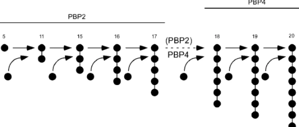
We suggest that protein interactions of PBP2 that occur via the N-terminal domain must be important for retaining PBP2 and/or other cell wall synthetic proteins in

Several of the 269 differen- tially expressed genes identified in this study encode for important proteins in oxidative pathways and liver metabo- lism and were expressed at

A total of 187 individual protein spots were detected, including spots of both resistant and susceptible strains, of which 58 proteins were found to be differentially expressed

In order to dissect the structural elements within the intrinsically disordered E7N domain, we recombinantly expressed or synthe- sized a series of proteins and peptide

Inclui os seguintes domínios: domínio físico (dor, energia e fadiga, atividade sexual, sono, descanso e funções sensoriais ótimas); domínio psicológico (sentimentos

Desta forma, diante da importância do tema para a saúde pública, delineou-se como objetivos do es- tudo identificar, entre pessoas com hipertensão arterial, os